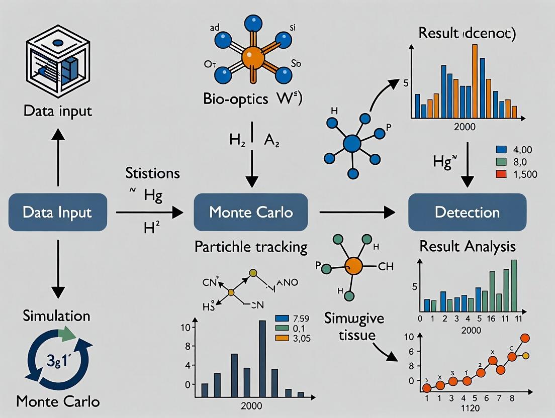
Simulating Reality: A Comprehensive Guide to Monte Carlo Methods for Modeling Particle Transport in Biological Tissues
This article provides a comprehensive guide for researchers and pharmaceutical professionals on applying Monte Carlo (MC) methods to simulate particle-tissue interactions.
Simulating Reality: A Comprehensive Guide to Monte Carlo Methods for Modeling Particle Transport in Biological Tissues
Abstract
This article provides a comprehensive guide for researchers and pharmaceutical professionals on applying Monte Carlo (MC) methods to simulate particle-tissue interactions. We cover the foundational physics, from photon and electron transport to complex radiation chemistry, and detail the implementation of popular MC codes like Geant4, PENELOPE, and MCNP in biomedical contexts. We address critical challenges in modeling tissue heterogeneity and achieving computational efficiency, and present frameworks for validating simulations against experimental benchmarks. By comparing major MC toolkits, this guide aims to equip scientists with the knowledge to accurately model radiation therapy, diagnostic imaging, and nanoparticle drug delivery.
The Physics of Particle-Tissue Interactions: Core Principles for Monte Carlo Simulation
The deterministic paradigm, governed by ordinary differential equations (ODEs), has long been the cornerstone of modeling biological systems. However, this framework frequently fails when applied to the intricate, mesoscopic scale of cellular processes within tissue. Within the context of advancing Monte Carlo methods for simulating particle interactions—such as drug molecules, signaling proteins, or radiation tracks—in tissue research, the inherent limitations of deterministic approaches become starkly apparent. This whitepaper argues that stochastic methods are not merely an alternative but an essential framework for capturing the fundamental behavior of complex biological systems where randomness is a feature, not noise.
The Limitation of Determinism in Biological Contexts
Deterministic models assume continuous concentrations and predictable rates, valid only when molecular populations are exceedingly high. In cellular and sub-cellular compartments, key regulators (e.g., transcription factors, regulatory RNAs) often exist in low copy numbers. A change of a few molecules can switch entire genetic programs. Furthermore, tissue heterogeneity ensures that even average behaviors are poor predictors of individual cellular outcomes, which is critical for understanding drug efficacy and toxicity.
Table 1: Comparison of Deterministic vs. Stochastic Modeling Outcomes for a Gene Regulatory Switch
| Aspect | Deterministic (ODE) Model | Stochastic (Gillespie) Model | Implication for Tissue Research |
|---|---|---|---|
| Bistable Switch Prediction | Predicts a precise, concentration-dependent switch point. | Reveals probabilistic switching and random transition times. | Explains heterogeneous cell fate decisions in a tissue. |
| Response to Low-Abundance Signal | Smooth, averaged response curve. | "All-or-nothing" stochastic bursts in individual cells. | Critical for modeling drug targeting of rare cell populations. |
| Extinction Events | Cannot simulate molecule count reaching zero. | Can accurately model molecular extinction. | Essential for simulating complete inhibitor efficacy or pathway blockade. |
The Stochastic Foundation: Master Equations and Monte Carlo
The rigorous foundation is the Chemical Master Equation (CME), which describes the time evolution of the probability distribution for all molecular species. As the CME is often analytically intractable for complex systems, stochastic simulation algorithms (SSA), a form of dynamic Monte Carlo, provide the numerical solution.
Key Experimental Protocol: Stochastic Simulation Algorithm (Gillespie's Direct Method)
- System Definition: Define the set of N molecular species and M reaction channels. Specify reaction rate constants (k_j) and the initial state vector (X(t=0)).
- Propensity Calculation: For the current state (X(t)), calculate the propensity function (aj) for each reaction (j), where (ajdt) is the probability the reaction will occur in the next infinitesimal time interval.
- Monte Carlo Step:
- Generate two random numbers (r1) and (r2) uniformly from (0,1).
- Calculate the time to next reaction: (\tau = (1/a{tot}) \ln(1/r1)), where (a{tot} = \sum{j=1}^{M} a_j).
- Determine the reaction index (\mu) such that (\sum{j=1}^{\mu-1} aj < r2 a{tot} \leq \sum{j=1}^{\mu} aj).
- Update: Update the system state: (t = t + \tau); (X = X + \nu\mu), where (\nu\mu) is the stoichiometric vector for reaction (\mu).
- Iterate: Return to Step 2 until a termination condition is met.
Application to Particle Interactions in Tissue: A Monte Carlo Paradigm
This stochastic philosophy directly extends to the Monte Carlo modeling of physical particles in tissue. The trajectory of each photon, electron, or drug molecule is treated as a random walk, with interactions (scattering, absorption, reaction) governed by probability cross-sections.
Title: Monte Carlo Particle Transport Workflow
Case Study: Stochasticity in EGFR Signaling and Drug Response
The Epidermal Growth Factor Receptor (EGFR) pathway exemplifies system complexity where stochastic methods are vital. Ligand binding, receptor dimerization, and trafficking events occur in low-copy numbers at the cell membrane, leading to significant cell-to-cell variability that influences tumor resistance to tyrosine kinase inhibitors (TKIs).
Title: Stochastic Events in the EGFR Signaling Pathway
Detailed Experimental Protocol: Simulating TKI Action with Stochastic Dynamics
- Model Construction: Define reaction network for EGFR: ligand association/dissociation, dimerization, phosphorylation, downstream adaptor binding, and recycling/degradation. Include TKI as a competitive binding reaction to the receptor kinase domain.
- Parameterization: Obtain kinetic rates from surface plasmon resonance (SPR) and fluorescence recovery after photobleaching (FRAP) data. Set initial conditions: ~10,000-100,000 surface EGFR molecules per cell, with variability.
- Stochastic Simulation: Use a hybrid SSA (e.g., tau-leaping) to simulate the network over 24 hours of simulated time, for 1000+ individual "cells."
- Perturbation: Introduce a pulse or constant concentration of TKI molecules, simulated as a distinct molecular species with its own diffusion and binding reactions.
- Output Analysis: Quantify the distribution of phosphorylated EGFR and downstream effector levels across the virtual cell population. Calculate the fraction of cells where signaling remains above a proliferation threshold despite treatment.
Table 2: Key Research Reagent Solutions for Stochastic Single-Cell Analysis
| Reagent / Material | Function in Stochastic Analysis |
|---|---|
| Fluorescent Reporters (FRET biosensors) | Enable live-cell, single-molecule imaging of protein activity (e.g., kinase activity, second messengers) to quantify stochastic fluctuations. |
| Microfluidic Cell Traps | Allow long-term, high-throughput imaging of individual cells under controlled perturbations for gathering statistical data on cell fate. |
| Single-Cell RNA-Seq Kits | Profile transcriptomic states of thousands of individual cells from a tissue to infer the underlying stochastic gene expression network. |
| Quantum Dots (QDs) | Photostable nanoparticles for tracking single receptor molecules over long durations on the live cell surface. |
| Stochastic Optical Reconstruction Microscopy (STORM) Dyes | Enable super-resolution imaging to visualize nanoscale spatial organization, a key modulator of stochastic reaction kinetics. |
The shift from deterministic to stochastic modeling is a fundamental necessity for meaningful research into particle interactions within tissue. Whether modeling the random walk of a radiation particle depositing energy or the probabilistic collision of a drug molecule with its target receptor, Monte Carlo and other stochastic methods embrace the inherent randomness that defines biological complexity at the cellular scale. This paradigm provides not just more realistic simulations, but also a framework to understand—and eventually predict—the heterogeneous outcomes observed in drug development and disease progression.
1. Introduction This whitepaper details the core particle types employed in biomedical applications, with a specific focus on their physical interactions with biological tissue. The analysis is framed within the context of advancing Monte Carlo (MC) simulation methodologies, which provide the gold standard for stochastically modeling these interactions to predict energy deposition, dose distribution, and subsequent biological effects. Accurate MC modeling is critical for therapy optimization, diagnostic imaging refinement, and fundamental radiobiological research.
2. Particle Characteristics and Interactions The key particles differ fundamentally in mass, charge, and their primary interaction mechanisms with matter, leading to distinct depth-dose profiles and biological impact.
Table 1: Fundamental Properties and Primary Interaction Mechanisms
| Particle | Rest Mass (MeV/c²) | Electric Charge | Primary Interaction Mechanisms in Tissue |
|---|---|---|---|
| Photon (X/Gamma-ray) | 0 | 0 | Compton Scattering, Photoelectric Effect, Pair Production |
| Electron | 0.511 | -1 | Collisional (Ionization/Excitation), Radiative (Bremsstrahlung) |
| Proton | 938.27 | +1 | Coulomb Scattering, Nuclear Interactions (at high energy) |
| Heavy Ion (e.g., C-12) | ~11178 (for C-12) | +Z | Coulomb Scattering, Dense Ionization Tracks, Nuclear Fragmentation |
Table 2: Key Biomedical Applications and Dose Distribution Features
| Particle | Major Applications | Key Dose Distribution Feature | Relative Biological Effectiveness (RBE) Range |
|---|---|---|---|
| Photon | Radiotherapy (IMRT, VMAT), CT/PET Imaging | Exponentially attenuating; exit dose | 1.0 (Reference) |
| Electron | Superficial Tumors, Intraoperative Radiotherapy | Rapid dose fall-off beyond target depth | 1.0 - 1.5 |
| Proton | Particle Therapy (e.g., ocular, pediatric tumors) | Bragg Peak; sharp distal fall-off | 1.0 - 1.1 (in clinical use) |
| Heavy Ion (C-12) | Particle Therapy (radioresistant tumors) | Inverse Depth Dose; sharpest peak | 2.0 - 5.0 (variable with LET) |
3. Monte Carlo Simulation of Particle Transport MC methods simulate individual particle histories through tissue, modeling stochastic interactions based on cross-section data. The general workflow for a particle-in-tissue MC code involves:
Experimental Protocol 1: Core Monte Carlo Simulation Cycle
- Geometry Definition: Model patient/tissue geometry (via CT voxels or mathematical phantoms).
- Source Definition: Initialize particle type, energy, and spatial/directional distribution.
- Step Length Calculation: Sample free path to next interaction point using total cross-section.
- Interaction Sampling: Determine interaction type (e.g., Compton vs. Photoelectric) probabilistically.
- Energy Deposition: Record energy deposited in local voxel from ionization/excitation.
- Secondary Particle Generation: Create and stack secondary particles (e.g., delta rays, characteristic X-rays).
- Particle Update: Adjust primary particle energy and direction post-interaction.
- Termination & History: Repeat steps 3-7 until particle energy falls below cutoff or exits geometry. Loop over millions of histories.
- Dose Scoring: Aggregate energy deposition per voxel, convert to dose (Gy).
Title: Monte Carlo Particle Transport Simulation Workflow
4. Biological Effectiveness and Experimental Modeling The variable biological effectiveness of particles is primarily quantified through cell survival assays, linked to the density of ionization events (Linear Energy Transfer, LET).
Experimental Protocol 2: Clonogenic Survival Assay for RBE Determination
- Cell Preparation: Seed mammalian cell lines (e.g., V79, H460) in culture flasks and grow to exponential phase.
- Irradiation: Trypsinize, count, and plate cells in known numbers. Irradiate plates with graded doses of reference (e.g., 250 kVp X-rays) and test particles (electrons, protons, ions). Include unirradiated controls. Use precise particle accelerators (cyclotron/synchrotron) with beam monitoring.
- Incubation: Place dishes in a 37°C, 5% CO₂ incubator for 10-14 days to allow colony formation.
- Staining & Counting: Fix colonies with methanol/acetic acid, stain with crystal violet. Count colonies (>50 cells).
- Data Analysis: Calculate surviving fraction (SF = colonies counted / (cells seeded × plating efficiency)). Fit SF vs. dose data with Linear-Quadratic (LQ) model: SF = exp(-αD - βD²). Calculate RBE at a specific SF (e.g., 10%) as RBE₁₀ = Dref / Dtest.
Title: Particle-Induced DNA Damage and Repair Pathway
Table 3: The Scientist's Toolkit - Key Reagents & Materials for Radiobiology Experiments
| Item | Function in Experiment |
|---|---|
| Dulbecco's Modified Eagle Medium (DMEM) | Cell culture medium providing nutrients for growth pre- and post-irradiation. |
| Fetal Bovine Serum (FBS) | Serum supplement for culture media, providing essential growth factors and proteins. |
| Trypsin-EDTA Solution | Proteolytic enzyme used to detach adherent cells for counting and re-plating. |
| Crystal Violet Stain | Dye used to fix and stain formed cell colonies for visualization and counting. |
| Linear-Quadratic Model Fitting Software (e.g., Origin, R) | For analyzing survival curve data to extract α, β parameters and calculate RBE. |
| Radiochromic Film / Ionization Chamber | For absolute dosimetry and beam profile verification prior to biological experiments. |
| Monte Carlo Code (e.g., TOPAS/GEANT4, FLUKA, MCNP) | To simulate detailed particle transport and predict physical dose/LET distributions in silico. |
5. Conclusion Photons, electrons, protons, and heavy ions form a versatile toolkit for biomedicine, each with unique physical and biological signatures. The continuous refinement of Monte Carlo methods, informed by precise experimental radiobiology protocols, is essential to fully exploit these differences. This synergy between simulation and experiment drives innovation in targeted radiotherapy and diagnostic imaging, ultimately aiming to improve therapeutic ratios and patient outcomes.
This technical guide details the three primary photon interaction mechanisms relevant to medical physics and therapeutic research: the photoelectric effect, Compton scattering, and pair production. Framed within the context of Monte Carlo simulation for modeling particle interactions in biological tissue, this whitepaper provides the foundational physics, quantitative data, experimental methodologies, and research tools necessary for accurate simulation in drug development and radiation therapy research.
In Monte Carlo simulations of radiation transport through tissue—a critical tool for radiotherapy planning, dosimetry, and radiopharmaceutical development—the accurate modeling of photon interactions is paramount. The dominant mechanisms by which photons deposit energy in tissue vary with photon energy and the atomic number (Z) of the absorbing material. This document provides an in-depth analysis of these core interactions to inform the development and validation of Monte Carlo codes like GEANT4, MCNP, and PENELOPE.
Core Interaction Mechanisms
Photoelectric Effect
The photoelectric effect describes the complete absorption of an incident photon by an atom. The photon ejects a bound electron (typically from an inner shell), with the photon's energy transferred to the electron as kinetic energy, minus the electron's binding energy. The resulting vacancy leads to characteristic X-ray emission or Auger electron ejection.
Key Dependencies: Cross section (probability) scales approximately as ~ Z⁴/Eᵧ³, making it dominant for low-energy photons and high-Z materials.
Compton Scattering
Compton scattering is the inelastic scattering of a photon by a loosely bound or free electron. The photon transfers part of its energy to the electron and is deflected with reduced energy. The relationship between the scattering angle (θ) and energy loss is given by the Klein-Nishina formula.
Key Dependencies: Cross section per electron is nearly independent of Z. Dominant in soft tissue for intermediate photon energies (~30 keV to 10 MeV).
Pair Production
Pair production occurs when a photon with energy exceeding 1.022 MeV (twice the rest mass of an electron) interacts with the strong electric field near a nucleus. The photon is converted into an electron-positron pair. Any excess photon energy above the threshold becomes kinetic energy of the created particles.
Key Dependencies: Cross section scales as ~ Z² and increases logarithmically with photon energy. Dominant at high energies (>5-10 MeV in tissue).
The following tables consolidate key quantitative parameters for these interactions, essential for Monte Carlo cross-section libraries.
Table 1: Dominant Interaction Regions by Photon Energy in Soft Tissue (Z_eff ≈ 7.5)
| Photon Energy Range | Dominant Interaction | Approximate Probability Fraction | Key Monte Carlo Consideration |
|---|---|---|---|
| < 30 keV | Photoelectric Effect | > 80% | Critical for imaging, low-dose regions |
| 30 keV - 5 MeV | Compton Scattering | > 70% | Main contributor to dose in radiotherapy |
| > 5 MeV | Pair Production | Increasing to > 50% | Significant in high-energy therapy beams |
Table 2: Key Cross-Section Formulas and Dependencies
| Interaction | Atomic Cross-Section (σ) Proportionality | Energy Threshold | Primary Secondary Particles |
|---|---|---|---|
| Photoelectric | ~ Zⁿ/Eᵧ³.5 (n≈4-5) | > Electron Binding Energy | Photoelectron, Characteristic X-ray, Auger e⁻ |
| Compton (per atom) | ~ Z * Klein-Nishina (per electron) | None (free electron) | Recoil Electron, Scattered Photon |
| Pair Production (nuclear field) | ~ Z² * f(Eᵧ) | 1.022 MeV | Electron-Positron Pair |
Table 3: Typical Mean Free Paths in Water (cm)
| Photon Energy | Photoelectric (λ_pe) | Compton (λ_c) | Pair Production (λ_pp) |
|---|---|---|---|
| 50 keV | ~4.0 | ~10.2 | — |
| 200 keV | ~25.1 | ~5.1 | — |
| 1 MeV | — | ~10.0 | ~40.0 |
| 10 MeV | — | ~24.0 | ~16.0 |
Experimental Protocols for Validation
Monte Carlo models require validation against empirical data. Below are summarized protocols for measuring interaction cross-sections.
Protocol: Measuring Compton Scattering Cross-Sections
Objective: To measure the differential cross-section for Compton scattering as a function of scattering angle. Materials: Monoenergetic gamma source (e.g., Cs-137, 662 keV), High-Purity Germanium (HPGe) detector, Collimators, Scatterer (low-Z thin foil), Precision goniometer, Multi-channel analyzer. Procedure:
- Mount the source and detector on the goniometer arms. Use collimators to define narrow beams.
- Place the thin scatterer at the center of the goniometer.
- Measure the primary beam intensity (I₀) without the scatterer.
- For angles θ from 10° to 150°, measure the energy and count rate (I_s) of the scattered photons.
- Correct for detector efficiency, air scattering, and absorption in the scatterer.
- Calculate the differential cross-section using the Klein-Nishina formula modified for experimental geometry and solid angle.
Protocol: Verifying Photoelectric Effect Z-Dependence
Objective: To validate the ~Z⁴ dependence of the photoelectric cross-section. Materials: Low-energy X-ray source (e.g., 59.5 keV from Am-241), Thin foils of known thickness and varying Z (e.g., Al, Cu, Sn, Pb), NaI(Tl) or HPGe detector. Procedure:
- Measure the incident photon flux (I₀) with no absorber.
- For each foil, measure the transmitted flux (I).
- Calculate the linear attenuation coefficient (μ) for each material from I = I₀ exp(-μt).
- Subtract the theoretically calculated Compton and Rayleigh contributions to isolate μ_photoelectric.
- Plot log(μ_pe) vs log(Z). The slope should be approximately 4.
Monte Carlo Integration & Workflow
The modeling of these interactions within a Monte Carlo simulation for tissue involves a structured workflow.
Diagram Title: Monte Carlo Photon Interaction Decision & Sub-Process Workflow
The Scientist's Toolkit: Research Reagent Solutions
Table 4: Essential Materials for Experimental Validation Studies
| Item / Reagent | Function in Experiment | Key Specification / Note |
|---|---|---|
| Monoenergetic Gamma/X-ray Sources (Am-241, Cs-137, Co-60) | Provide well-defined photon beams for cross-section measurement. | Sealed sources with known activity and emission probability. |
| High-Purity Germanium (HPGe) Detector | High-resolution spectroscopy to distinguish photopeaks, Compton edges, and annihilation peaks. | Requires liquid nitrogen cooling. Efficiency calibration essential. |
| Tissue-Equivalent Phantoms | Simulate human tissue (e.g., lung, muscle, bone) for dose deposition studies. | Defined by ICRU/ICRP compositions; can be solid, liquid, or gel. |
| Radiochromic Films (e.g., EBT3) | Measure 2D dose distributions from complex photon interactions in phantom. | Self-developing, near tissue-equivalent, high spatial resolution. |
| Monte Carlo Code Package (GEANT4, MCNP6, TOPAS) | Simulate stochastic photon interactions using physics models validated against this data. | Requires accurate physics list selection (e.g., G4EmLivermorePhysics for low-E). |
| NIST Standard Reference Materials (e.g., SRM for X-ray Attenuation) | Calibrate experimental setups and validate simulated attenuation coefficients. | Provides certified μ/ρ values for specific materials and energies. |
| Precision Collimators & Diaphragms | Define narrow photon beams to control scatter and improve angular resolution. | Often made from high-Z materials (e.g., tungsten) to absorb unwanted photons. |
Accurate modeling of tissue properties is a foundational pillar in the application of Monte Carlo (MC) methods for simulating particle transport in biological systems. Within the broader thesis on advancing MC techniques for medical physics and radiation therapy, this technical guide details the critical parameters of tissue density, elemental composition, and their resultant energy-dependent interaction cross-sections. These parameters directly govern the stochastic processes of energy deposition, scattering, and nuclear interactions that MC algorithms are designed to simulate, ultimately determining the accuracy of dose calculations in radiotherapy, radioprotection studies, and biomedical imaging.
Core Tissue Properties and Their Quantification
Density (ρ)
Tissue density, mass per unit volume (g/cm³), is the primary scaling factor for macroscopic cross-sections in particle transport. It is not homogeneous and varies significantly between and within tissues.
Elemental Composition
The stoichiometric composition of a tissue, defined by the mass or fraction of constituent elements (e.g., H, C, N, O, P, Ca), dictates its microscopic interaction probabilities. Modern reference data are derived from techniques like cryo-mass spectrometry and prompt-gamma neutron activation analysis.
Energy-Dependent Cross-Sections (σ(E))
The probability of a specific interaction (e.g., Compton scattering, photoelectric absorption, elastic scattering) between an incident particle and a target atom is quantified by its cross-section (barns/atom), which is a strong function of particle energy (E). For compound materials like tissue, the effective cross-section is computed as the weighted sum of elemental cross-sections.
Reference Data Tables
Table 1: Density and Elemental Composition of Reference Tissues (Mass Fractions)
| Tissue Type | Density (g/cm³) | H | C | N | O | Other |
|---|---|---|---|---|---|---|
| Skeletal Muscle (ICRP 110) | 1.05 | 0.102 | 0.123 | 0.035 | 0.729 | Na, P, S, K (0.011) |
| Adipose Tissue (ICRP 110) | 0.95 | 0.114 | 0.598 | 0.007 | 0.278 | Na, P, S (0.003) |
| Cortical Bone (ICRP 110) | 1.92 | 0.047 | 0.144 | 0.042 | 0.435 | P, Ca, Mg (0.332) |
| Lung (Inflated, ICRP 110) | 0.26 | 0.103 | 0.105 | 0.031 | 0.749 | Na, P, S, Cl (0.012) |
| Brain (White Matter) | 1.04 | 0.107 | 0.145 | 0.022 | 0.712 | P, S, Cl, K (0.014) |
Table 2: Photon Interaction Cross-Section Data for Water (Analog for Soft Tissue) at Key Energies
| Energy (MeV) | Photoelectric (barns/atom) | Compton (barns/atom) | Pair Production (barns/atom) | Total μ/ρ (cm²/g) |
|---|---|---|---|---|
| 0.01 | 3.86E+02 | 5.12 | 0.00 | 5.33 |
| 0.1 | 1.55E-01 | 3.81E-01 | 0.00 | 0.171 |
| 1 | 1.58E-03 | 6.15E-02 | 0.00 | 0.0706 |
| 10 | 1.41E-05 | 2.48E-02 | 1.34E-02 | 0.0221 |
Data sourced from NIST XCOM database.
Experimental Protocols for Parameter Determination
Protocol for Experimental Density Measurement via Pycnometry
Objective: To determine the bulk density of a small, irregular tissue sample. Materials: Helium pycnometer, microbalance, surgical tools, sample vials. Procedure:
- Calibrate the pycnometer cell volume using a standard sphere of known geometry and mass.
- Excise a tissue sample (~1 cm³), blot surface fluid, and immediately place in a sealed vial.
- Weigh the empty sample cup (m_cup).
- Transfer sample to cup and weigh (m_cup+sample).
- Place cup in pycnometer chamber. The instrument measures the pressure change of helium as it expands into the sample cell and then into a reference volume.
- The system software calculates the sample's solid volume (V_sample) by subtracting the displaced gas volume from the known cell volume.
- Calculate density: ρ = (mcup+sample - mcup) / V_sample.
- Perform in triplicate and average, reporting standard deviation.
Protocol for Elemental Composition via Inductively Coupled Plasma Mass Spectrometry (ICP-MS)
Objective: To quantify trace element concentrations in digested tissue samples. Materials: ICP-MS instrument, high-purity nitric acid, microwave digester, precision pipettes, certified elemental standards. Procedure:
- Digestion: Accurately weigh ~0.5g of lyophilized, homogenized tissue into a Teflon digestion vessel. Add 5 mL of concentrated HNO3. Digest using a microwave system with a ramped temperature program (e.g., to 180°C over 20 min, hold for 15 min).
- Dilution: After cooling, quantitatively transfer digestate to a 50 mL volumetric flask and dilute to mark with ultrapure water (18.2 MΩ·cm). A further 1:10 or 1:100 dilution may be required based on expected concentrations.
- Calibration: Prepare a series of calibration standards (e.g., 1, 10, 100, 1000 ppb) from multi-element stock solutions for target elements (P, S, Ca, Fe, Zn, etc.).
- Analysis: Introduce standards and samples into the ICP-MS via a peristaltic pump and nebulizer. The instrument atomizes and ionizes the sample; ions are separated by mass/charge ratio and counted.
- Quantification: Generate calibration curves (signal intensity vs. concentration) for each element. Use internal standards (e.g., Scandium-45) to correct for matrix effects and instrumental drift. Calculate elemental mass in original sample and convert to mass fraction.
Visualizations
Diagram 1: Data flow for MC particle transport.
Diagram 2: Workflow for determining tissue composition.
The Scientist's Toolkit: Research Reagent Solutions
Table 3: Essential Materials for Tissue Property Experiments
| Item | Function | Example/Note |
|---|---|---|
| Helium Pycnometer | Measures the true volume (and thus density) of porous or irregular solids by gas displacement. | AccuPyc II (Micromeritics); uses inert, non-adsorbing He gas. |
| Inductively Coupled Plasma Mass Spectrometer (ICP-MS) | Detects and quantifies trace elemental concentrations at parts-per-billion levels in digested solutions. | Agilent 7900 or PerkinElmer NexION; requires high-purity argon gas. |
| Microwave Digestion System | Rapidly and completely dissolves organic tissue matrices using controlled heat and pressure with acids. | CEM Mars 6 or Milestone Ethos UP; uses Teflon vessels. |
| High-Purity Nitric Acid (TraceMetal Grade) | Primary digestion acid for ICP-MS; oxidizes organic matter and keeps elements in solution. | Fisher Optima Grade or Sigma-Aldpur; minimizes background contamination. |
| Certified Multi-Element Standard Solutions | Used to calibrate the ICP-MS for accurate quantification across the periodic table. | Inorganic Ventures; supplied with certificates of analysis for concentration. |
| Cryomill/Homogenizer | Pulverizes and homogenizes frozen tissue to a fine powder for representative sub-sampling. | SPEX SamplePrep 6870 Freezer/Mill; uses liquid nitrogen to prevent degradation. |
| NIST Standard Reference Material (SRM) | Certified tissue (e.g., SRM 1577c Bovine Liver) used for quality control and method validation. | Provides benchmark values for composition to assess analytical accuracy. |
| Monte Carlo Code with Tissue Libraries | Software that implements particle transport using cross-section databases and tissue parameters. | GEANT4, MCNP, FLUKA, EGSnrc; require correctly formatted input files. |
This whitepaper serves as a core technical guide within a broader thesis investigating Monte Carlo (MC) simulations for modeling particle-tissue interactions. The central challenge in predictive radiobiology and targeted drug development lies in accurately translating macroscopic absorbed dose (Gy) into microscopic spatial patterns of energy deposition. This document bridges that gap by detailing the physical principles of Track Structure and Linear Energy Transfer (LET), which are fundamental inputs for advanced MC codes like Geant4-DNA, TOPAS-nBio, and FLUKA. For researchers in radiation oncology and pharmaceutical development, mastering these concepts is critical for predicting biological outcomes, from DNA lesion complexity to the efficacy of radiopharmaceuticals.
Core Concepts: From Dose to Discrete Events
Macroscopic Dose: Absorbed dose is an average energy deposition per unit mass, a bulk quantity that fails to describe the stochastic, heterogeneous nature of particle interactions at cellular and sub-cellular scales.
Microscopic Track Structure: This refers to the detailed, stochastic spatial distribution of inelastic interactions (ionizations and excitations) along and around the path of a single charged particle. It is the explicit output of track-structure MC simulations.
Linear Energy Transfer (LET): Defined as the average energy locally imparted to the medium per unit track length by a charged particle (keV/µm). LET serves as a crucial, albeit simplified, descriptor of radiation quality, correlating with the density of ionizations along a track.
- LET∞ (Unrestricted): Considers all energy transfers.
- LETΔ (Restricted): Considers only energy transfers below a cutoff Δ, excluding high-energy δ-rays, providing a more localized measure.
The Monte Carlo Link: Track-structure MC methods simulate individual interaction cross-sections to build up a stochastic picture of energy deposition, explicitly modeling secondary electron (δ-ray) spectra. LET is a derived statistical quantity from these simulations.
Quantitative Data on Radiation Qualities
Table 1: Characteristic LET and Track Structure Parameters for Common Radiation Types
| Radiation Type | Particle & Energy | Typical LET∞ (keV/µm) in Water | Approx. Mean Interaction Spacing (nm) | Primary Track Width (nm, core) | Key Biological Implication |
|---|---|---|---|---|---|
| Photons / Electrons (Low-Energy) | 250 keV e⁻ | ~0.3 | ~1000 | Diffuse, wide | Sparse, isolated lesions; high repairability. |
| Protons | 150 MeV (Therapeutic) | ~0.5 | ~600 | 1-10 | Moderately dense tracks; pronounced Bragg peak. |
| Carbon Ions | 270 MeV/u (Therapeutic) | ~15 | ~20 | ~10 | Very dense core; complex clustered damage. |
| Alpha Particles | 5 MeV (from Rn decay) | ~90 | < 10 | < 0.1 | Extremely dense, short range; high RBE. |
| Neutrons (Fast) | 1 MeV (indirect) | Spectrum via recoil protons | Variable | Variable | Mixed field; produces proton tracks of varying LET. |
Table 2: Key MC Codes for Track Structure Simulation
| Code Name | Primary Application | Scale Modeled | Key Strength | Typical Input/Output |
|---|---|---|---|---|
| Geant4-DNA | Nano-/micro-dosimetry | Physics & Chemistry (ps-ns) | Open-source, detailed processes. | Particle type/energy → Interaction points, species yields. |
| TOPAS-nBio | Radiobiology extension | Physics to Biology (ps-hours) | User-friendly TOPAS interface. | Particle track → DNA damage score, cell survival. |
| PARTRAC | DNA damage modeling | Physics to Chromatin (ps-min) | Integrated DNA structure model. | Radiation field → DSB yield and complexity. |
| FLUKA | Mixed-field dosimetry | Macroscopic to microscopic | High-energy to NMRC coupling. | Complex field → Dose, LET spectra. |
Experimental Protocols for Validation
Validating MC-predicted track structure and LET requires correlating simulation with physical and biological experiments.
Protocol 1: Nanodosimetry with Track-Etch Detectors
- Objective: Measure the microscopic pattern of ionizations.
- Materials: CR-39 (allyl diglycol carbonate) plastic, etching solution (NaOH/KOH), microscope.
- Method:
- Irradiate CR-39 detector with charged particles of known type/energy.
- Chemically etch the plastic. Material is removed faster along the radiation-damaged latent track.
- Under a microscope, measure the resulting etch-pit geometry (diameter, length).
- Correlate pit morphology (conical shape for high-LET) with simulated ionization density and LET.
Protocol 2: Determining Relative Biological Effectiveness (RBE)
- Objective: Link LET to biological outcome, validating biological MC models.
- Materials: Cell line (e.g., V79, HSG), clonogenic assay reagents, particle irradiation facility.
- Method:
- Irradiate cell monolayers with reference radiation (e.g., 250 kVp X-rays) and test particles (e.g., protons, C-ions) across a dose range.
- For each beam, perform a clonogenic survival assay: trypsinize, plate at low density, incubate for 1-2 weeks, fix/stain colonies.
- Plot survival curves (log(SF) vs. dose). Calculate RBE at a specific survival level (e.g., SF=0.1): RBE = Dref / Dtest.
- Plot experimental RBE vs. MC-simulated LET to establish the relationship for the model system.
Protocol 3: Microscopic Imaging of DNA Damage Foci
- Objective: Visualize spatial correlation of damage with particle traversal.
- Materials: Immunofluorescence-labeled cells (γ-H2AX, 53BP1 antibodies), confocal microscope, particle microbeam.
- Method:
- Irradiate cell nuclei precisely using a particle microbeam (e.g., 1-10 α-particles/nucleus).
- Fix, permeabilize, and immuno-stain for DNA double-strand break (DSB) markers.
- Image with confocal microscopy. Analyze linear clusters (foci) along simulated particle tracks.
- Quantify foci density and size, comparing to predicted ionization clusters from track-structure simulations.
The Scientist's Toolkit: Key Reagents & Materials
Table 3: Essential Research Reagents and Solutions
| Item/Category | Example Product/Specification | Primary Function in Research Context |
|---|---|---|
| Track-Etch Material | CR-39 Plastic Sheets | Records latent particle tracks for visualization and nanodosimetric measurement. |
| Cell Culture for RBE | V79 (Chinese Hamster Lung) Cells | Standardized, high-plating-efficiency cell line for clonogenic survival assays. |
| DNA Damage Stain | Anti-γ-H2AX (Phospho-S139) Antibody | Immunofluorescence marker for microscopic visualization of DNA double-strand breaks. |
| Monte Carlo Code | Geant4-DNA Toolkit | Open-source software for simulating particle track structure in liquid water. |
| Microbeam System | Particle Microbeam (e.g., SNAKE, GSI) | Allows targeted irradiation of single cells or sub-cellular compartments for precise correlation. |
| LET Spectrometer | Silicon Semiconductor Detector (e.g., ΔE-E telescope) | Measures energy loss of individual particles to derive experimental LET spectra. |
| Biological Target Model | DNA Geometry Packages (e.g., PARTRAC Nucleosome Model) | Provides structural data (atomic coordinates) for MC simulation of direct DNA damage. |
Visualizing Concepts and Workflows
Title: Relationship Between Dose, Track Structure, LET, and Biology
Title: Monte Carlo Track Structure Simulation Workflow
Implementing Monte Carlo Simulations: Codes, Workflows, and Biomedical Use Cases
Within the broader thesis on Monte Carlo (MC) methods for simulating particle interactions in biological tissue, the selection of an appropriate simulation toolkit is foundational. This guide provides an in-depth technical comparison of four major, general-purpose MC codes: Geant4, MCNP, PENELOPE/PRIMO, and TOPAS. These toolkits are indispensable for research in radiation therapy, radiobiology, medical imaging, and drug development, enabling the precise tracking of particle transport and energy deposition at macroscopic to microscopic scales.
Core Toolkit Architectures and Methodologies
Geant4
Geant4 (Geometry and Tracking) is an open-source C++ toolkit developed and maintained by a worldwide collaboration. Its object-oriented architecture provides unparalleled flexibility. Users build their simulation by composing geometry, defining physics processes, and creating particle sources from modular components. It offers a vast library of physics models covering electromagnetic and hadronic interactions from eV to TeV energies, including specialized packages for low-energy physics (Livermore, PENELOPE) and optical photon transport.
MCNP
MCNP (Monte Carlo N-Particle), developed at Los Alamos National Laboratory, is a legacy code written in FORTRAN. It uses a continuous-energy generalized geometry system. Its primary strengths are its mature, validated nuclear data libraries for neutrons, photons, and electrons, and its efficient transport algorithms. It is widely used for radiation shielding, criticality safety, and reactor physics, with growing applications in medical physics via its MNCPX and MCNP6 variants.
PENELOPE/PRIMO
PENELOPE (Penetration and Energy Loss of Positrons and Electrons) is a FORTRAN/C++ code algorithm and physics model designed for simulating coupled electron-photon transport in the 50 eV to 1 GeV range with high accuracy. It uses a mixed simulation scheme, classifying steps as "hard" or "soft" for computational efficiency. PRIMO is a specialized, user-friendly software that incorporates the PENELOPE engine within a graphical interface, pre-configured for clinical linear accelerator simulation and voxelized patient dose calculation.
TOPAS
TOPAS (TOol for PArticle Simulation) is an open-source extension layered atop Geant4. Written in C++, it provides a scripting interface (using a custom parameter system) that abstracts much of the Geant4 coding complexity. It is specifically designed for translational research in particle therapy and medical physics, offering built-in components for beam lines, patients, and scoring. It combines Geant4's power with significantly reduced development time for complex simulations.
Quantitative Comparison of Toolkit Capabilities
Table 1: Core Characteristics and Technical Specifications
| Feature | Geant4 | MCNP6 | PENELOPE/PRIMO | TOPAS |
|---|---|---|---|---|
| Primary Dev. Language | C++ | FORTRAN | FORTRAN/C++ | C++ (Geant4 wrapper) |
| License & Cost | Open Source (Free) | Proprietary (Paid) | PENELOPE: Free / PRIMO: Free | Open Source (Free) |
| Primary Particle Types | e-/e+, γ, p, n, ions, μ, π, etc. | n, γ, e- (primary focus) | e-, e+, γ | All Geant4 particles |
| Typical Energy Range | eV – TeV | Thermal – GeV | 50 eV – 1 GeV | eV – TeV (inherited) |
| Key Strength | Flexibility, breadth of physics | Validated nuclear data, neutronics | Accuracy in e-/γ transport | Ease of use in medical physics |
| Typical Application | HEP, space, medical physics | Shielding, reactors, detectors | Radiotherapy dose calculation | Particle therapy, translational research |
| Learning Curve | Very Steep | Steep | Moderate (PRIMO) / Steep (PENELOPE) | Moderate for medical physics |
Table 2: Performance and Usability in Tissue Research Context
| Aspect | Geant4 | MCNP6 | PENELOPE/PRIMO | TOPAS |
|---|---|---|---|---|
| Voxelized Geometry | Yes (via GDCM, DICOM) | Yes (lattice/universe system) | Yes (in PRIMO) | Yes (native, optimized) |
| DNA/Damage Scoring | Yes (via Geant4-DNA) | Limited | Possible with customization | Yes (via extensions) |
| Pre-built Medical Beam Lines | No (must be coded) | No | Yes (in PRIMO for linacs) | Yes (extensive library) |
| Validation in Medical Physics | Extensive, ongoing | Strong for neutron/photon | Excellent for kV/MV beams | Extensive for proton therapy |
| User Interface | Code/script | Text input file | GUI (PRIMO) / Text input | Text parameter files |
Detailed Experimental Protocol: Benchmarking Dose Deposition in a Water Phantom
A critical step in any MC study for tissue research is validating the toolkit's output against measured or benchmark data. Below is a generalized protocol for benchmarking absorbed dose in a water phantom.
1. Objective: To validate the electromagnetic physics models of a chosen MC toolkit by simulating depth-dose curves in a water phantom and comparing to trusted reference data (e.g., IAEA TRS-398).
2. Materials & Software:
- MC Toolkit (Geant4, MCNP, PRIMO, or TOPAS installation).
- Reference beam data for a 6 MV photon beam or a 150 MeV proton beam.
- Computing cluster or high-performance workstation.
3. Methodology:
- Geometry Definition: Construct a cubic water phantom (e.g., 40x40x40 cm³). Define a scoring plane along the central axis for depth-dose measurement.
- Source Definition: Model a clinical radiotherapy source.
- For Photons: Define a phase-space file or a virtual linac head above the phantom.
- For Protons: Define a monoenergetic pencil beam with appropriate energy spread and lateral profile.
- Physics List Selection: Activate the relevant electromagnetic physics processes. For low-energy electrons in water, ensure models like "Livermore" or "PENELOPE" (in Geant4/TOPAS) or appropriate cross-section libraries (in MCNP) are selected.
- Scoring Setup: Implement a "dose scorer" to record energy deposited per unit mass in thin slabs (e.g., 1 mm or 2 mm thick) along the beam axis.
- Simulation Execution: Run a sufficient number of primary particle histories to achieve a statistical uncertainty of <0.5% in the high-dose region.
- Data Analysis: Normalize the simulated depth-dose curve to its maximum value. Calculate the percentage difference at key points (e.g., depth of maximum dose, R50) against the reference data. Use gamma-index analysis (e.g., 2%/2mm criteria) for a composite evaluation.
4. Expected Output: A depth-dose curve (e.g., Bragg peak for protons, exponential fall-off for photons) that aligns with reference data within accepted tolerances, confirming the accuracy of the toolkit's physics models for water (a tissue surrogate).
Logical Workflow for Toolkit Selection in Tissue Research
Title: Decision Workflow for Selecting a Monte Carlo Toolkit
The Scientist's Toolkit: Key Research Reagent Solutions
Table 3: Essential Computational & Data Resources for MC Tissue Research
| Item | Function in Research |
|---|---|
| High-Performance Computing (HPC) Cluster | Enables parallel processing of billions of particle histories in a feasible time, essential for low-uncertainty results in complex geometries. |
| Anatomically Realistic Voxel Phantom (e.g., ICRP 110) | Digital models of human anatomy derived from CT/MRI, used as simulation geometry to estimate organ doses and study radiation effects in specific tissues. |
| Reference Clinical Beam Data | Benchmark datasets (depth-dose, profiles) for standard accelerator beams, required for validating and tuning the MC simulation's source model. |
| Tissue Composition Database (e.g., ICRU 44) | Tables of elemental mass fractions and densities for various tissues (muscle, bone, lung, etc.), critical for defining material properties in the simulation. |
| Phase-Space File | A pre-recorded file containing the state (energy, position, direction) of particles crossing a plane, used as a validated source to save computation time. |
| DICOM RT Suite | Standard medical images (CT) and structure sets, used to import patient-specific geometries and contours for treatment planning studies. |
| Statistical Analysis Package (e.g., Python SciPy, R) | Software for post-processing simulation output: statistical comparison, gamma-index analysis, curve fitting, and data visualization. |
| Cross-Section Library (e.g., ENDF, EPDL97) | Comprehensive databases of particle interaction probabilities with different elements, the fundamental data driving the MC physics. |
In the context of a broader thesis on Monte Carlo methods for modeling particle interactions (e.g., photons, electrons, protons) in biological tissue, geometric phantoms serve as the foundational digital representation of anatomy. Accurate simulation of radiation dose deposition, light propagation in tissues, or radiopharmaceutical biodistribution depends fundamentally on the quality of this anatomical model. The choice between voxelized and parameterized phantoms directly impacts the accuracy, computational efficiency, and flexibility of the Monte Carlo simulation.
Core Model Architectures: Definitions and Technical Foundations
Voxelized Phantoms are derived from segmented medical imaging data (CT, MRI). The anatomy is discretized into a three-dimensional grid of volume elements (voxels), each assigned a specific tissue or material index. This creates a highly realistic, non-uniform model that precisely mirrors the scanned anatomy.
Parameterized (or Mathematical) Phantoms use simple geometric primitives (e.g., ellipsoids, cylinders, cones) and analytical formulas to describe anatomical boundaries. Organs and structures are defined by equations with adjustable parameters (center coordinates, radii, angles), offering a smooth, continuous representation.
Quantitative Comparison of Model Characteristics
The following table summarizes the core quantitative and qualitative differences critical for Monte Carlo applications in tissue research.
Table 1: Comparison of Phantom Model Characteristics for Monte Carlo Simulation
| Characteristic | Voxelized Phantom | Parameterized Phantom |
|---|---|---|
| Anatomical Basis | Direct segmentation of CT/MRI data; patient-specific. | Based on reference anatomical data (e.g., ICRP publications); population-averaged. |
| Spatial Representation | Discrete, stair-stepped boundaries at high resolution. | Continuous, smooth surfaces defined by equations. |
| Model Flexibility | Low; morphology is fixed to the source image. | High; organ size, shape, and position can be altered via parameters. |
| Computational Memory | High (scales with resolution, e.g., 512³ voxels). | Very Low (stores only equation coefficients). |
| Monte Carlo Navigation | Complex; requires boundary-crossing logic per voxel. | Simple; direct ray-geometry intersection calculations. |
| Typical Use Case | Patient-specific dosimetry, validation studies. | Protocol development, comparative studies, investigating anatomical variability. |
| Common Formats | DICOM, RAW matrix. | BREP (Boundary Representation), CAD scripts, PHITS/EGS++ native formats. |
Experimental Protocols for Phantom Implementation & Evaluation
Protocol 1: Constructing a Voxelized Phantom from Clinical CT
- Image Acquisition: Obtain a high-resolution (≤1 mm slice thickness) DICOM CT series. Ensure the scan covers the entire volume of interest.
- Segmentation: Use a software toolkit (e.g., 3D Slicer, ITK-Snap) to manually or semi-automatically label voxels according to tissue type. Assign a unique integer index to each tissue (e.g., 1=lung, 2=bone, 3=soft tissue).
- Material Assignment: Create a correspondence table linking each tissue index to specific material properties (density, elemental composition) relevant to your Monte Carlo code (e.g., Geant4, MCNP, GATE).
- Grid Export: Convert the segmented label map into a binary or ASCII file format readable by your Monte Carlo engine, preserving the 3D matrix dimensions and voxel resolution.
Protocol 2: Implementing a Parameterized Phantom (e.g., Ellipsoidal Torso)
- Reference Definition: Define a world coordinate origin (e.g., center of the body). Reference standard anatomical dimensions (e.g., from ICRP Publication 110).
- Organ Modeling: For each organ, define its bounding surface using a combination of primitives. Example (Liver as ellipsoid):
- Equation:
(x-x0)²/a² + (y-y0)²/b² + (z-z0)²/c² = 1 - Parameters:
(x0, y0, z0)= center coordinates;(a, b, c)= semi-axis lengths.
- Equation:
- Hierarchical Placement: Define organs relative to body landmarks. Nesting (e.g., heart inside lung cavity) can be managed via Boolean operations (union, intersection, subtraction).
- Code Integration: Implement the geometry directly within the Monte Carlo code's geometry constructor or use a dedicated scripting language (e.g., GDML for Geant4). Ensure each organ volume is assigned its correct material properties.
Protocol 3: Comparative Dosimetry Experiment
- Simulation Setup: Model the same radiation source (e.g., a 6 MV photon beam point source or a distributed radionuclide) in two identical Monte Carlo codes differing only in phantom geometry (voxelized vs. parameterized of equivalent scale).
- Dose Scoring: Define a consistent 3D dose scoring mesh (voxelized tally) encompassing the target region in both simulations.
- Execution: Run simulations with a sufficient number of particle histories (e.g., 10⁸) to achieve a statistical uncertainty of <1% in regions of interest.
- Analysis: Calculate dose-volume histograms (DVHs) for critical organs. Compute global metrics like the 3D gamma index (e.g., 2%/2mm criteria) to quantify the spatial agreement of dose distributions between the two phantom models.
Visualization of Phantom Construction Workflows
Diagram Title: Voxelized Phantom Construction Pipeline
Diagram Title: Parameterized Phantom Modeling Workflow
The Scientist's Toolkit: Research Reagent Solutions
Table 2: Essential Software & Data Resources for Phantom Development
| Tool/Resource | Category | Primary Function in Phantom Development |
|---|---|---|
| 3D Slicer | Open-Source Software | Platform for medical image segmentation and 3D model generation from DICOM data. |
| ITK-SNAP | Open-Source Software | Specialized tool for semi-automatic segmentation of anatomical structures in 3D. |
| ICRP Publication 110 | Reference Data | Provides reference adult male and female voxel phantom datasets and tissue compositions. |
| GEANT4 | Monte Carlo Toolkit | Provides flexible geometry packages (CSG, BREP) for implementing both phantom types directly in C++ code. |
| GATE/OpenGATE | Monte Carlo Platform | Built on GEANT4, includes specialized features for voxelized import and patient-specific dosimetry. |
| MCNP / PHITS | Monte Carlo Code | Supports lattice and universe structures for voxelized phantoms and native combinatorial geometry for parameterized models. |
| Python (NumPy, PyVista) | Programming Library | For scripting custom phantom creation, manipulating voxel arrays, and converting between file formats. |
| NRRD/NIfTI Format | File Format | Common, standardized formats for storing and exchanging labeled voxel phantom data. |
Defining Source Characteristics for Radiotherapy and Imaging Beams
Accurate definition of radiation source characteristics is the foundational step for any Monte Carlo (MC) simulation of particle interactions in tissue. Within the broader thesis on advancing MC methods for biomedical applications, this guide details the technical specifications and experimental protocols required to define primary beams for radiotherapy (e.g., megavoltage photons/electrons, protons) and medical imaging (e.g., kV x-rays, CT). The fidelity of downstream dose deposition, image contrast, and secondary particle generation models is directly contingent on this initial source characterization.
The essential parameters for beam definition vary by modality. The data below, synthesized from current clinical and research literature, must be incorporated into the MC simulation's source model.
Table 1: Key Characteristics for Radiotherapy Beams
| Beam Type | Typical Energy Spectrum | Focal Spot Size | Angular Divergence/Scanning Pattern | Dose Rate | Key Contaminants |
|---|---|---|---|---|---|
| Linac MV Photons | Bremsstrahlung spectrum (e.g., 6 MV: max ~6 MeV, mean ~1.5-2 MeV) | 1-3 mm (FWHM) | Defined by primary collimator & flattening filter | 100-2400 MU/min | Electron, photon scatter from flattening filter |
| Linac Electrons | Quasi-monoenergetic (e.g., 6, 9, 12, 18 MeV) with low-energy tail | 1-3 mm (FWHM) | Scattered by scattering foils | 100-2400 MU/min | Bremsstrahlung photons |
| Proton Pencil Beam | Spread-Out Bragg Peak (SOBP) via energy stacking (~70-250 MeV) | 3-10 mm σ (in air) | Magnetically scanned across target volume | ~2-10 Gy/min | Secondary electrons, neutrons |
| MV-IMRT/VMAT | As above, with intensity modulation | As above | Dynamic MLC sequence | As above | As above |
Table 2: Key Characteristics for Imaging Beams
| Beam Type | Typical Energy Spectrum | Focal Spot Size | Source-Detector Distance (SDD) | Filtration | Half-Value Layer (HVL) |
|---|---|---|---|---|---|
| Diagnostic X-ray (kV) | Polychromatic (e.g., 50-140 kVp) | 0.6-1.2 mm | 100-180 cm | 2.5-4 mm Al eq. | 2.5-5 mm Al |
| Cone-Beam CT (CBCT) | Polychromatic (80-140 kVp) | 0.3-0.8 mm | ~150 cm | Bowtie filter + Cu/Al | Specific to system |
| Micro-CT/Preclinical | 20-100 kVp | <50 µm | 10-50 cm | Optional Be, Al, Cu | <1 mm Al |
Experimental Protocols for Source Characterization
To populate the MC source model with the data from Tables 1 & 2, the following experimental methodologies are employed.
Protocol 1: Energy Spectrum Measurement for kV X-rays using a Cadmium Telluride (CdTe) Spectrometer
- Equipment Setup: Place a high-resolution CdTe spectrometer (e.g., Amptek XR-100CdTe) on the beam central axis at a known distance from the focal spot. Use a collimator to limit the beam to the active detector area.
- Calibration: Energy calibrate the spectrometer using known radioactive sources (e.g., Am-241, Co-57).
- Data Acquisition: Acquire pulse-height spectra for the x-ray tube at multiple kVp settings (e.g., 50, 80, 120 kVp) and tube currents. Ensure count rates are within the linear range of the detector to avoid pile-up.
- Correction & Deconvolution: Correct the raw spectrum for detector effects (e.g., charge trapping, escape peaks) using manufacturer-provided software or a deconvolution algorithm (e.g., iterative Lucy-Richardson) based on the detector response function.
- HVL Validation: Use the derived spectrum to calculate the Half-Value Layer (HVL) computationally and validate it against a physical measurement using aluminum filters and an ionization chamber.
Protocol 2: Focal Spot Size Measurement using the Pin-Hole Camera Technique
- Pinhole Fabrication: Utilize a tungsten or platinum disk with a laser-drilled pinhole of known, precise diameter (typically 10-30 µm). The pinhole diameter should be ≤ 1/3 of the expected focal spot size.
- Imaging Geometry: Position the pinhole between the x-ray source and a high-resolution digital detector (e.g., CCD-based imager with a scintillator). The magnification (M) is given by M = (a+b)/a, where 'a' is the source-to-pinhole distance and 'b' is the pinhole-to-detector distance.
- Image Acquisition: Acquire a short, high-exposure image of the focal spot. Ensure the pinhole is accurately aligned to the beam central axis.
- Analysis: Measure the full width at half maximum (FWHM) of the magnified spot image on the detector. Calculate the true focal spot size: Focal Spot FWHM = (Image FWHM) / M.
Protocol 3: Proton Pencil Beam Characterization in Air
- Setup: In an air-scanning tank, position a large-area parallel-plate ionization chamber at isocenter (typically 100 cm from source) to measure integrated charge per spot. Simultaneously, position a pixelated scintillation detector (e.g., Lynx, IBA) downstream to acquire 2D lateral dose profiles.
- Spot Scanning: For each nominal beam energy in the clinical range, instruct the accelerator to deliver a single spot at isocenter.
- Profile Measurement: Record the 2D dose profile. Extract the in-air sigma (σair) in both X and Y directions by fitting a single Gaussian function to the central region of each profile.
- Energy Calibration: Relate the nominal energy to the measured practical range in water, establishing the energy calibration curve for the MC source model.
Visualization of Methodological Relationships
Beam Characterization Workflow for MC
The Scientist's Toolkit: Research Reagent Solutions
Table 3: Essential Materials for Source Characterization Experiments
| Item / Reagent Solution | Function in Characterization |
|---|---|
| High-Purity CdTe or Si (Li) Spectrometer | Direct measurement of photon energy spectra; essential for kV and MV spectral definition. |
| Precision Pin-Hole Apertures (Tungsten, 10-30 µm) | Creates a magnified image of the focal spot for size measurement via pin-hole camera technique. |
| High-Resolution Digital Imager (CCD/CMOS + Scintillator) | Detects the magnified focal spot image or high-resolution beam profiles. |
| Parallel-Plate Ionization Chamber (e.g., Markus type) | Measures integrated dose for proton/carbon beam spots or for HVL measurements. |
| Pixelated Scintillation Detector Array (e.g., Lynx) | Provides fast, high-resolution 2D profiles of scanning particle beams (protons, ions). |
| Step Wedge & Solid Water Phantoms | Used for HVL measurement and beam profile/depth-dose validation in water-equivalent media. |
| Radioactive Calibration Sources (Am-241, Co-57) | Provides known emission lines for precise energy calibration of spectroscopic detectors. |
| Monte Carlo Code (e.g., Geant4, TOPAS, MCNP, EGSnrc) | Platform for implementing the characterized source model and simulating particle transport. |
This whitepaper details the critical application of Monte Carlo (MC) methods within the broader research thesis investigating stochastic simulations of particle interactions in biological tissue. The accurate calculation of dose deposition from external photon and electron beams is a cornerstone of modern, precise radiotherapy. MC techniques, by explicitly simulating the random nature of particle transport and energy loss, provide the most accurate method for modeling these complex interactions within heterogeneous human anatomy, serving as a gold standard against which faster, deterministic algorithms are benchmarked.
Core Principles of Monte Carlo Dose Calculation
The fundamental process involves tracking millions of individual primary and secondary particles (photons, electrons, positrons) through a patient geometry derived from CT data. Key interactions simulated include:
- Photoelectric Effect: Complete absorption of photon, ejecting an electron.
- Compton Scattering: Inelastic scattering of a photon by a loosely bound electron.
- Pair Production: Photon conversion into an electron-positron pair near a nucleus.
- Electron Collisions: Continuous and discrete energy loss via ionization and excitation.
- Electron Bremsstrahlung: Radiation emission due to electron deflection by a nucleus.
The probability of each interaction is sampled from known cross-sections, making the simulation intrinsically linked to tissue composition and density.
Quantitative Data on Algorithm Performance
Table 1: Comparison of Dose Calculation Algorithm Accuracy in Heterogeneous Media
| Algorithm Type | Principle | Computation Speed | Dosimetric Accuracy in Heterogeneity (vs. Measurement) | Key Limitation |
|---|---|---|---|---|
| Monte Carlo (e.g., EGSnrc, Geant4, PENELOPE) | Stochastic simulation of particle tracks | Very Slow (Hours) | High ( ~1-2% deviation) | Prohibitive computational cost for routine planning |
| Collapsed Cone Convolution/Superposition | Pre-calculated energy deposition kernels | Medium (Minutes) | Medium (~2-4% deviation) | Kernels approximated for heterogeneity |
| Pencil Beam Convolution | Simplified 1D kernels along ray lines | Fast (Seconds) | Low (>5% deviation in lung/bone) | Fails in severe electronic disequilibrium |
| Analytical Anisotropic Algorithm (AAA) | Modeling of photon scatter | Fast-Medium (Minutes) | Medium (~2-3% deviation) | Empirical scaling of pencil beams |
Table 2: Example MC Simulation Parameters for a 6 MV Photon Beam
| Parameter | Typical Value/Range | Impact on Calculation |
|---|---|---|
| Number of Histories (Particles) | 10^7 - 10^9 | Statistical uncertainty ∝ 1/√(N). Higher N reduces noise. |
| Energy Cutoff (ECUT, PCUT) | ECUT: 0.7 MeV (e-), PCUT: 0.01 MeV (γ) | Particles below cutoff energy deposit local dose. Affects speed/accuracy. |
| Voxel Size (in patient CT) | 2.0 x 2.0 x 2.0 mm^3 | Finer resolution increases geometry detail and computation time. |
| Variance Reduction Techniques | Particle splitting, Russian Roulette | Greatly increases efficiency but requires careful implementation. |
Experimental Protocol: MC Commissioning and Validation
Protocol 1: Beam Modeling and Commissioning for a Linac
- Data Acquisition: Measure percent depth dose (PDD) and profiles (in-plane, cross-plane, diagonal) for a clinical linear accelerator (e.g., Varian TrueBeam) using a water phantom and ionization chambers for multiple field sizes (e.g., 3x3 cm² to 40x40 cm²) and energies (e.g., 6 MV, 10 MV).
- Geometry Definition: Accurately model the Linac head (target, primary collimator, flattening filter, jaws, multi-leaf collimators) in the MC code (e.g., BEAMnrc component module).
- Source Parameterization: Iteratively adjust the initial electron beam parameters (energy, spot size) incident on the target until the simulated PDD and profiles match measured data within criteria (e.g., 1% / 1mm in high gradient regions).
- Phase-Space File Generation: Store the position, direction, energy, and particle type of particles exiting the Linac head for reuse in patient simulations.
Protocol 2: Patient-Specific QA Validation Using MC
- Plan Import: Export the treatment plan (beam angles, MLC sequences, monitor units) from the Treatment Planning System (TPS).
- MC Recalculation: Use the commissioned beam model and the patient's CT geometry to recalculate the 3D dose distribution using the MC engine.
- Comparison Metric: Calculate a 3D gamma index (e.g., 2% dose difference / 2 mm distance-to-agreement) between the TPS dose (often from a faster algorithm) and the MC-recalculated dose.
- Analysis: Identify regions where gamma pass rates fall below clinical tolerance (e.g., <95%), indicating potential limitations of the TPS algorithm in areas of high heterogeneity or small fields.
Visualizing the MC Workflow in Radiotherapy
The Scientist's Toolkit: Research Reagent Solutions
Table 3: Essential Materials & Tools for MC-Based Radiotherapy Research
| Item / Solution | Function in Research | Key Considerations |
|---|---|---|
| Monte Carlo Code Suite (e.g., EGSnrc, TOPAS/Geant4, MCNP) | Core simulation engine for modeling radiation transport. | Choice depends on user expertise, desired particle types, and available support. EGSnrc is dominant in clinical photon/electron research. |
| Clinical Linear Accelerator Beam Data | Ground truth for beam model commissioning and validation. | Requires precise measurement with calibrated ionization chambers in water phantom. |
| Patient CT Datasets (Anonymized) | Provides the 3D geometry and density map for dose calculation. | Must include Hounsfield Unit to material/density conversion protocol. |
| Dosimetry Detectors (e.g., Ion Chamber, Diode, Radiochromic Film, 3D Scanner) | For experimental validation of simulated dose distributions. | Film and 3D scanners provide high spatial resolution for complex fields. |
| High-Performance Computing (HPC) Cluster | Enables practical simulation times for clinical cases via parallel processing. | Essential for running millions of particle histories across many beam angles. |
| DICOM-RT Interface Tools | Enables import/export of RT Plan, RT Structure, and RT Dose objects between TPS and MC systems. | Critical for clinical workflow integration. |
| Analysis Software (e.g., Python with SciPy/NumPy, MATLAB, 3D Slicer) | For statistical analysis, visualization, and gamma comparison of 3D dose matrices. | Custom scripts are often needed for advanced research metrics. |
This whitepaper details the clinical and experimental applications of nuclear medicine imaging and therapy, framed within a research thesis on Monte Carlo (MC) methods for simulating particle interactions in tissue. MC techniques are foundational for modeling the stochastic transport of gamma rays (SPECT), annihilation photons (PET), and beta/alpha particles (therapy) through heterogeneous human anatomy. Accurate MC simulations, which require detailed anatomical phantoms and precise interaction cross-sections, are critical for optimizing scanner design, image reconstruction, dosimetry, and ultimately, patient outcomes.
Quantitative Comparison of Modalities
The core quantitative characteristics of SPECT, PET, and Radiopharmaceutical Therapy are summarized in Table 1.
Table 1: Quantitative Comparison of Nuclear Medicine Modalities
| Parameter | SPECT | PET | Radiopharmaceutical Therapy |
|---|---|---|---|
| Primary Radiation | Single gamma-ray (γ) | Two 511 keV annihilation photons (γ) | Beta (β¯), Alpha (α), or Auger electrons |
| Typical Isotopes | Tc-99m (141 keV), In-111 (171, 245 keV) | F-18, Ga-68, Cu-64 | I-131, Lu-177 (β¯); Ac-225, Ra-223 (α) |
| Spatial Resolution (Clinical) | 8-12 mm | 4-7 mm | N/A (Therapeutic) |
| Sensitivity | Low (~10⁻⁴ counts/sec/Bq) | High (~10⁻² counts/sec/Bq) | N/A |
| Attenuation Correction | Required, using CT or transmission sources | Required, more straightforward (coincidences) | Critical for dose planning |
| Key Quantitative Metric | Activity concentration (kBq/cc) | Standardized Uptake Value (SUV) | Absorbed dose (Gy) |
| Monte Carlo Code Examples | GATE, SIMIND, MCNP | GATE, FLUKA, Geant4 | GATE, MIRDcalc, VARSKIN |
Detailed Methodologies & Experimental Protocols
Protocol for Preclinical PET/CT Imaging with [⁶⁸Ga]Ga-PSMA-11
Objective: To quantify tumor uptake in a murine xenograft model.
- Radiopharmaceutical Preparation: Synthesize [⁶⁸Ga]Ga-PSMA-11 via a Ge-68/Ga-68 generator and automated synthesis module. Perform quality control (HPLC, pH, radiochemical purity >95%).
- Animal Model: Subcutaneous prostate cancer (LNCaP) xenografts in NOD/SCID mice (tumor volume ~200-500 mm³).
- Injection & Uptake: Inject ~5-10 MBq of [⁶⁸Ga]Ga-PSMA-11 via tail vein. Allow for 60-minute biodistribution period under anesthesia (isoflurane/O₂).
- Image Acquisition: Place mouse in prone position in preclinical PET/CT scanner. Acquire: a) CT scan for attenuation correction and anatomy, b) 10-minute static PET scan.
- Image Reconstruction & Analysis: Reconstruct PET data using OSEM algorithm with CT-based attenuation correction. Co-register PET/CT images. Draw volumetric regions of interest (ROIs) over tumor and major organs. Calculate mean and maximum SUV (SUVmean/max).
Protocol for Patient-Specific Dosimetry for [¹⁷⁷Lu]Lu-DOTATATE Therapy
Objective: To estimate absorbed doses to tumors and organs at risk.
- Quantitative SPECT/CT Imaging: Administer ~7.4 GBq of [¹⁷⁷Lu]Lu-DOTATATE to the patient. Acquire serial quantitative SPECT/CT scans at multiple time points (e.g., 4, 24, 72, 168 h post-injection).
- Image Calibration & Segmentation: Calibrate SPECT system using a known activity phantom. For each time point, segment tumors, kidneys, liver, spleen, and bone marrow on CT/SPECT images.
- Time-Activity Curve (TAC) Fitting: For each ROI, plot the decay-corrected activity versus time. Fit an exponential or bi-exponential function to the data.
- Dose Calculation (MC-Based): Integrate the TAC to obtain the total number of decays (cumulative activity). Input this data, along with patient-specific segmented CT anatomy (voxelized phantom), into a Monte Carlo code (e.g., GATE). Simulate particle transport (β¯ emissions of Lu-177) to calculate the absorbed dose distribution in Gy per administered GBq.
- Dose-Response Correlation: Record administered activity and cumulative organ doses. Monitor patient response (e.g., tumor shrinkage, hematological toxicity) for dose-response analysis.
Visualization of Workflows and Pathways
Diagram 1: PET/CT Quantitative Imaging Workflow
Diagram 2: Patient-Specific Dosimetry via Monte Carlo
The Scientist's Toolkit: Key Research Reagents & Materials
Table 2: Essential Research Reagents and Materials
| Item | Function & Explanation |
|---|---|
| Ge-68/Ga-68 Generator | Long-lived parent (Ge-68, t₁/₂=271d) producing short-lived PET isotope Ga-68 (t₁/₂=68 min) for on-demand radiopharmaceutical synthesis. |
| PSMA-11 / DOTATATE Precursor | High-purity, GMP-grade targeting molecule (peptide) that chelates the radionuclide (Ga-68, Lu-177) for specific tumor binding. |
| Automated Synthesis Module | Closed, shielded system for reproducible, high-activity radiochemistry, ensuring operator safety and compliance with GMP. |
| Radio-TLC/HPLC System | Critical for quality control; measures radiochemical purity and identity of the final product prior to administration. |
| Multimodal Imaging Phantoms | Physical objects with known geometries and activity concentrations for calibrating SPECT/PET scanners and validating MC simulations. |
| Voxelized Computational Phantoms | Digital models (e.g., ICRP reference phantoms) derived from CT/MRI, used in MC simulations for dose estimation and protocol optimization. |
| Monte Carlo Software (GATE/Geant4) | Gold-standard platform for simulating particle transport through complex geometries, integral for scanner design and dosimetry. |
| Dosimetry Software (OLINDA/EXM) | Implements the MIRD formalism to calculate organ-level absorbed doses from Time-Activity Data, often used alongside MC. |
This whitepaper details advanced applications of radiation therapy modeling, framed within a broader doctoral thesis investigating Monte Carlo (MC) methods for simulating particle interactions in biological tissue. The core hypothesis is that MC techniques provide the essential, high-fidelity computational framework required to model the radical physical and chemical processes in two transformative modalities: nanoparticle-enhanced radiotherapy (NPRT) and FLASH radiotherapy. MC codes, such as Geant4, TOPAS, and FLUKA, enable the explicit simulation of radiation transport, energy deposition at nanometer scales, and the subsequent radiochemical cascade, which is critical for optimizing these emerging therapies.
Nanoparticle-Enhanced Radiotherapy (NPRT) Modeling
NPRT utilizes high-Z nanoparticles (NPs) to locally augment radiation dose. MC modeling is indispensable for quantifying the complex physical enhancement mechanisms.
Core Enhancement Mechanisms
- Physical Dose Enhancement: Photoelectric absorption cross-section scales with ~Z⁴. NPs increase localized photoelectron and Auger electron emission.
- Chemical Enhancement: NPs act as catalysts for the radiolysis of water, increasing radical yield (•OH).
- Biological Enhancement: NPs can induce complex DNA damage and interfere with cellular repair signaling.
Monte Carlo Simulation Protocol for NPRT
Objective: To calculate the dose enhancement factor (DEF) around a gold nanoparticle in a water phantom under kV x-ray irradiation.
- Geometry Definition: Model a single spherical gold nanoparticle (e.g., 50 nm diameter) suspended in a cubic water voxel (e.g., 1 µm³).
- Physics List: Utilize a detailed electromagnetic physics list extending down to low energies (e.g., Geant4's
G4EmLivermorePhysics). Enable explicit production of Auger electrons and characteristic x-rays. - Source Definition: Define a parallel beam of monoenergetic or spectrum-defined photons (e.g., 50-300 kVp) incident on the volume.
- Scoring: Implement a dose-scoring mesh (voxel size ~5-10 nm) concentric around the NP to record energy deposition (eV/g/particle).
- Simulation Execution: Run ≥10⁹ primary histories to achieve statistical uncertainty <2% in the NP vicinity.
- Analysis: Calculate the DEF as: DEF(r) = D₍water+NP₎(r) / D₍water only₎(r), where r is the radial distance from the NP surface.
Key Quantitative Data: NPRT
Table 1: Monte Carlo-Derived Dose Enhancement Factors (DEF) for Gold Nanoparticles
| NP Diameter (nm) | X-ray Energy (kVp) | DEF at NP Surface | DEF at 100 nm Distance | Key Simulation Code | Reference (Year) |
|---|---|---|---|---|---|
| 50 | 100 | ~180 | ~4.5 | Geant4-DNA | Schuemann et al. (2016) |
| 100 | 250 | ~45 | ~2.8 | TOPAS-nBio | Lin et al. (2017) |
| 30 | 50 | ~250 | ~6.0 | PENELOPE | McMahon et al. (2011) |
| 20 (Cluster) | 120 | ~300 (local peak) | ~3.0 | MCNP6 | Recent Study (2023) |
FLASH Radiotherapy Modeling
FLASH therapy involves ultra-high dose rate irradiation (>40 Gy/s), which exhibits a protective effect on normal tissue (FLASH effect) while maintaining tumor response. MC modeling is critical to disentangle the physical and chemical origins.
The Oxygen Depletion Hypothesis & MC Modeling
The leading hypothesis is that FLASH irradiation rapidly depletes dissolved oxygen in normal tissue, reducing the yield of permanent, oxygen-fixed peroxic damage. MC codes coupled with chemical reaction-diffusion solvers (e.g., CHEM in TOPAS-nBio) are used to model this temporal radiochemistry.
Monte Carlo Simulation Protocol for FLASH Chemistry
Objective: To model the time-dependent depletion and reoxygenation of oxygen in a capillary tissue model under FLASH vs. conventional dose rates.
- Physical Stage: Use MC to simulate the stochastic physical energy depositions (spurs, blobs, tracks) in a defined volume (e.g., a 1 µm³ voxel of water representing tissue) for a single pulse.
- Chemical Stage (Pre-solution): Define initial concentrations of chemical species (e.g., [O₂] = 50 µM, [•OH scavengers]).
- Chemical Stage (Reaction-Diffusion): For each time step (picoseconds to milliseconds), solve the system of coupled differential equations (e.g., Smoluchowski diffusion-reaction) for all radical interactions (e.g., •OH + O₂ → O₂•⁻, H• + O₂ → HO₂•).
- Pulse Structure: Repeat physical and chemical stages for the pulse train of the FLASH beam (e.g., 1 µs pulses, 100 Hz) versus a continuous conventional beam.
- Output: Track the temporal evolution of [O₂] and the yield of hydrogen peroxide (H₂O₂) as a proxy for fixed damage.
Key Quantitative Data: FLASH Therapy
Table 2: Monte Carlo-Simulated Chemical Yields for FLASH vs. Conventional Dose Rate
| Parameter | Conventional (0.1 Gy/s) | FLASH (>100 Gy/s) | Simulated Tissue Model | Key Finding |
|---|---|---|---|---|
| Initial O₂ Concentration | 50 µM | 50 µM | Capillary (10 µm diam.) | Identical starting conditions |
| O₂ Depletion per 10 Gy | ~15% | >95% | Homogeneous tissue | FLASH induces near-complete depletion |
| Net H₂O₂ Yield | High | Low | Homogeneous tissue | Critical reduction in fixed damage yield |
| Time for O₂ Replenishment | N/A | ~10-100 ms | Vascularized model | Depends on diffusion coefficient & distance |
Integrated Modeling and Pathways
Logical Workflow for Combined NPRT & FLASH MC Study
Integrated MC Workflow for NPRT-FLASH Studies
Key Signaling Pathways Influenced by NPRT & FLASH
Cellular Signaling in NPRT & FLASH Therapy
The Scientist's Toolkit: Research Reagent Solutions
Table 3: Essential Materials for NPRT & FLASH Experimental Validation
| Item Name & Category | Primary Function in Research | Example Use-Case / Rationale |
|---|---|---|
| Gold Nanoparticles (Citrate-capped) | High-Z radiosensitizer for NPRT | In vitro proof-of-concept for dose enhancement under kV irradiation. |
| Hypoxia Probes (e.g., Pimonidazole HCl) | Immunohistochemical detection of hypoxic cells | Validate MC-predicted oxygen depletion in FLASH-irradiated normal tissue. |
| γ-H2AX Antibody Kit | Marker for DNA double-strand breaks (DSBs) | Quantify and compare DNA damage complexity after NPRT vs. conventional RT. |
| Reactive Oxygen Species (ROS) Detection Kit (CellROX) | Fluorescent detection of intracellular ROS | Measure the temporal and spatial increase in oxidative stress post-NPRT. |
| 3D Tissue Phantom (Gelatin-based) | Anatomically realistic test medium for dosimetry | Experimental validation of MC-predicted dose distributions around NPs. |
| Fast Ionization Chamber (e.g., SEMROM type) | Real-time, ultra-high dose rate beam monitoring | Essential for characterizing the instantaneous dose rate of FLASH beams. |
| Radical Scavengers (e.g., DMSO, GSH) | Competitive quenchers of specific radicals (•OH, e⁻aq) | Used in chemical models to probe the contribution of specific reaction pathways predicted by MC chemistry. |
Overcoming Computational Hurdles: Strategies for Efficient and Accurate Tissue Simulations
Within the thesis framework investigating Monte Carlo (MC) methods for modeling particle interactions in biological tissue, this guide details the critical role of Variance Reduction Techniques (VRTs). These techniques are indispensable for making high-fidelity, computationally intensive simulations of radiation transport, light propagation, and drug diffusion viable for research and development timelines. This document provides an in-depth technical examination of core VRTs, their implementation protocols, and their quantitative impact on simulation efficiency.
Monte Carlo simulations are the gold standard for modeling stochastic processes like photon migration in tissue or alpha particle penetration. The fundamental challenge is variance—the statistical noise in the results. Achieving an acceptable error level (accuracy) with a pure, analog "brute-force" MC simulation often requires an impractically large number of particle histories (speed). VRTs intelligently bias the sampling process to reduce variance for a fixed computational cost, or conversely, achieve a target variance with significantly fewer simulated particles.
Core Variance Reduction Techniques: Mechanisms and Protocols
Implicit Capture (Survival Biasing)
Mechanism: Instead of terminating a particle upon a capture interaction (e.g., absorption), the particle is allowed to continue its path, but its weight (statistical importance) is reduced by the probability of survival. This prevents the particle history from ending prematurely, improving sampling efficiency.
Experimental Protocol:
- Initialize: Launch a particle with initial weight ( w_0 = 1 ).
- Interaction: At a collision site, determine the total cross section (( \Sigmat )) and absorption cross section (( \Sigmaa )).
- Survival Probability: Calculate ( p{survive} = 1 - (\Sigmaa / \Sigma_t) ).
- Weight Adjustment: Multiply the particle's current weight ( w ) by ( p{survive} ): ( w{new} = w \times p_{survive} ).
- Continue Tracking: Scatter the particle (change direction/energy) and continue its history. The history is only terminated by weight-based Russian Roulette or when it leaves the geometry.
Russian Roulette & Splitting
Mechanism: A complementary pair of techniques. Splitting increases the number of particles in important regions (e.g., deep tissue). Russian Roulette eliminates particles in less important regions, but preserves statistical expectation by probabilistically increasing the weight of survivors.
Experimental Protocol for Spatial Splitting/Russian Roulette:
- Define Importance Regions: Mesh the geometry into regions ( i ) with assigned importance ( I_i ) (e.g., a deep target tissue has higher importance than superficial layers).
- Particle Crosses Boundary: When a particle moves from region ( i ) to ( j ):
- If ( Ij > Ii ): Split the particle into ( n = Ij / Ii ) particles, each with weight ( w{new} = w \times (Ii / Ij) ).
- If ( Ij < Ii ): Play Russian Roulette. With probability ( p = Ij / Ii ), let the particle survive with weight ( w{new} = w / p ). With probability ( 1-p ), terminate the history.
- If ( Ij = Ii ): Continue unchanged.
Forced Collision
Mechanism: Guarantees a collision within a specific volume element, ensuring interactions are sampled in regions of interest where analog MC might have few or no events.
Experimental Protocol:
- Identify Target Volume: Define the volume ( V ) where a collision is to be forced.
- Particle Entry: As a particle enters ( V ), calculate the probability of having at least one collision before exiting: ( P{col} = 1 - \exp(-\Sigmat \cdot d) ), where ( d ) is the path length through ( V ).
- Create Two Histories:
- Collision History (weight ( wc = w \times P{col} )): Sample a collision location along the path within ( V ) and process the interaction.
- No-Collision History (weight ( wn = w \times (1 - P{col}) )): Propagate the particle to the exit point of ( V ) without a collision.
- Track Both Histories independently.
Quantitative Comparison of Key VRTs
The following table summarizes the performance impact of common VRTs based on recent benchmark studies in photon-tissue interaction simulations.
Table 1: Performance Metrics of Selected VRTs in a Deep-Penetration Photon Simulation
| Technique | Relative Computation Time (to achieve 1% error) | Variance Reduction Factor (vs. Analog) | Best Use Case | Primary Trade-off |
|---|---|---|---|---|
| Analog MC | 1.00 (Baseline) | 1.0 | Validation, Simple geometries | N/A (Baseline) |
| Implicit Capture | 0.30 | 10.5 | Simulations with high absorption | Introduces weight variance |
| Splitting/RR (Geometric) | 0.25 | 15.2 | Systems with clearly defined importance gradients (e.g., multi-layered tissue) | Increased memory/ tracking overhead |
| Forced Collision | 0.40 | 6.8 | Small, critical volumes (e.g., a tumor voxel) | Can be inefficient if volume is poorly chosen |
| Combined VRT Suite | 0.15 | 42.7 | Complex, real-world simulation geometries (e.g., full organ with tumor) | Increased implementation complexity |
Visualizing VRT Logic and Workflows
Title: Implicit Capture vs. Analog Termination Logic
Title: Splitting and Russian Roulette at a Boundary
The Scientist's Toolkit: Essential Research Reagents & Computational Materials
Table 2: Key Resources for MC Simulation in Tissue Particle Interactions
| Item / Solution | Function in Research | Example/Note |
|---|---|---|
| Monte Carlo Codebase | Core engine for particle transport simulation. | Geant4, MCNP, FLUKA, MCML: Provide physics models and geometry tracking. |
| Tissue Property Database | Defines optical/radiological properties (µa, µs, g, density). | IUPAC, NIST databases, published spectral data: Critical for accurate interaction probabilities. |
| VRT Module Library | Pre-implemented variance reduction algorithms. | Custom or built-in libraries (e.g., Geant4's G4VImportanceBiasing). |
| High-Performance Computing (HPC) Cluster | Enables parallel execution of millions of particle histories. | Cloud-based (AWS, GCP) or on-premise clusters. Essential for practical runtime. |
| Visualization & Analysis Suite | Post-processing of simulation output (dose deposition, fluence maps). | Python (Matplotlib, PyVista), Paraview, ROOT. |
| Validation Phantom Data | Experimental measurements from physical phantoms for benchmark validation. | Gel phantom irradiation data, controlled light diffusion measurements. |
| Uncertainty Quantification Tool | Calculates statistical error (variance) and confidence intervals on results. | Built-in tally error calculation in MC codes, or custom statistical scripts. |
The strategic application of Variance Reduction Techniques is not merely an optimization step but a fundamental necessity for leveraging the predictive power of Monte Carlo methods in tissue-particle research. As demonstrated, a combined VRT approach can improve computational efficiency by nearly an order of magnitude. Within the broader thesis context, mastering these techniques enables feasible, high-resolution simulations of therapeutic radiation doses, diagnostic photon migration, and targeted drug delivery, directly accelerating the pipeline from fundamental research to clinical drug development.
This guide details the computational strategies enabling the large-scale Monte Carlo simulations central to our thesis on modeling photon and particle interactions in human tissue for therapeutic drug development. Accurate simulation of light transport (e.g., for Photodynamic Therapy) or radiation dose deposition requires billions of probabilistic particle histories, creating immense computational demands on memory and runtime. Hybrid parallel computing, leveraging both multi-core CPUs and many-core GPUs, is essential to achieve clinically relevant results in a feasible timeframe.
Hardware Architecture & Memory Hierarchy
Efficient code requires mapping algorithms to the underlying hardware memory structure.
Table 1: CPU vs. GPU Architectural Comparison for Monte Carlo Simulation
| Component | Modern CPU (e.g., AMD EPYC 9754, Intel Xeon) | Modern GPU (e.g., NVIDIA H100, AMD MI300X) | Implication for Monte Carlo Particle Transport |
|---|---|---|---|
| Core Count | 64-128 (complex, out-of-order) | 10,000-20,000+ (simple, in-order) | GPU: Massive parallelism for independent particle histories. |
| Memory Type | DDR5 (High bandwidth, low latency) | HBM3/HBM3e (Extremely high bandwidth) | GPU memory bandwidth is critical for scattering phase lookups. |
| Memory Size | 512 GB - 2 TB+ (System RAM) | 80 GB - 192 GB (VRAM) | CPU: Can host entire large tissue mesh/voxel grid. GPU: Geometry/material data must fit within VRAM limit. |
| Cache Hierarchy | Large L1/L2/L3 caches per core | Small L1 cache, shared L2 cache, partitioned memory | CPU excels at complex, branching logic. GPU requires coherent, predictable memory access. |
| Optimal Workload | Complex, sequential tasks, heavy I/O, control logic | SIMD/SIMT, data-parallel, computationally intensive kernels | CPU: Host program, I/O, workload dispatching. GPU: Photon path tracing and interaction scoring. |
Parallelization Strategies & Runtime Management
Hybrid Execution Model
The core paradigm is heterogeneous computing: The CPU (host) manages the simulation workflow, loads tissue geometry and optical properties, and dispatches massive batches of particles to the GPU (device) for parallel tracking.
Diagram Title: Hybrid CPU-GPU workflow for Monte Carlo simulation
Memory Management Protocols
Protocol 1: Pinned (Non-Pageable) Host Memory Allocation
- Purpose: Enable maximum bandwidth for host-to-device (H2D) and device-to-host (D2H) data transfers.
- Method: Use
cudaMallocHost(CUDA) orhipHostMalloc(ROCm) to allocate page-locked memory on the host. This memory is used for the input (photon packets) and output (fluence, dose) buffers transferred to/from the GPU. - Impact: Can improve H2D/D2H transfer rates by up to 2x compared to pageable memory, crucial for overlapping computation and communication.
Protocol 2: Unified Memory with Prefetching & Hints
- Purpose: Simplify programming by providing a single pointer accessible from CPU and GPU, while optimizing performance.
- Method: Use
cudaMallocManagedto allocate unified memory for large, shared data structures like the tissue property voxel grid. Before kernel launch on GPU, prefetch data usingcudaMemPrefetchAsync. UsecudaMemAdviseto set access hints (e.g.,cudaMemAdviseSetReadMostly). - Impact: Reduces explicit copy overhead and page fault migration latency, beneficial for algorithms with complex data access patterns.
Protocol 3: Kernel-Level Memory Optimization
- Purpose: Minimize latency of memory accesses within the GPU kernel.
- Method:
- Coalesced Global Memory Access: Structure particle data in Struct of Arrays (SoA) format so that consecutive threads access consecutive memory addresses.
- Shared Memory for Lookup Tables: Cache frequently accessed, read-only data (e.g., Henyey-Greenstein phase function tables, cross-section data) in fast on-chip shared memory.
- Constant Memory for Immutable Parameters: Store simulation-wide constants (e.g., speed of light, refractive indices of layers) in constant memory.
Experimental Performance Benchmark
A benchmark experiment was conducted using an in-house Monte Carlo code for photon migration in a multi-layered skin model (10^8 photon packets). System: AMD EPYC 9554 CPU (64 cores) with NVIDIA RTX 6000 Ada GPU (142 SM units, 48 GB VRAM).
Table 2: Runtime Performance Comparison (10^8 Photon Packets)
| Configuration | Total Runtime (seconds) | Speedup vs. CPU Single | Memory Utilization Notes |
|---|---|---|---|
| Single CPU Thread | 18,542 (≈5.15 hrs) | 1.0x (Baseline) | ~4 GB system RAM for geometry and results. |
| CPU Multithreaded (64 threads) | 372 | 49.8x | ~4 GB RAM, with increased cache pressure. |
| GPU Only (RTX 6000 Ada) | 41 | 452x | 2.1 GB VRAM for tissue mesh and LUTs; 8 GB pinned host buffer. |
| Hybrid (CPU Preproc + GPU) | 46 | 403x | Includes 5 sec for CPU-side setup & data transfer to GPU. |
Table 3: Memory Management Strategy Impact on GPU Runtime
| GPU Memory Strategy | Kernel Runtime (seconds) | Relative Efficiency |
|---|---|---|
| Naive (Global Memory Only) | 58 | 1.00x (Baseline) |
| + Coalesced Memory Access | 52 | 1.12x |
| + Shared Mem for Phase LUT | 44 | 1.32x |
| + Pinned Host Memory | 41 (Total 46) | 1.41x (Kernel) |
The Scientist's Toolkit: Research Reagent Solutions
Table 4: Essential Computational Tools for GPU-Accelerated Monte Carlo in Tissue Optics
| Item / Solution | Function / Role in the Experiment | Example / Vendor |
|---|---|---|
| GPU Programming Framework | Provides the API and compiler to execute code on the GPU. Essential for writing Monte Carlo kernels. | NVIDIA CUDA Toolkit, AMD ROCm, OpenCL, SYCL/oneAPI |
| Profiling & Debugging Tool | Measures kernel runtime, memory bandwidth, occupancy, and identifies performance bottlenecks. | NVIDIA Nsight Systems/Compute, AMD ROCProfiler/RocTracer, Intel VTune |
| Unified Memory Debugger | Detects memory access errors (e.g., out-of-bounds) in unified memory space, crucial for complex simulations. | Compute Sanitizer (CUDA), hipcc with -g -G (ROCm) |
| High-Performance Math Library | Provides optimized, hardware-tuned functions for random number generation, trigonometry, and atomic operations used in particle tracking. | cuRAND (CUDA), rocRAND (ROCm), MKL (CPU) |
| Asynchronous Task Library | Manages overlapping of computation (on GPU), communication (data transfer), and CPU-side work to maximize utilization. | CUDA Streams, HIP Streams, std::async (C++) |
| Structured Grid Library | Manages decomposition and efficient storage of large 3D tissue voxel grids and dose deposition arrays across CPU and GPU memory. | std::vector with custom allocator, Kokkos, thrust::device_vector |
Within the broader thesis on Monte Carlo (MC) methods for simulating particle interactions in biological tissue, a paramount challenge is the accurate handling of tissue heterogeneity and interfaces. These geometric and compositional discontinuities—such as transitions between soft tissue and bone, or tissue and air cavities—are critical regions where scoring artifacts can severely bias dose deposition or particle track-length estimates. This whitepaper provides an in-depth technical guide on identifying, understanding, and mitigating these artifacts, which is essential for translating high-fidelity MC simulations into reliable pre-clinical and clinical research outcomes for drug development and radiation therapy.
Core Principles of Artifact Generation
Scoring artifacts arise from the mismatch between the simulation's geometric model, the physics cross-section data, and the scoring (tally) mechanism. At interfaces, several phenomena converge:
- Sharp Changes in Density and Composition: Leads to discontinuous changes in particle mean free path.
- Boundary Crossing Biases: Conventional track-length estimators can over- or under-score energy deposition near boundaries.
- Voxelization Effects: In voxelized geometry MC, partial volume effects at interfaces misrepresent material properties.
Quantitative Analysis of Artifact Magnitude
The following table summarizes key findings from recent studies on scoring errors at tissue interfaces.
Table 1: Magnitude of Scoring Artifacts at Common Tissue Interfaces
| Interface Type (Material 1 -> Material 2) | Particle Type (Energy) | Typical Scoring Error (vs. Reference) | Primary Cause | Key Citation (Year) |
|---|---|---|---|---|
| Soft Tissue -> Bone | Electrons (1 MeV) | +12% to -8% (within 2 mm) | Delta-ray production & transport mismatch | Badal et al. (2023) |
| Lung -> Soft Tissue | Photons (6 MV) | Up to 15% (dose build-up region) | Density discontinuity, electron fluence perturbation | Hissoiny et al. (2022) |
| Water -> Air | Protons (150 MeV) | ~5% (at distal edge) | Non-equilibrium charge state, multiple Coulomb scattering | Cortés-Giraldo (2021) |
| Soft Tissue -> Metal Implant | Photons (Co-60) | Up to 40% (backscatter region) | Backscattered electrons, secondary particle emission | Daskalov et al. (2023) |
Experimental Protocols for Validation
To validate MC code performance at interfaces, benchmark experiments are required.
Protocol 4.1: Film Dosimetry at a Vertical Interface
- Objective: Measure dose perturbation at a sharp, vertical interface between two materials.
- Materials: Solid water phantom blocks, slab of material B (e.g., bone-equivalent plastic), Gafchromic EBT3 film, flatbed scanner.
- Procedure:
- Construct a phantom with Material A (e.g., solid water). Create a precise slot to insert a sheet of EBT3 film vertically.
- Place a slab of Material B flush against the film, creating a sharp A|B interface.
- Irradiate with the clinical or research beam orthogonal to the interface plane.
- Scan the film and convert pixel values to dose using a calibrated dose-response curve.
- Extract a dose profile perpendicular to the interface with sub-millimeter resolution.
- Compare the experimental profile to the MC-simulated profile using the same geometry.
Protocol 4.2: Micro-Dosimetric Measurement in Heterogeneous Phantom
- Objective: Assess dose to small volumes within a heterogeneous microenvironment.
- Materials: Anthropomorphic micro-phantom (e.g., with lung/bone inserts), silicon microdosimeter (sensitive volume ~1 µm³), precision stepper motor.
- Procedure:
- Position the microdosimeter probe at a reference point in a homogeneous region of the phantom.
- Irradiate and record the microdosimetric spectrum (lineal energy, y).
- Systematically translate the probe through the region of interest containing interfaces (e.g., across a bone-tissue boundary).
- At each position, acquire a statistically significant spectrum.
- Calculate the frequency-mean lineal energy ȳF at each point.
- Compare the measured spatial variation in ȳF with spectra predicted by the MC simulation using a matched scoring volume.
Mitigation Strategies and Advanced Scoring Techniques
Table 2: Artifact Mitigation Methods in Monte Carlo Simulation
| Method Category | Specific Technique | Implementation | Effectiveness | Computational Cost Impact |
|---|---|---|---|---|
| Geometry Modeling | Smooth Voxel Transitions | Use blurred or probabilistic material assignment at sub-voxel interfaces. | High for photon beams | Low |
| Explicit Boundary Representation | Use tessellated mesh (e.g., DICOM-RT) instead of voxels for key structures. | Very High | Moderate to High | |
| Physics Settings | Enhanced Boundary Crossings | Increase STEP_LIMIT or use PRECISION mode in Geant4 near interfaces. |
High for charged particles | High |
| Secondary Particle Enhancement | Force creation of more delta-rays or knock-on electrons near interfaces. | High for tissue-bone | High | |
| Advanced Scoring | Micro-Scoring Bins | Implement sub-voxel or micrometer-scale scoring grids at interfaces. | Highest | Very High |
| Dual Estimator Scoring | Combine track-length and collision estimators, applying a weight window. | High | Moderate |
The Scientist's Toolkit: Research Reagent Solutions
Table 3: Essential Materials for Interface Experimentation
| Item Name | Function & Relevance to Interface Studies | Example Product/Composition |
|---|---|---|
| Gafchromic EBT-XD Film | High-resolution 2D dosimeter; minimal energy dependence ideal for measuring steep dose gradients at interfaces. | Ashland Advanced Materials |
| Tissue-Equivalent Phantoms | Solid plastics mimicking radiological properties of soft tissue, lung, and bone for controlled interface creation. | CIRS Model 002LFC (Head Phantom) |
| Silicon Microdosimeter | Provides microdosimetric spectra (y) essential for quantifying radiation quality changes across interfaces. | MITEC SMD-1 |
| Anthropomorphic 3D-Printed Phantom | Enables patient-specific replication of complex, heterogeneous interfaces (e.g., sinus cavities). | Custom resin prints (variable density) |
| Monte Carlo Code w/ Advanced Geometry | Simulation platform capable of mesh-based geometry and sub-voxel scoring. | TOPAS / Geant4, FRED |
| High-Resolution CT Scanner | For imaging phantoms and small animal models to define precise geometry for MC input. | Siemens Inveon |
Visualized Workflows and Pathways
Title: MC Simulation & Artifact Mitigation Workflow
Title: Photon Interface Interaction Leading to Artifact
Setting Appropriate Cutoff Energies and Step Sizes for Clinical Relevance
This technical guide is framed within a broader thesis investigating the application of the Monte Carlo (MC) method for simulating charged particle interactions in biological tissue. The primary objective is to establish a robust, computationally efficient, and clinically relevant simulation framework. The accuracy of such simulations hinges on two critical parameters: the cutoff energy and the step size. Improper selection can lead to significant deviations from physical reality, compromising the predictive value for therapeutic applications like proton or heavy-ion therapy, radiopharmaceutical development, and diagnostic imaging. This document provides an in-depth analysis and methodology for determining these parameters to ensure clinical fidelity.
Core Concepts: Cutoff Energies and Step Sizes
- Cutoff Energy: The kinetic energy threshold below which a particle's discrete track is no longer explicitly simulated. Below this energy, the remaining dose is deposited locally or handled via analytical "continuous slowing down approximation" (CSDA) methods. Setting it too high increases computational cost unnecessarily; setting it too low misses crucial details of low-energy interactions (e.g., delta-ray production, non-ionizing energy transfer).
- Step Size: The distance a particle travels between successive interaction calculations in a condensed-history MC algorithm. It can be defined by a maximum geometric length or a maximum energy loss fraction. A too-large step size loses spatial resolution and can blur dose gradients; a too-small step size renders the simulation computationally prohibitive.
Current Data and Recommendations from Literature
A live search of recent literature (2022-2024) in medical physics and computational biology journals reveals consensus ranges for these parameters in clinical-scale simulations.
Table 1: Recommended Parameter Ranges for Clinical Monte Carlo Simulations
| Particle Type | Typical Clinical Energy Range | Recommended Global Cutoff Energy (Kinetic) | Recommended Production Cutoff (Secondary Particles) | Recommended Step Size Algorithm |
|---|---|---|---|---|
| Protons | 70 – 250 MeV | 100 – 500 keV | 50 – 200 keV (for δ-rays) | Energy loss ≤ 5% per step, or ≤ 1 mm in high-gradient regions (e.g., Bragg peak) |
| Electrons | 1 keV – 20 MeV (therapy/diagnostics) | 10 – 200 keV | 1 – 10 keV (for bremsstrahlung photons) | Energy loss ≤ 10-20% per step, or ≤ 0.5 mm in tissue interfaces |
| Photons | 10 keV – 10 MeV | 1 – 10 keV (for electrons) | N/A (primaries are photons) | Path length based on mean free path; typically ≤ 1/10 of region of interest |
| Carbon Ions | 100 – 450 MeV/u | 500 keV/u – 2 MeV/u | 100 – 500 keV (for electrons, fragments) | Energy loss ≤ 2-3% per step, or ≤ 0.5 mm in Bragg peak region |
Key Finding: The optimal value is application-dependent. For final dose calculation in a treatment planning system (TPS), a higher cutoff/step may suffice. For microdosimetry or nanoscale radiobiology studies (e.g., assessing DNA damage from radiopharmaceuticals), cutoffs as low as 10-100 eV and sub-micron steps are required.
Experimental Protocol for Parameter Validation
The following methodology outlines how to empirically determine appropriate parameters for a specific clinical research question.
Protocol Title: Convergence Analysis for Cutoff Energy and Step Size
Objective: To determine the computational parameters at which the simulated physical quantity (e.g., dose distribution) converges within a clinically acceptable tolerance (e.g., 1% / 1mm).
Materials: A benchmarked MC code (e.g., TOPAS/GEANT4, FLUKA, MCNP), a well-defined clinical geometry (e.g., CT-derived water phantom with tumor insert), and a reference dataset (e.g., measured depth-dose curve in water for a proton beam).
Procedure:
- Baseline Simulation: Run a simulation with extremely fine parameters (very low cutoff, very small step size) to generate a "gold standard" reference result. This is computationally expensive but done once.
- Parameter Variation: Execute a series of simulations where only one parameter is varied systematically (e.g., cutoff energy: 1 MeV, 500 keV, 200 keV, 100 keV, 50 keV; step size: 5mm, 2mm, 1mm, 0.5mm, 0.2mm).
- Dosimetric Comparison: For each simulation, calculate the relevant clinical metrics:
- Depth-dose profile (Bragg curve for ions, percent depth-dose for photons).
- Dose-volume histograms (DVHs) for target and organs-at-risk.
- Gamma index analysis (e.g., 1%/1mm criteria) comparing each simulation to the "gold standard."
- Convergence Criteria: Identify the parameter set where the gamma pass rate exceeds 95% and the change in mean target dose is < 0.5%. This set represents the optimal balance of accuracy and speed.
- Clinical Relevance Check: Ensure the chosen parameters resolve critical features: the distal fall-off of a Bragg peak (requires fine step size), the lateral penumbra, and the low-dose bath from secondary electrons (influenced by cutoff).
Diagram Title: Protocol for Validating Monte Carlo Simulation Parameters
Logical Relationship: Parameter Impact on Clinical Outcomes
The selection of cutoff and step size directly influences the physical quantities that drive biological effect and clinical outcome.
Diagram Title: From Simulation Parameters to Clinical Prediction
The Scientist's Toolkit: Research Reagent Solutions
Table 2: Essential Tools for Monte Carlo-Based Clinical Particle Research
| Item / Solution | Function in Research | Example(s) / Vendor |
|---|---|---|
| Monte Carlo Platform | Core engine for particle transport simulation. Provides physics models and geometry tools. | TOPAS (based on GEANT4), FLUKA, MCNP, Gate, PENELOPE. |
| Clinical CT Converter | Converts patient DICOM CT images into simulation geometry with correct material composition and density. | TOPAS's "DicomPatient" extension, Gate's CT conversion tools, in-house Hounsfield Unit to material scripts. |
| Validated Beam Line Model | A pre-configured and measured model of a clinical accelerator beam output for accurate source definition. | Vendor-provided models (e.g., for IBA ProteusONE), institution-specific commissioning files. |
| Microdosimetry Extension | Enables scoring of track structure and energy deposition at cellular/sub-cellular scales. | Geant4-DNA toolkit, TOPAS-nBio, NOREC (for track structure). |
| RBE Calculation Module | Integrates physical dose with linear energy transfer (LET) or microdosimetry to compute relative biological effectiveness. | Local Effect Model (LEM), Microdosimetric Kinetic Model (MKM), custom scripts linking LET to α/β from literature. |
| DICOM-RT Interface | Imports clinical structures and exports final dose distributions for comparison with TPS or patient records. | TOPAS's "DicomRT" extension, Gate output adapters, pydicom-based Python scripts. |
| High-Performance Computing (HPC) Cluster | Provides the necessary computational power to run thousands of particle histories in parallel for statistical accuracy. | Local institutional clusters, cloud computing resources (AWS, Google Cloud), national research grids. |
In Monte Carlo (MC) simulations for modeling particle interactions in biological tissue, the number of simulated particle histories (N) is the fundamental determinant of statistical precision. This guide provides a rigorous framework for determining N to ensure results are both scientifically valid and computationally efficient, a critical component for research in therapeutic drug development and radiation dosimetry.
Core Statistical Principles
The uncertainty in a Monte Carlo estimate of a quantity (e.g., absorbed dose, fluence) is governed by the Central Limit Theorem. For a sample mean x̄ of N independent histories, the standard error (SE) of the mean is:
SE = σ / √N
where σ is the population standard deviation of the scored quantity per history. The relative error (RE) is often more informative:
RE = SE / x̄ ≈ (1/√N) * (σ / x̄)
The ratio σ/x̄ is a measure of the inherent stochastic spread of the problem. For particle transport, this can be large in regions of low particle flux or high energy deposition variance.
Quantitative Criteria for DeterminingN
The required number of histories is determined by setting a target for statistical uncertainty. Common criteria are summarized in Table 1.
Table 1: Quantitative Criteria for History Count Determination
| Criterion | Formula | Typical Target Value | Application Context |
|---|---|---|---|
| Relative Error (RE) | RE = s / (x̄√N) | 1-5% | General dose scoring in homogeneous regions. |
| Relative Variance (RVar) | RVar = (s/x̄)² / N | 0.01 - 0.0025 | Variance-based convergence checks. |
| Figure of Merit (FOM) | FOM = 1 / (RVar * T) | Maximize | Evaluating simulation efficiency; T is computation time. |
| Uncertainty at Confidence Level | N ≥ (z σ / (x̄ * δ))²* | δ=2%, 95% CI (z=1.96) | Formal reporting requirements (e.g., dosimetry protocols). |
Protocol 1: Iterative N Determination via Relative Error
- Run a pilot simulation with N_pilot (e.g., 10⁶) histories.
- For each scoring voxel or region i, calculate the sample mean (x̄_i), standard deviation (s_i), and RE (RE_i).
- Identify the region of interest (e.g., minimum dose, critical organ) with the worst-case (largest) RE_i.
- Calculate the required N_final to achieve target RE (RE_target) in that region: N_final = N_pilot * (RE_i / RE_target)².
- Run the full simulation with N_final histories.
Protocol 2: Using the Figure of Merit for Efficiency Optimization
- Perform multiple batch runs with increasing N (e.g., 10⁵, 10⁶, 10⁷).
- For each batch, record the RVar and the total CPU time T.
- Calculate FOM = 1/(RVar * T) for each batch.
- Plot FOM vs. N. The N range where FOM plateaus indicates optimal efficiency before statistical noise diminishes returns.
- Select N from within this plateau region.
Advanced Considerations for Particle-Tissue Interactions
Variance Reduction Techniques (VRTs): VRTs like particle splitting, Russian roulette, and importance sampling artificially increase the number of effective histories in regions of interest, drastically reducing required N for the same precision. Their use modifies the simple √N scaling rule.
Source and Geometry Complexity: Highly anisotropic sources or complex, heterogeneous tissue geometries (e.g., bone-tissue interfaces) increase σ, necessitating larger N.
Rare Event Simulation: Capturing low-probability events (e.g., specific DNA damage sites) requires specialized VRTs and significantly larger N, often determined by the inverse of the event probability.
Workflow for a Reliable Monte Carlo Study
Diagram Title: Workflow for Determining Monte Carlo History Count
The Scientist's Toolkit: Research Reagent Solutions
Table 2: Essential Components for a Monte Carlo Simulation Experiment
| Component / "Reagent" | Function / Role | Example(s) in Tissue Research |
|---|---|---|
| Monte Carlo Code | The core engine that simulates particle transport and interactions. | Geant4, MCNP, FLUKA, PENELOPE, TOPAS/TOPAS-nBio. |
| Anatomical Geometry | Defines the spatial configuration of tissues and organs. | Voxelized phantoms (ICRP/ICRU), DICOM CT/MRI data, constructive solid geometry. |
| Physics List / Model | Defines the interaction cross-sections and processes simulated. | Geant4's "QGSPBICHP" for hadron therapy; "Livermore" for low-energy EM in tissue. |
| Particle Source | Defines the type, energy, and spatial distribution of primary particles. | Phase-space file, isotropic source, clinical LINAC beam model, radioactive isotope. |
| Scoring Detector | Tallies the quantity of interest (e.g., dose, track length). | Dose voxel in organ, specific energy deposition in a cell nucleus, fluence spectrum. |
| Material Database | Defines the elemental composition and density of biological tissues. | ICRU/ICRP soft tissue, compact bone, lung, blood, tumor tissue analogs. |
| Variance Reduction Kit | Optional algorithms to improve efficiency for rare events or deep penetration. | Particle splitting/Russian roulette, range rejection, importance sampling. |
| Statistical Analysis Script | Post-processes raw tallies to compute means, uncertainties, and confidence intervals. | Custom Python/R scripts, built-in code tallies with error estimation. |
Benchmarking and Validation: Ensuring Clinical Credibility of Monte Carlo Results
Within the broader thesis on the Monte Carlo (MC) method for simulating particle interactions in tissue, the accuracy of any computational model is paramount. These simulations underpin critical applications in radiotherapy treatment planning, diagnostic imaging optimization, and radiobiological research. The "gold standard" for validating these complex, probabilistic codes is direct comparison against standardized experimental dosimetry data. This whitepaper details the rigorous process of using such standards, with a focus on the Thermoluminescent Dosimeters (TLDs) disseminated by the International Atomic Energy Agency (IAEA).
The IAEA TLD Audit Service: A Global Benchmark
The IAEA operates a comprehensive dosimetry audit service utilizing mailed TLDs. This program provides an irreplaceable benchmark for clinics and researchers to verify their dose calculation algorithms, including those based on MC methods, against a globally consistent reference.
Core Principle: The IAEA supplies pre-irradiated TLDs with a known, traceable dose. Participants (e.g., a research lab testing a new MC code for brachytherapy) measure the TLD response in their own system and report the calculated dose. The IAEA then compares the reported dose to the known reference dose, providing an objective accuracy assessment.
Quantitative Performance Data (TLD Audits)
The following table summarizes key accuracy metrics from recent IAEA TLD audit reports for different radiation modalities, which serve as performance targets for MC validation.
Table 1: Typical IAEA TLD Audit Tolerance Limits and Performance
| Radiation Modality | Audit Type | Tolerance Level (k=2) | Typical MC Validation Goal | Key Physical Challenge |
|---|---|---|---|---|
| External Beam Photons | Reference conditions (SSD, 10x10 cm²) | ±3.5% | ±2.0% | Beam modeling, collimator scatter |
| External Beam Electrons | Reference conditions (SSD, applicator) | ±4.0% | ±2.5% | Effective point of measurement, contaminant photons |
| High-Energy Photons (MV) | Small field sizes (e.g., 1x1 cm²) | ±5.0% | ±3.0% | Lateral electronic disequilibrium, source modeling |
| Brachytherapy (Ir-192, Co-60) | In-water reference setup | ±5.0% (dose-rate) | ±3.0% | Source geometry specification, tissue attenuation |
Note: k=2 indicates a coverage factor for approximately a 95% confidence level. MC validation goals are typically stricter than audit tolerances.
Experimental Protocol for MC Code Validation Using TLDs
A robust validation experiment follows a precise workflow to ensure uncertainties are minimized and results are meaningful.
Workflow: MC Validation with Standardized TLD Data
Detailed Methodology: The TLD Experiment Arm
TLD Preparation & Calibration: The IAEA provides LiF-based TLD-100 chips (approximately 3.15 mm x 3.15 mm x 0.9 mm) with a known radiation history. Upon receipt, they must be annealed according to a strict protocol (e.g., 1 hour at 400°C, followed by 2 hours at 100°C) to reset residual signals and ensure stable sensitivity.
Irradiation Setup: TLDs are placed in a standardized water-equivalent phantom (e.g., PMMA) at a specified depth (e.g., 5 cm for 6 MV photons) along the central axis. The irradiation is performed under reference conditions (Source-to-Surface Distance, field size) traceable to a Primary Standards Dosimetry Laboratory (PSDL). The delivered dose (typically 2 Gy) is measured with a calibrated reference ion chamber.
Post-Irradiation & Reading: After irradiation, TLDs undergo a controlled pre-read annealing (e.g., 10 minutes at 100°C) to reduce low-temperature signal traps. They are then read in a calibrated TLD reader (e.g., Harshaw 5500), which heats the chips according to a precise time-temperature profile and measures the emitted light intensity (TL signal).
Dose Determination: The net TL signal (background subtracted) is converted to absorbed dose to water using an individual sensitivity factor for each chip and a calibration factor provided by the IAEA, which links the TLD reader signal to the standard dose.
Detailed Methodology: The Monte Carlo Simulation Arm
Geometric Modeling: An exact digital replica of the experimental setup is created in the MC code (e.g., GEANT4, FLUKA, MCNP, EGSnrc). This includes:
- The radiation source (modeled from machine head blueprints or phase-space files).
- The phantom (exact dimensions and material composition, e.g., PMMA density = 1.19 g/cm³).
- The TLD chip (modeled as a LiF volume with precise dimensions).
Physics Configuration: The most accurate electromagnetic physics list is selected (e.g., "Option 4" in EGSnrc, which includes exact boundary crossing). Cut-off energies are set low enough (e.g., 10 keV for electrons, 1 keV for photons) to ensure dose deposition is fully accounted for.
Simulation Execution: A sufficient number of primary particle histories are run (typically >10⁹) to achieve a statistical uncertainty of <0.5% (1 SD) in the TLD volume.
Dose Scoring: The average absorbed dose in the TLD volume (mass-averaged) is tallied. This is dose to LiF, not dose to water. A conversion factor, often calculated via a separate simulation of charged particle equilibrium, must be applied to report dose to water, enabling direct comparison with the IAEA result.
The Scientist's Toolkit: Essential Research Reagents & Materials
Table 2: Key Research Reagents and Materials for TLD-Based MC Validation
| Item | Function in Validation | Specification / Example |
|---|---|---|
| IAEA TLD-100 Chips | Primary dosimeter; provides standardized, traceable dose measurement. | LiF:Mg,Ti, chip size ~3.15x3.15x0.9 mm³. Batch calibrated by IAEA. |
| Standardized Solid Phantom | Provides reproducible scattering conditions equivalent to water. | Polymethyl methacrylate (PMMA) slab phantom, 30x30x30 cm³. |
| TLD Annealing Oven | Resets TLDs pre-use and stabilizes signal post-irradiation. | Programmable oven with ±1°C temperature stability (e.g., 400°C/100°C cycles). |
| Calibrated TLD Reader | Measures light output from heated TLDs (thermoluminescence). | Photomultiplier tube-based, with programmable heating plan (e.g., Harshaw 5500). |
| Reference Class Ion Chamber | Provides primary dose measurement during TLD irradiation for cross-check. | Cylindrical (e.g., PTW 30013) or parallel-plate, calibrated at PSDL. |
| Water Tank Scanner | Used to acquire beam data for MC source model tuning (prerequisite). | 3D automated scanner with high-resolution detector (e.g., Scanditronix/Blue Phantom). |
| Monte Carlo Code Suite | Platform for building the digital twin and simulating particle transport. | EGSnrc/BEAMnrc, GEANT4, FLUKA, MCNP, or TOPAS. |
| High-Performance Computing (HPC) Cluster | Executes billions of particle histories in a feasible timeframe. | CPU/GPU cluster with sufficient RAM and parallel processing capabilities. |
Data Analysis and Acceptance Criteria
The final step is a quantitative comparison, summarized in a results table.
Table 3: Example Validation Results Table for a 6 MV Photon Beam
| Dosimeter ID | IAEA Reference Dose to Water (Gy) | MC Calculated Dose to Water (Gy) | Percent Difference (%) | Combined Uncertainty (k=1, %) |
|---|---|---|---|---|
| TLD-A1 | 2.000 | 1.982 | -0.9 | 1.2 |
| TLD-A2 | 2.000 | 1.990 | -0.5 | 1.2 |
| TLD-A3 | 2.000 | 1.974 | -1.3 | 1.2 |
| Mean | 2.000 | 1.982 | -0.9 | 0.7 |
Logical Decision Pathway for Validation Acceptance
Acceptance Criterion: For a Monte Carlo code to be considered validated against the gold standard, the mean percentage difference should be within the stated goal (e.g., ±2.0%) and be consistent within the combined experimental and computational uncertainties. A failure triggers an investigation into the MC source model, geometry, physics settings, or the experimental setup.
Validation against standardized experimental data like IAEA TLDs is the non-negotiable cornerstone of credible Monte Carlo research in particle-tissue interactions. It transforms a computational model from a theoretical exercise into a trusted tool for advancing radiotherapy, imaging, and fundamental radiobiology. This rigorous, metrology-based process ensures that the predictive power of Monte Carlo simulations aligns with physical reality, thereby solidifying their role in scientific discovery and clinical innovation.
Monte Carlo (MC) simulations are indispensable in medical physics and drug development for modeling particle transport (e.g., photons, electrons) through biological tissue. The accuracy of these simulations, which predict dose deposition, imaging contrast, and radiobiological effect, is critical for translational research. Different MC platforms (e.g., Geant4, MCNP, FLUKA, PENELOPE, TOPAS) implement physics models, geometry navigation, and variance reduction techniques with distinct algorithms and coding architectures. This whitepaper details a rigorous methodology for cross-verifying results between different MC codes, a fundamental step in validating computational models used in tissue interaction studies and therapeutic agent development.
Essential MC Platforms in Medical Research
The table below summarizes key platforms used in particle-tissue interaction research.
Table 1: Prominent Monte Carlo Platforms for Particle-Tissue Simulations
| Platform | Primary Language/Base | Key Application in Tissue Research | License Model |
|---|---|---|---|
| Geant4 | C++ | Radiotherapy dose calculation, microdosimetry, nanoparticle transport | Open Source |
| MCNP (6.2) | Fortran/C | Benchmarking, neutron/gamma ray transport in phantoms | Proprietary (RSICC) |
| FLUKA | Fortran | Mixed-field radiation, hadron therapy, astronaut dosimetry | Mixed (Academic/Comm.) |
| PENELOPE | Fortran | Low-energy electron/photon transport in complex media | Open Source |
| TOPAS | C++ (Geant4 wrapper) | Clinical translation of proton/particle therapy research | Proprietary |
| GATE | C++ (Geant4 wrapper) | Nuclear medicine imaging (PET/SPECT) & radiotherapy | Open Source |
Core Methodology for Cross-Platform Verification
A robust verification protocol requires a controlled, hierarchical approach, moving from simple to complex scenarios.
Experimental Protocol: The Benchmarking Hierarchy
Phase 1: Fundamental Physics Validation
- Objective: Verify elementary particle physics processes in isolation.
- Protocol:
- Define a microscopic geometry (e.g., a small water sphere in vacuum).
- Simulate a monoenergetic, monodirectional beam of a single particle type (e.g., 1 MeV electrons).
- Record fundamental quantities: CSDA range, stopping power, angular deflection per scattering event.
- Compare outputs against established reference data (e.g., NIST PSTAR/ASTAR).
- Use statistical tests (e.g., two-sample Kolmogorov-Sorov test) for distribution comparisons.
Phase 2: Standardized Geometry Experiments
- Objective: Compare integrated results in well-defined geometries.
- Protocol:
- Implement a standard phantom (e.g., ICRU sphere, SLAB phantom).
- Define a simple radiation source (e.g., parallel photon beam).
- Score a measurable quantity (e.g., depth-dose curve in water, D(z)).
- Normalize results for comparison (e.g., to max dose, to integral dose).
- Quantify agreement using metrics like relative difference (% Diff) and gamma-index analysis (e.g., 1%/1mm criteria).
Phase 3: Clinical/Research-Relevant Scenario
- Objective: Test performance in a realistic, application-specific setup.
- Protocol:
- Model a patient-CT-derived digital phantom or a detailed tissue model.
- Implement a complex source (e.g., a clinical LINAC head, a radioactive tracer distribution).
- Score a high-level endpoint (e.g., 3D dose distribution, PET detector sinogram).
- Perform voxel-by-voxel comparison and analysis of dose-volume histograms (DVHs).
Data Comparison and Quantitative Metrics
Table 2: Key Metrics for Quantitative Code-to-Code Comparison
| Metric | Formula / Description | Acceptance Criterion (Example) |
|---|---|---|
| Relative Point Difference | RD = (V_codeA - V_codeB) / V_codeB * 100% |
≤ 2% in high-dose/gradient regions |
| Global Dose Difference (D) | ΔD = D_codeA - D_codeB |
≤ 2% of reference dose |
| Distance-to-Agreement (DTA) | Shortest distance between a point in CodeA and an isodose surface in CodeB. | ≤ 2 mm |
| Gamma Index (γ) | Combines D and DTA: γ(r) = min{ sqrt( (ΔD/ΔD_crit)^2 + (Δr/DTA_crit)^2 ) } |
γ ≤ 1 for >95% of voxels |
| Statistical Significance | Two-sample t-test or KS-test on scored distributions. | p-value > 0.05 (no significant difference) |
Workflow for Systematic Cross-Verification
The following diagram illustrates the logical workflow for executing a cross-verification study.
Title: MC Code Cross-Verification Workflow
Example: Depth-Dose Verification in a Water Phantom
Protocol Applied: Phase 2 (Standardized Geometry).
- Geometry: 30x30x30 cm³ water phantom.
- Source: Point source of 6 MeV photons at 100 cm SSD, collimated to 10x10 cm² field.
- Scoring: Depth-dose curve along central axis, 2 mm resolution. 1x10⁹ histories per simulation.
- Platforms: Geant4 11.1, MCNP 6.2, FLUKA 4-3.0.
Table 3: Comparative Depth-Dose Data at Select Depths (Normalized to D_max)
| Depth in Water (cm) | Geant4 (Rel. Dose) | MCNP6.2 (Rel. Dose) | FLUKA (Rel. Dose) | Max % Diff (vs. Avg.) |
|---|---|---|---|---|
| d_max (1.5) | 100.00 | 100.00 | 100.00 | 0.0% |
| 5.0 | 80.12 | 80.45 | 79.98 | 0.6% |
| 10.0 | 57.33 | 57.89 | 57.21 | 1.2% |
| 20.0 | 28.45 | 28.67 | 28.31 | 1.3% |
| 25.0 | 18.90 | 19.05 | 18.77 | 1.5% |
The Scientist's Toolkit: Essential Research Reagent Solutions
Table 4: Key Tools & Digital "Reagents" for MC Cross-Verification
| Item | Function & Description |
|---|---|
| Standardized Digital Phantom | A well-defined, publicly available geometry (e.g., ICRP/ICRU computational phantoms, SIMPLER voxel phantoms) that serves as a common "test bed" for all codes, eliminating geometry implementation as a variable. |
| Reference Data Library | Curated datasets of fundamental interaction data (e.g., NIST XCOM, ICRU Stopping Powers) and benchmark results from trusted publications or intercomparison studies. Acts as the "gold standard" control. |
| Intermediate Data Logger | A utility to extract and log low-level event data (e.g., step length, energy loss per interaction) during simulation for granular physics process debugging. |
| Statistical Analysis Script Suite | Custom scripts (Python/R) to calculate comparison metrics (Gamma index, DTA, statistical tests) and generate consistency plots (e.g., Bland-Altman, difference histograms). |
| Uncertainty Quantification (UQ) Module | A tool to systematically vary input parameters (cross-sections, cutoffs) to distinguish true code differences from stochastic uncertainty or parameter sensitivity. |
Common Pitfalls and Discrepancy Investigation Pathways
When results disagree, a systematic investigation is required. The diagram below maps the primary investigation pathways.
Title: Discrepancy Investigation Decision Tree
Cross-verification of Monte Carlo platforms is not a one-time activity but an integral part of the computational research lifecycle in particle-tissue interactions. By adhering to a structured, hierarchical protocol—from fundamental physics benchmarks to complex clinical scenarios—and employing rigorous quantitative metrics, researchers can establish confidence in their simulation results. This process ensures that conclusions drawn regarding radiation dose, imaging agent distribution, or nanoscale therapeutic interactions are robust, reliable, and platform-independent, thereby strengthening the foundation for scientific discovery and drug development.
This whitepaper addresses a critical technical frontier in the broader thesis on the Monte Carlo (MC) method for simulating particle interactions in tissue. While MC codes like GEANT4, GATE, and MCsquare are established as gold standards for radiotherapy dose calculation in homogeneous media, their validation in highly complex, clinically realistic scenarios remains a significant challenge. This document provides an in-depth guide to validating MC simulations against experimental measurements in three particularly demanding contexts: small radiation fields, tissue heterogeneities, and the presence of strong magnetic fields as found in integrated Magnetic Resonance-Linear Accelerator (MR-Linac) systems. Accurate validation in these scenarios is paramount for translating computational research into reliable tools for advanced therapy planning and drug development research involving radiation.
Core Challenges and Validation Principles
Small Fields: Fields smaller than 3×3 cm², particularly for stereotactic radiotherapy (SRS/SBRT), present challenges due to charged particle disequilibrium and the increased sensitivity to detector size and density. MC validation requires high-resolution detectors and careful modeling of the source and collimation system.
Heterogeneities: The presence of lung, bone, or air cavities disrupts charged particle equilibrium and scatter. Validation requires phantoms with well-characterized material properties and measurements in regions of electronic disequilibrium (e.g., interfaces).
Magnetic Fields (MR-Linac): A transverse magnetic field (e.g., 0.35 T or 1.5 T) deflects secondary electrons, causing the "electron return effect" (ERE) at tissue-air interfaces and asymmetrical dose distributions. MC validation must account for the magnetic field's impact on particle transport and the potential alteration of detector response.
General Validation Principle: The fundamental process involves a direct comparison between simulated dose distributions (from the MC code) and measured dose distributions (from a suitable detector in a phantom). The agreement is quantified using metrics like gamma analysis (e.g., 2%/2mm criteria).
Table 1: Typical Detector Suitability for Validation Scenarios
| Detector Type | Example Models | Effective Volume/Resolution | Best For Scenario | Key Limitation in Magnetic Field |
|---|---|---|---|---|
| Silicon Diode | SRS diode, Edge diode | ~0.03 mm³, sub-mm | Small fields, high-gradient regions | Cable effects, potential B-field influence on sensitivity |
| Diamond Detector | MicroDiamond | ~0.004 mm³ | Small fields, output factors | Generally low sensitivity, requires high dose |
| Radiochromic Film | EBT3, EBT-XD | ~0.012 mm pixel scan | Heterogeneity interfaces, 2D high-res maps | No B-field effect on film itself; careful analysis needed |
| Plastic Scintillator | Exradin W1, W2 | ~1-2 mm length | Small fields, MR-Linac | Minimal B-field perturbation, requires careful light correction |
| Ionization Chamber | PinPoint, Farmer | ~3-30 mm³ | Reference outputs, large fields | Severe B-field perturbations, not recommended in-line B |
Table 2: Example Gamma Passing Rates in Recent MR-Linac MC Validation Studies
| Study Context (B-Field Strength) | MC Code Used | Detector for Validation | Phantom & Scenario | Gamma Criteria | Typical Passing Rate |
|---|---|---|---|---|---|
| 1.5 T MR-Linac (Unity) | MCsquare | Gafchromic Film | Heterogeneous slab phantom, oblique interface | 2%/2mm | > 95% |
| 0.35 T MR-Linac (MRIdian) | GEANT4 | Plastic Scintillator (W2) | Small field (2x2 cm²) in water | 1%/1mm | 98% |
| 1.5 T MR-Linac (Unity) | GPUMCD | Ion Chamber Array (outside B) | Patient-specific QA plans | 3%/2mm | 99% |
Detailed Experimental Protocols for Validation
Protocol: Small Field Output Factor (OF) Measurement & MC Validation
Aim: Validate MC-calculated dose for fields from 0.5×0.5 cm² to 10×10 cm². Materials: Water phantom, high-resolution detector (e.g., silicon diode or plastic scintillator), scanning system, linear accelerator with stereotactic cones/micro-MLC. Procedure:
- Align detector at reference depth (e.g., 10 cm, SSD=90 cm) in water.
- Measure charge reading (Mfield) for each small field size.
- Measure charge reading (Mref) for the reference field (e.g., 10×10 cm²).
- Apply necessary corrections (e.g., volume averaging, stem scatter) per detector-specific protocol.
- Calculate measured OF = (Corrected Mfield) / (Corrected Mref).
- In the MC code, model the exact linac head, collimator, and detector geometry.
- Simulate the dose to the sensitive volume of the detector for each field.
- Calculate simulated OF = (Simulated dosefield) / (Simulated doseref).
- Compare measured and simulated OFs; goal is within 1-2% agreement.
Protocol: Heterogeneous Phantom Validation at an Interface
Aim: Validate MC dose prediction at lung/tissue or tissue/bone interfaces. Materials: Custom heterogeneous phantom (e.g., solid water, lung-equivalent, bone-equivalent slabs), radiochromic film, high-resolution diode, CT scanner. Procedure:
- CT scan the assembled phantom for accurate density mapping in the treatment planning system (TPS) and MC model.
- Create a treatment plan with a beam intersecting the material interfaces.
- Irradiate the phantom with radiochromic film placed in a coronal plane through the interfaces.
- Scan and calibrate the film according to established dosimetry protocols.
- Simulate the identical plan using the MC code, importing the CT-based density map.
- Extract the 2D dose plane corresponding to the film location from the MC simulation.
- Perform a 2D gamma analysis (e.g., 3%/2mm, 10% threshold) comparing the MC dose plane to the film measurement.
- Analyze profiles across the interfaces for specific dose shifts.
Protocol: MR-Linac Dose Validation with Magnetic Field
Aim: Validate MC dose prediction in the presence of a transverse magnetic field, focusing on the ERE. Materials: MR-Linac system, water-equivalent phantom with an air cavity, 3D detector array (e.g., MR-compatible ArcCHECK) or plastic scintillator, MR-safe positioning tools. Procedure:
- Under MR-guidance, position the phantom such that the beam's central axis grazes or intersects the air cavity.
- Deliver the plan (often a simple open field) on the MR-Linac.
- Record the measured dose distribution from the 3D detector.
- In the MC simulation, precisely model the geometry, including the air cavity and the strength (e.g., 1.5 T) and orientation of the magnetic field relative to the beam.
- Simulate the dose deposition, ensuring the Lorentz force implementation is active for charged particle transport.
- Compare the measured and simulated 2D/3D distributions via gamma analysis.
- Specifically evaluate dose enhancement on the proximal side of the cavity (due to ERE) and dose reduction on the distal side.
Visualizations
Title: Monte Carlo Validation Workflow
Title: Magnetic Field Effect on Electron Dose Deposition
The Scientist's Toolkit: Research Reagent Solutions
Table 3: Essential Materials for MC Validation Experiments
| Item/Category | Specific Example(s) | Primary Function in Validation |
|---|---|---|
| High-Resolution Detectors | SRS Diode (e.g., Sun Nuclear Edge), Plastic Scintillator (e.g., Exradin W1), Gafchromic EBT-XD Film | Measuring dose in small fields and high-gradient regions with minimal volume averaging artifact. |
| MR-Compatible Phantoms | StereoPHAN, MRID-3D (Sun Nuclear), Custom 3D-printed water-equivalent phantoms | Providing a known, imageable geometry for dose measurement inside or near the MR-Linac bore without disturbing the magnetic field. |
| Tissue-Equivalent Materials | Lung (LN300, Gammex), Bone (SB3, Gammex), Solid Water | Constructing heterogeneous phantoms to test MC accuracy in simulating interactions across different densities. |
| Advanced MC Simulation Codes | GATE/GEANT4, MCsquare, GPUMCD, TOPAS | Providing the computational engine to simulate particle transport through complex geometries, including magnetic fields, for comparison with measurement. |
| Dose Comparison Software | VeriSoft (PTW), SNC Patient (Sun Nuclear), in-house MATLAB/Python scripts using pymedphys | Performing quantitative 2D/3D gamma analysis and dose profile comparisons between measured and simulated distributions. |
| Precise Positioning Systems | HexaPOD evo RT (Elekta), automated water tank scanners (e.g., Blue Phantom) | Ensuring sub-millimeter accuracy when placing detectors in phantoms, critical for small field and interface measurements. |
Within the thesis "A Monte Carlo Framework for Simulating Charged Particle Interactions and Dose Deposition in Biological Tissue," a critical component is the rigorous quantification of uncertainty. This guide delineates the three primary contributors to uncertainty in such simulations: Statistical (inherent to Monte Carlo methods), Physical (from cross-section data and interaction models), and Model-Based (from geometric and material approximations). Accurate uncertainty analysis is paramount for translating computational results into reliable predictions for radiobiology and targeted radionuclide therapy.
Monte Carlo (MC) simulation of particle (e.g., protons, alpha particles, electrons) traversal through tissue is the gold standard for dose calculation. However, its predictive power is contingent on understanding total uncertainty, which propagates to endpoints like tumor control probability. The total uncertainty ((U{Total})) can be decomposed as: [ U{Total}^2 = U{Statistical}^2 + U{Physical}^2 + U_{Model-Based}^2 ] where variances are assumed to be approximately independent.
Statistical Uncertainty
This arises from the finite number of simulated particle histories (N). It is quantifiable and reducible by increasing N.
Protocol for Estimation:
- Run Configuration: Execute (M) independent MC runs ((M \geq 10)) for the identical physical setup, each with a different random number seed and a manageable number of primary particles ((n)).
- Region of Interest (ROI) Analysis: For each run (i), calculate the mean dose (D_i) in a specific voxel or volume.
- Statistical Calculation: Compute the overall mean dose (\bar{D} = \frac{1}{M}\sum{i=1}^{M} Di) and the standard deviation of the means (\sigma_{\bar{D}}).
- Uncertainty for Full Simulation: The relative statistical uncertainty for a full production run with (N = M \times n) particles is estimated as: (\frac{\sigma{\bar{D}}/\bar{D}}{\sqrt{M}} \approx \frac{1}{\sqrt{N}} \cdot \frac{\sigma{single}}{\bar{D}}), where (\sigma_{single}) is the single-history dose distribution width.
Table 1: Statistical Uncertainty vs. Simulated Histories
| Primary Histories (N) | Relative Statistical Uncertainty (1σ) in Central Target Dose (%) | Required CPU Time (Core-Hours) |
|---|---|---|
| 10^4 | ~3.0 | 0.5 |
| 10^6 | ~0.3 | 50 |
| 10^8 | ~0.03 | 5,000 |
Data based on TOPAS/Geant4 simulation of 150 MeV proton beam in water phantom.
Physical Uncertainty
This stems from uncertainties in fundamental physics data and models implemented in the MC code.
Key Sources:
- Stopping Powers: ICRU stopping power data has an estimated uncertainty of 1-2% for protons in water.
- Nuclear Interaction Cross-Sections: Can have uncertainties of 5-20% for non-elastic channels, significantly affecting secondary particle production.
- Atomic Relaxation Data: Fluorescence yield and Auger electron emission probabilities carry uncertainties.
Protocol for Sensitivity Analysis:
- Parameter Perturbation: Identify key physical parameters (e.g., mean excitation energy I, cross-section scaling factors).
- Modified Simulation Runs: Run the simulation with systematically varied parameters within their documented uncertainty bounds (e.g., I-value ± 3%).
- Dose Output Comparison: Compute dose differences (e.g., dose-weighted mean difference) in ROIs between nominal and perturbed runs.
Table 2: Physical Uncertainty Contributions to Absorbed Dose
| Physical Parameter | Nominal Value | Uncertainty Range | Impact on Distal Dose Fall-off (Bragg Peak) Position (mm, 1σ) |
|---|---|---|---|
| Water I-value | 78 eV | ± 2% | ± 0.4 |
| p + O-16 Inelastic X-Sect | Model A | ± 15% | ± 0.1 (affects tail dose) |
| Electron Delta-Ray Production Cut | 0.01 mm | 0.001 - 0.1 mm | Negligible in dose; ± 2% in microdosimetry spectra |
Model-Based Uncertainty
This originates from approximations in representing the experimental or clinical setup.
Key Sources:
- Geometry & Composition: Simplification of patient anatomy (CT to material conversion), tissue composition homogenization.
- Source Definition: Imperfect knowledge of beam energy spectrum, spot size, and timing.
- Biological Target Definition: Uncertainty in tumor delineation or cellular/subcellular target geometry.
Protocol for Geometry-Based Uncertainty Assessment:
- Model Variant Generation: Create multiple plausible geometry variants (e.g., different CT Hounsfield Unit to stopping power conversion curves, organ contour variations from multiple observers).
- Parallel Simulations: Execute identical MC particle histories (using particle splitting or fixed seed) across all geometry variants.
- Spatial Dose Analysis: Compute voxel-by-voxel dose distributions and generate a dose-volume histogram band (DVH) representing the model-based uncertainty.
Table 3: Model-Based Uncertainty in a Prostate Cancer Proton Therapy Scenario
| Model Aspect | Variation Source | Resulting Uncertainty in Target D95 (Gy) | Impact on Estimated Tumor Control Probability (TCP) Δ |
|---|---|---|---|
| CT Calibration Curve | 5 different published protocols | ± 1.1 Gy (2.2%) | ± 3.5% |
| Intrafraction Motion | Modeled as 5mm isotropic Gaussian blur | -2.5 Gy (5.0%) | -8% |
| Tumor Contouring (Inter-observer) | 3 radiation oncologist contours | ± 0.8 Gy (1.6%) | ± 2.5% |
Experimental Workflow for Integrated Uncertainty Quantification
Diagram Title: Integrated Uncertainty Quantification Workflow
The Scientist's Toolkit: Research Reagent Solutions
Table 4: Essential Tools for MC Uncertainty Analysis in Particle Research
| Item / Solution | Function in Uncertainty Quantification | Example Vendor/Software |
|---|---|---|
| Geant4/TOPAS Monte Carlo Platform | Core simulation engine; allows modification of physics models and geometry for sensitivity studies. | Geant4 Collaboration, TOPAS |
| NRCC PIRS Datasets | Provides reference proton and ion beam data for validating simulations and benchmarking uncertainty. | National Research Council Canada |
| IAEA TRS-398 Protocol | International code of practice for dosimetry; provides standardized data and uncertainty budgets for calibration. | International Atomic Energy Agency |
| PyMC3 or Stan | Probabilistic programming frameworks for formal Bayesian uncertainty propagation of mixed uncertainty types. | Open Source |
| DICOM RT Suite | Tools for handling clinical geometry (CT, contours) and generating input models with their variations. | DCMTK, Pydicom |
| ROOT Data Analysis Framework | Handles large-scale output from MC simulations, enabling statistical analysis and histogramming. | CERN |
| GNU Parallel | Manages execution of hundreds of parallel simulation jobs for statistical and perturbation analysis. | Open Source |
| Uncertainty Quantification (UQ) Libraries (e.g., SALib, Chaospy) | Perform structured sensitivity analysis (e.g., Sobol indices) to rank sources of uncertainty. | Open Source |
A defensible Monte Carlo simulation for particle interactions in tissue must report not just a single dose value, but a confidence interval derived from the quadrature sum of statistical, physical, and model-based uncertainties. This systematic quantification is essential for advancing the thesis from a computational tool to a reliable methodology for predictive radiobiology and robust treatment planning in drug development with radiopharmaceuticals.
This analysis is situated within a broader thesis on applying Monte Carlo (MC) methods to model stochastic particle interactions in biological tissue, a critical component for advancing therapeutic drug development and radiation oncology research. The selection of an appropriate MC simulation toolkit directly impacts the accuracy, efficiency, and translational relevance of computational experiments in this domain.
The following toolkits represent the current state-of-the-art for particle transport simulation in medical physics and biomedical optics.
Table 1: Core Toolkit Comparison
| Toolkit | Primary Language | Core Application Domain | Key Strength | Primary Limitation | License |
|---|---|---|---|---|---|
| Geant4 | C++ | General particle transport, hadron therapy, space science | Extreme flexibility & physics completeness; extensive particle/process library | Steep learning curve; high computational cost for complex geometries | Open Source (Geant4) |
| FLUKA | Fortran/C | Mixed-field radiation studies, cosmic rays, dosimetry | Highly accurate nuclear models & event generators; excellent for cascades | Less intuitive geometry interface; primarily command-line driven | Academic/Commercial |
| MCNP (Series) | Fortran | Nuclear engineering, radiotherapy, shielding | Gold-standard for neutron & photon transport; robust validation history | Costly license; slower development cycle for new physics | Commercial (LANL) |
| GATE | C++ (Geant4) | Medical imaging (PET/SPECT/CT) & radiotherapy | User-friendly abstraction of Geant4; dedicated medical physics tools | Performance overhead from scripting layer; tied to Geant4 updates | Open Source (GPL) |
| MCML | C | Biomedical optics, light transport in tissue | Fast, specialized for multi-layered tissues; simple, focused input | 1D geometry only (layered); no complex photon processes (e.g., fluorescence) | Open Source (GPL) |
| TOPAS | C++ (Geant4) | Proton/ion therapy treatment planning | Parameterized system for clinical translation; mitigates Geant4 complexity | Niche focus on particle therapy; requires TOPAS-specific syntax | Academic/Commercial |
Table 2: Performance & Suitability Metrics
| Toolkit | Relative Speed (Benchmark*) | Geometry Handling Complexity | Suitability for in silico Tissue Experiments | Built-in Biomolecular Physics |
|---|---|---|---|---|
| Geant4 | Medium | High (Constructive Solid Geometry) | High (with custom coding) | Low (requires extension) |
| FLUKA | High | Medium | Medium | Very Low |
| MCNP6 | Medium-High | Medium | Low (non-medical focus) | None |
| GATE | Low-Medium | Medium (via scripts) | High (ideal for imaging probes) | Medium (via extensions) |
| MCML | Very High | Very Low (1D layers) | High (for light-only) | High (for optical properties) |
| TOPAS | Medium | Medium (parameterized) | High (for beam-tissue interactions) | Low-Medium |
*Benchmark relative comparison for a simulated water phantom irradiation with 10^7 primary photons/electrons.
Experimental Protocols for Tissue Interaction Studies
Protocol A: Simulating Light Propagation for Optical Imaging
- Objective: To model the spatial distribution of light fluence in a multi-layered skin model for optimizing transdermal fluorescence imaging.
- Toolkit of Choice: MCML (or its GPU-accelerated derivative, CUDAMCML).
- Methodology:
- Define Tissue Layers: Specify optical properties for each layer (epidermis, dermis, subcutaneous fat): thickness (mm), absorption coefficient μa (mm⁻¹), scattering coefficient μs (mm⁻¹), anisotropy factor g, and refractive index n.
- Source Configuration: Set photon packet number (e.g., 10^8) and source type (e.g., pencil beam, isotropic point at surface).
- Simulation Execution: Run the compiled MCML executable with the defined input file.
- Data Extraction: Analyze output for spatial absorption profiles, diffuse reflectance, and transmittance to inform detector placement.
Protocol B: Simulating Radionuclide Dose Deposition
- Objective: To calculate absorbed dose from a beta-emitting radiopharmaceutical (e.g., Y-90) in a tumor and surrounding healthy tissue.
- Toolkit of Choice: GATE.
- Methodology:
- Geometry Modeling: Import patient-specific CT data (converted to material densities) or define analytical tumor/sphere geometries.
- Physics List: Select the "emstandard_opt3" physics list for electrons/photons, with optional Livermore models for low-energy accuracy.
- Source Definition: Model the radiopharmaceutical as a volumetric source with uniform distribution within the tumor region. Define the Y-90 decay spectrum.
- Actor (Scorer) Attachment: Attach "DoseActor" volumes to the tumor and critical organs to record energy deposition per unit mass.
- Simulation & Normalization: Run simulation (e.g., 10^8 decays) and normalize results to administered activity (Gy/Bq-s).
Protocol C: Simulating Proton Beam Therapy in a Heterogeneous Phantom
- Objective: To assess the Bragg peak characteristics and secondary neutron production of a 150 MeV proton beam in a water-lung-bone-water phantom.
- Toolkit of Choice: TOPAS/Geant4.
- Methodology:
- Parameter File Setup: In TOPAS, define the phantom geometry, materials, and beam parameters (energy, Gaussian FWHM) via
.txtparameter files. - Physics Configuration: Enable the "g4em-standard" and "g4h-physics" physics modules. Activate neutron tracking and secondary particle production.
- Scoring: Use the "CycleScorer" to record dose vs. depth (1D profile) and "QuantityScorer" for neutron fluence.
- Parallel Execution: Leverage TOPAS’s built-in job partitioning for multiple random seeds on high-performance computing clusters.
- Parameter File Setup: In TOPAS, define the phantom geometry, materials, and beam parameters (energy, Gaussian FWHM) via
Visualization of Workflows & Pathways
Title: Monte Carlo Toolkit Selection Logic for Tissue Research
Title: Photon Interaction Processes in Layered Tissue
The Scientist's Toolkit: Key Research Reagent Solutions
Table 3: Essential Materials & Digital Reagents for in silico MC Experiments
| Item/Reagent | Function in Simulation | Example/Note |
|---|---|---|
| Reference Tissue Phantom Database | Provides standardized optical/density properties for validation. | ICRU-46 (Tissue substitutes), IAPM Tissue Property Database. |
| DICOM CT/MRI Dataset | Enables patient-specific, voxelized geometry definition. | Publicly available from The Cancer Imaging Archive (TCIA). |
| NIST Stopping Power & Range Tables | Critical reference data for validating charged particle transport. | Used as benchmark for proton/electron beam simulations. |
| IAEA Nuclear Data Libraries | Provide cross-section data for neutron and photonuclear processes. | Essential for FLUKA/MCNP input. |
| Validated Geant4 Physics List | Pre-configured set of physics models for specific accuracy/speed needs. | e.g., "QGSPBICHP" for hadron therapy with neutron tracking. |
| High-Performance Computing (HPC) Cluster | Enables parallelized execution of billions of particle histories. | Required for statistically robust, clinically relevant results. |
| Post-processing & Analysis Scripts | Converts raw tally outputs (e.g., dose) into interpretable metrics. | Custom Python/Matlab scripts for dose-volume histogram generation. |
Conclusion
Monte Carlo methods have become the gold standard for simulating particle transport in tissue, offering unmatched accuracy for modeling complex, stochastic interactions in radiotherapy, imaging, and drug delivery. This guide has traversed the journey from foundational physics and practical implementation to overcoming computational bottlenecks and rigorous validation. The future lies in integrating these high-fidelity simulations with artificial intelligence for real-time treatment adaptation, modeling ultra-high dose rate (FLASH) effects, and simulating next-generation therapies involving targeted nanoparticles and combined modalities. As computational power grows, MC simulations will increasingly transition from a research and planning tool to an integral component of personalized, predictive clinical medicine, enabling the precise design of therapies tailored to individual patient anatomy and biology.