Precision 3D Optic Nerve Head Morphometry: Advanced OCT Protocols for Research and Drug Development
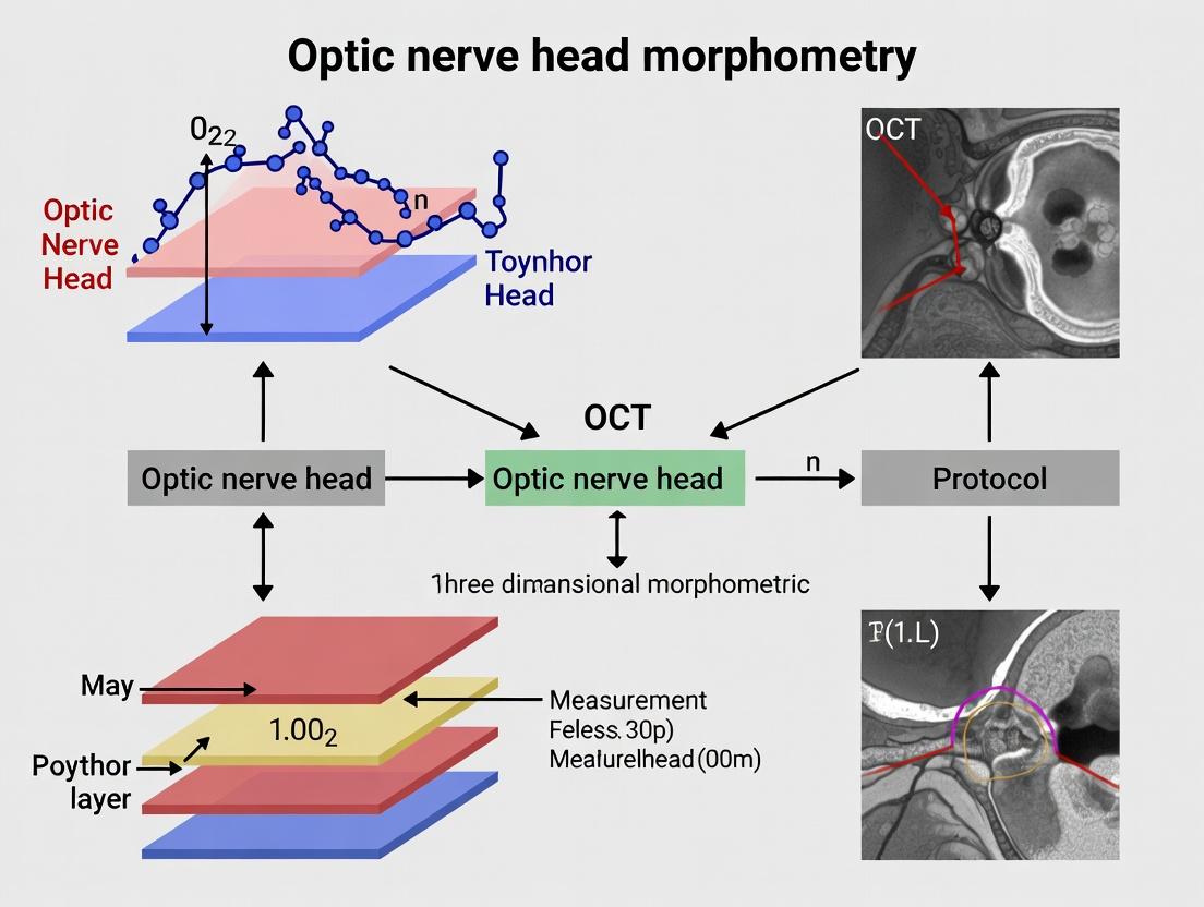
This article provides a comprehensive guide to Optical Coherence Tomography (OCT) protocols for three-dimensional morphometric analysis of the optic nerve head (ONH).
Precision 3D Optic Nerve Head Morphometry: Advanced OCT Protocols for Research and Drug Development
Abstract
This article provides a comprehensive guide to Optical Coherence Tomography (OCT) protocols for three-dimensional morphometric analysis of the optic nerve head (ONH). Tailored for researchers and drug development professionals, it explores the anatomical and pathophysiological rationale for 3D ONH metrics in glaucoma and neurodegenerative research. We detail current acquisition and segmentation methodologies, identify common technical challenges and optimization strategies, and validate protocols through comparisons with histology and other imaging modalities. The synthesis aims to establish standardized, reproducible approaches for quantifying ONH structure as critical biomarkers in preclinical and clinical trials.
The 3D Optic Nerve Head: Anatomic Rationale and Clinical Significance in Biomedical Research
Application Notes
This document details the application of Optical Coherence Tomography (OCT) for the three-dimensional (3D) morphometric analysis of the optic nerve head (ONH). Quantitative measurement of key ONH structures is pivotal for understanding glaucoma pathophysiology, assessing neurodegeneration, and evaluating therapeutic efficacy in clinical trials.
Bruch's Membrane Opening (BMO)
The BMO is the innermost point of Bruch's membrane at the ONH margin. It serves as the primary anatomical landmark and reference plane for defining other ONH structures. Unlike the clinically used optic disc margin, BMO is visible on high-resolution OCT and provides a consistent, stable reference.
Neuroretinal Rim (NRR)
The NRR is the tissue between the BMO and the internal limiting membrane (ILM). It contains the retinal ganglion cell axons and is a primary site of damage in glaucoma. Its minimum distance width (BMO-MRW) and regional volume are critical, three-dimensional measures of neural tissue loss, more sensitive than conventional two-dimensional clinical disc assessment.
Lamina Cribrosa Depth (LCD)
LCD measures the anterior surface of the lamina cribrosa (LC) relative to a reference plane (typically BMO). Posterior LC migration and increased depth are indicators of structural change and biomechanical strain within the ONH, representing a direct measure of connective tissue remodeling in glaucoma.
Cup Volume (CV)
CV quantifies the 3D volume of the cup, defined as the space beneath the ILM-BMO reference plane and above the ONH tissues (rim, cup floor). It provides a global volumetric assessment of ONH excavation, correlating with functional vision loss.
Table 1: Quantitative Normative Ranges and Clinical Significance of Key ONH Parameters
| Parameter | Typical Normative Range (Mean ± SD) | Glaucomatous Change | Primary Clinical/Research Significance |
|---|---|---|---|
| BMO Area | ~1.8 - 2.4 mm² | Generally stable | Defines reference plane for all other measurements. |
| Minimum BMO-MRW | 250 - 350 µm | Decreases (>15-20% loss is significant) | Earliest and most sensitive structural indicator of axon loss. |
| Lamina Cribrosa Depth | 300 - 500 µm (from BMO plane) | Increases (posterior bowing) | Marker of biomechanical compliance and tissue remodeling. |
| Cup Volume | 0.1 - 0.4 mm³ | Increases substantially | Global measure of ONH excavation; correlates with VF loss. |
Table 2: Comparison of OCT Technologies for ONH Morphometry
| OCT Technology | Axial/Transverse Resolution | Key Advantage for ONH | Primary Limitation |
|---|---|---|---|
| Spectral-Domain OCT (SD-OCT) | ~5 µm / ~15 µm | High scan speed, reduced motion artifact. | Limited depth penetration for deep LC visualization. |
| Swept-Source OCT (SS-OCT) | ~6 µm / ~20 µm | Deeper penetration, better visualization of deep LC and sclera. | Generally higher cost. |
| Enhanced Depth Imaging (EDI-OCT) | Similar to SD-OCT | Improves LC visualization on SD-OCT platforms. | Requires specific software/protocols. |
Experimental Protocols
Protocol 1: High-Density Radial OCT Scan Acquisition for ONH Morphometry
Objective: To acquire volumetric ONH data for precise definition of BMO, NRR, LC, and cup. Equipment: Commercial SS-OCT or SD-OCT with EDI capability, chin rest, internal fixation target. Software: Device-native acquisition software.
Procedure:
- Patient/Subject Preparation: Dilate pupil (>6 mm). Position subject at chin rest with forehead against strap.
- Scan Pattern Selection: Select a high-density, volumetric scan pattern centered on the ONH.
- Recommended: 256 B-scans x 512 A-scans over a 6x6 mm area, or a dense radial pattern (e.g., 24-48 lines, 12-15 mm length).
- Alignment & Focus: Use real-time en face IR image to center ONH. Adjust focus knob for sharpest B-scan visualization.
- Signal Optimization: Adjust Z-offset to position ONH within imaging window. Use EDI mode if available and targeting LC.
- Acquisition: Instruct subject to fixate on the internal target. Perform multiple volume scans (≥2) to allow for averaging.
- Quality Control: Immediately review B-scans for centration, signal strength (≥7/10), and absence of motion artifact (e.g., vessel duplication).
Protocol 2: Semi-Automated Segmentation and BMO Definition
Objective: To delineate BMO points and the ILM for all subsequent measurements. Equipment: Workstation with FDA-cleared or research-grade ONH analysis software (e.g., Heidelberg Eye Explorer, Iowa Reference Algorithms, OCTARA). Software: Segmentation/analysis module.
Procedure:
- Data Import: Load the acquired OCT volume into analysis software.
- Initial Automated Segmentation: Run the built-in layer segmentation algorithm for the ILM and RPE/Bruch's membrane complex.
- BMO Point Identification (Manual Correction): a. On each radial B-scan, manually identify the termination of the RPE/Bruch's membrane layer on either side of the ONH. b. Place a marker at the exact end-point. This is one BMO point. c. Repeat for all radial scans (e.g., 24-48 points total).
- BMO Reference Plane Construction: The software will fit a geometric plane (e.g., best-fit plane, BMO reference plane) through all identified BMO points.
Protocol 3: Measurement of Neuroretinal Rim (BMO-MRW) and Cup Volume
Objective: To compute the minimum rim width and cup volume based on the BMO reference plane. Software: ONH analysis software with BMO-MRW and cup volume algorithms.
Procedure:
- ILM Segmentation Verification: Ensure the ILM is accurately segmented across the entire volume. Manually correct any errors, particularly near vessel shadows.
- Parameter Calculation: a. The software calculates the minimum distance from each BMO point to the ILM along the local BMO plane normal. This generates the BMO-MRW map and global minimum. b. Cup Volume is computed as the total volume of space between the ILM surface and the BMO reference plane within the BMO area.
- Data Export: Export global metrics (e.g., global BMO-MRW, sectoral values, cup volume) and topographic maps for statistical analysis.
Protocol 4: Lamina Cribrosa Depth Measurement
Objective: To measure the anterior LC surface position relative to the BMO plane. Software: Software capable of manual or semi-automated LC segmentation.
Procedure:
- LC Identification: On each radial B-scan, identify the hyperreflective anterior surface of the LC.
- LC Point Placement: Manually place points along the anterior LC surface.
- Depth Calculation: The software calculates the perpendicular distance from each anterior LC point to the BMO reference plane. The average of all points is the mean LCD.
- Alternative (Grid-based): Measure LCD at predefined locations (e.g., superior, inferior, nasal, temporal) and calculate an average.
Mandatory Visualizations
Title: OCT ONH Morphometry Analysis Workflow
Title: Anatomic Relationship of Key ONH Parameters
The Scientist's Toolkit
Table 3: Essential Research Reagent Solutions for ONH Morphometry Studies
| Item | Function in ONH Research | Example/Notes |
|---|---|---|
| Swept-Source OCT (SS-OCT) Device | Provides the volumetric scan data. Essential for deep penetration imaging of the lamina cribrosa. | e.g., Topcon DRI OCT Triton, Zeiss PLEX Elite 9000. |
| Enhanced Depth Imaging (EDI) Software | Optimizes SD-OCT scans to improve visualization of deep ONH structures like the LC. | Built-in option on Heidelberg Spectralis. |
| FDA-Cleared ONH Analysis Software | Provides standardized, validated algorithms for BMO-MRW and rim volume calculation. | Heidelberg Glaucoma Module (Heidelberg Eye Explorer). |
| Research-Grade Segmentation Software | Allows manual correction and custom analysis beyond FDA-cleared parameters (e.g., LC depth). | Iowa Reference Algorithms, 3D Slicer with custom modules, OCTARA. |
| Statistical Analysis Software | For data aggregation, normative database comparison, and longitudinal change analysis. | R, Python (Pandas, SciPy), SAS, SPSS. |
| Pupil Dilation Drops | Ensures a large pupil diameter (>6mm) for optimal scan quality and peripheral BMO visualization. | Tropicamide 1%, Phenylephrine 2.5%. |
| Chin Rest & Fixation Target | Stabilizes subject position to minimize motion artifact during high-density scan acquisition. | Integrated into OCT device. |
| Digital Phantom/Test Object | Validates device axial and transverse measurement scale accuracy for longitudinal studies. | e.g., USP 1964A Ophthalmic Biometer Test Standard. |
This document provides detailed application notes and experimental protocols to support a doctoral thesis focused on developing and validating a standardized OCT protocol for three-dimensional (3D) morphometric measurements of the optic nerve head (ONH). The core thesis posits that precise 3D ONH parameter quantification is not merely correlative but is a direct, in vivo reflection of underlying glaucomatous neurodegeneration and axonal loss. These protocols are designed to establish a robust, reproducible framework for linking structural decay to pathophysiological mechanisms, thereby serving as essential tools for researchers, scientists, and drug development professionals in biomarker discovery and therapeutic efficacy assessment.
Key Pathophysiological Mechanisms & Quantitative Biomarkers
The deformation of the ONH architecture in glaucoma results from a complex interplay of biomechanical stress and biological vulnerability. Key quantitative parameters measurable via 3D OCT are direct surrogates for these pathological processes.
Table 1: 3D ONH Morphometric Parameters and Their Pathophysiological Correlates
| 3D ONH Parameter | Typical Glaucomatous Change | Quantitative Range (Example Data) | Pathophysiological Link & Interpretation |
|---|---|---|---|
| Minimum Rim Width (MRW) | Thinning | Normal: ~350-400 µm; Glaucoma: Can be <200 µm | Reflects direct loss of retinal ganglion cell (RGC) axons. 1 µm of thinning approximates loss of ~1200-1400 axons. |
| Bruch's Membrane Opening (BMO) Area | Expansion | Normal: ~1.8-2.5 mm²; Glaucoma: Increase of 0.1-0.4 mm² | Indicates permanent, connective tissue remodeling from chronic mechanical strain (IOP-related or independent). |
| Lamina Cribrosa (LC) Depth/Curvature | Posterior bowing, increased depth | Posterior displacement can exceed 150-300 µm vs. baseline | A biomarker of translaminar pressure gradient and direct mechanical insult to axonal bundles. |
| Pre-Laminar Tissue Thickness | Thinning | Variable; significant reduction vs. age-matched controls | Represents prelaminar neural tissue loss, including astrocytes and microglia activation preceding gross axonal loss. |
| Peripapillary Retinal Nerve Fiber Layer (ppRNFL) Thickness | Focal & diffuse thinning | Global average: Normal >90 µm; Glaucoma: <80 µm (severe: <60 µm) | Standard correlate of axonal loss; 3D mapping reveals focal defects corresponding to neuroretinal rim loss. |
Core Experimental Protocols
Protocol 2.1: Standardized 3D OCT Acquisition for ONH Morphometry
Objective: To acquire high-resolution, isotropic 3D volumetric scans of the ONH suitable for BMO-based planimetric and depth measurements.
- Participant Preparation & Positioning: Dilate pupil (>6 mm). Align patient chin and forehead on the rest. Use internal fixation target.
- Device & Settings: Use a spectral-domain (SD) or swept-source (SS) OCT with a dedicated ONH scanning protocol.
- Scan Pattern: Dense, radial, or volumetric raster scan. Recommended: 256x256 or 512x512 A-scans over a 6x6 mm area.
- Depth Resolution: ≤ 5 µm (axial).
- Averaging: Enable enhanced depth imaging (EDI) and use eye-tracking with multiple-frame averaging (≥ 4-8 B-scans per location) to improve LC visibility.
- Quality Control: Post-acquisition, check signal strength (≥ 7/10), absence of motion artifacts, and full visualization of BMO margins and LC.
Protocol 2.2: BMO-MRW and LC Morphometry Analysis
Objective: To derive key 3D parameters from acquired volumes using validated segmentation software.
- Data Export: Export raw volumetric OCT data in vendor-neutral format (e.g., .vol/.fda for Heidelberg, .img for Zeiss).
- Reference Plane Definition: Import data into analysis software (e.g., Heidelberg Eye Explorer with ONH Module, Iowa Reference Algorithms).
- Automatically or manually identify 40-50 points around the BMO in all relevant B-scans.
- The software fits a plane (BMO reference plane) through these points. This is the fundamental plane for all subsequent measurements.
- Automated Segmentation & Measurement:
- The software segments the internal limiting membrane (ILM) and BMO points in 3D.
- MRW Calculation: For each radial sector, the minimum distance from the BMO to the ILM is computed perpendicular to the BMO reference plane.
- LC Surface Depth: The anterior surface of the LC is segmented. Its depth is measured as the perpendicular distance from the BMO reference plane at each point.
- Output: Review segmentation accuracy manually. Export numerical data (global and sectoral MRW, BMO area, LC curvature indices) and color-coded topographic maps.
Protocol 2.3: Longitudinal Change Analysis (Progression Detection)
Objective: To detect significant change in 3D parameters over time, indicative of ongoing neurodegeneration.
- Baseline and Follow-up Registration: Use software capable of 3D image registration (e.g., Guided Progression Analysis for ONH or research-based tools like 3D OCTOR).
- Voxel-to-Voxel Comparison: Align follow-up volume to baseline using retinal blood vessels and BMO as landmarks.
- Change Analysis: Compute difference maps for MRW and LC depth. Apply statistical noise thresholds (e.g., 95% confidence interval based on test-retest variability) to distinguish true change from measurement noise.
- Progression Definition: A cluster of significant change (e.g., ≥ 20 contiguous superpixels showing thinning beyond threshold) in a consistent anatomical location over ≥ 2 consecutive visits is suggestive of progression.
Visualization of Pathophysiological Pathways & Workflows
Title: Pathophysiology of Glaucoma & 3D ONH Changes
Title: 3D ONH Morphometry Experimental Workflow
The Scientist's Toolkit: Key Research Reagent Solutions
Table 2: Essential Materials for 3D ONH Research
| Item / Reagent Solution | Function in ONH Research | Example / Notes |
|---|---|---|
| High-Resolution OCT System | Acquisition of 3D volumetric data. | Spectralis (Heidelberg Eng.), Cirrus (Zeiss), or DRI/OCT-1 (Topcon) with ONH cube and EDI capabilities. |
| BMO-MRW Analysis Software | Standardized segmentation and quantification of key 3D parameters. | Heidelberg Eye Explorer ONH Module, Iowa Reference Algorithms, or equivalent research-grade software. |
| 3D Image Registration Tool | Alignment of longitudinal scans for precise change detection. | Custom software (3D OCTOR), or commercial progression modules with 3D capability. |
| Normative Database | Age-adjusted reference ranges for identifying abnormality. | Device-specific databases (e.g., Heidelberg, Zeiss) or large population studies (e.g., UK Biobank). |
| Phantom Eye / Test Target | Calibration and monitoring of instrument performance and scan geometry. | Used to validate scan dimensions and ensure measurement consistency across sites in multi-center trials. |
| Statistical Analysis Package | Modeling longitudinal change and correlating structure with function. | R, SAS, or Python (with pandas, SciPy) for advanced mixed-effects models and progression analysis. |
Optical Coherence Tomography (OCT) has become an indispensable, non-invasive imaging tool in translational ophthalmic and neurological research. Its core utility lies in providing high-resolution, in vivo cross-sectional and three-dimensional images of retinal layers and the optic nerve head (ONH). In the context of a thesis focused on 3D morphometric measurement of the ONH, OCT serves as the critical bridging technology that enables direct comparison of structural endpoints across the translational pipeline. For neuroprotection studies—aimed at halting or slowing neuronal cell death in diseases like glaucoma, optic neuritis, and Alzheimer's—OCT-derived metrics such as retinal nerve fiber layer (RNFL) thickness, ganglion cell complex (GCC) volume, and ONH parameters (rim area, cup volume) are primary outcome measures. These quantitative biomarkers allow researchers to track disease progression and treatment efficacy from preclinical animal models through to human clinical trials with remarkable consistency.
Table 1: Core OCT Biomarkers for Translational Neuroprotection Studies
| Biomarker | Preclinical Model (e.g., Mouse/Rat) | Human Clinical Trial Application | Measured Impact of Neuroprotection |
|---|---|---|---|
| Retinal Nerve Fiber Layer (RNFL) Thickness | Mean global thickness: ~40-50 µm (rodent). Reduction of 10-20% in injury models. | Mean global thickness: ~90-100 µm (human). Slowing of atrophy rate (e.g., from -2.0 µm/yr to -0.5 µm/yr) indicates efficacy. | Primary endpoint. Stabilization or reduced rate of thinning is the gold-standard evidence of protection. |
| Ganglion Cell Complex (GCC) Thickness | Combined thickness of RNFL+GCL+IPL. Highly sensitive to ganglion cell soma loss. | Standard macular scan parameter. Detection of early, localized loss before RNFL changes. | Preservation of GCC volume/thickness indicates direct protection of neuronal cell bodies. |
| Optic Nerve Head (ONH) Morphometry | 3D parameters: Cup Volume, Rim Area, Linear Cup-to-Disc Ratio. Requires high-resolution OCT. | Standardized disc cube scan. Rim area loss correlates with axon count. | Stabilization of neuroretinal rim area indicates halting of overall axonal loss at the disc. |
| Total Retinal Thickness | Less specific, but used in models of generalized retinal degeneration. | Used in trials for diseases like inherited retinal degenerations. | An ancillary measure; specific layer analysis is preferred for neuroprotection. |
Table 2: Comparative OCT Specifications for Translational Stages
| Parameter | Preclinical OCT (Animal Models) | Clinical OCT (Human Trials) |
|---|---|---|
| Central Wavelength | ~850 nm or ~1050-1300 nm (for better choroid/sclera penetration). | ~840 nm (spectral-domain) or ~1050 nm (swept-source). |
| Axial/Transverse Resolution | 1-3 µm axial / 3-5 µm transverse (adaptive optics optional). | 5-7 µm axial / 10-20 µm transverse. |
| Scan Pattern for ONH | Dense radial scans or 3D volume cubes (e.g., 1.4 mm x 1.4 mm, 1000 A-scans/B-scan). | 6x6 mm or 4.5x4.5 mm disc-centered cube (e.g., 200x200 or 500x500 A-scans). |
| Key 3D Analysis | Custom segmentation for rodent ONH & lamina cribrosa. Manual landmark setting often required. | Automated segmentation of RNFL/GCC, and ONH topography (e.g., Zeiss Glaucoma Module, Heidelberg HEYEX). |
| Primary Challenge | Alignment and standardization due to small eye size and curvature. | Image quality (media opacity), patient fixation, and scan-to-scan variability. |
Detailed Experimental Protocols
Protocol 1: Longitudinal OCT Imaging for Neuroprotection Assessment in a Rodent Glaucoma Model
Objective: To quantitatively assess the efficacy of a novel neuroprotective agent (NPA) by measuring changes in RNFL thickness and ONH morphology over time in a laser-induced ocular hypertensive (OHT) rat model.
Materials:
- In vivo high-resolution spectral-domain OCT system (e.g., Bioptigen/Leica Envisu R2200, or similar).
- Anesthesia system (isoflurane vaporizer).
- Rodent alignment stage with head and eye positioning aids.
- Topical mydriatic (e.g., 0.5% tropicamide) and lubricating eye gel.
- Laser photocoagulation system for OHT induction.
- Custom or commercial software for rodent retinal layer segmentation (e.g., DOCTRAP, InVivoVue).
Procedure:
- Baseline Imaging (Day -7): Anesthetize animal. Dilate pupil. Apply lubricant. Position animal on stage. Acquire 3D volume scans centered on the optic disc (e.g., 1.6 mm x 1.6 mm, 100 B-scans x 1000 A-scans). Acquire 4 radial line scans (0°, 45°, 90°, 135°), each averaged 10 times.
- Disease Model Induction (Day 0): Induce unilateral OHT via laser photocoagulation of the trabecular meshwork. The contralateral eye serves as control.
- Treatment Regimen (Day 1+): Administer NPA or vehicle systemically or intravitreally according to study design.
- Longitudinal Imaging (Days 7, 14, 28, 56): Repeat OCT imaging as in Step 1 at defined intervals. Maintain consistent anesthesia, pupil dilation, and scan positioning.
- Image Processing & Analysis:
- 3D ONH Morphometry: Register all volume scans to baseline. Manually define the Bruch's Membrane Opening (BMO) points in each B-scan of the baseline scan. Apply automated or semi-automated segmentation to identify the internal limiting membrane (ILM) and anterior scleral surface. Compute 3D parameters: BMO area, minimum rim width (BMO-MRW), and cup volume.
- RNFL Thickness: Use automated segmentation algorithms to identify the RNFL boundaries in radial or circular scans. Calculate average global and quadrant-specific RNFL thickness.
- Statistical Analysis: Compare rates of change in RNFL thickness and ONH parameters between NPA-treated and vehicle-treated OHT eyes using mixed-effects models.
Protocol 2: OCT Biomarker Integration in a Phase II Neuroprotection Clinical Trial
Objective: To utilize standardized OCT imaging protocols as primary endpoints in a randomized, double-masked, placebo-controlled trial of a neuroprotective drug for glaucoma.
Materials:
- Certified clinical spectral-domain or swept-source OCT device (e.g., Heidelberg Spectralis, Zeiss Cirrus, Topcon Maestro).
- Validated and locked device software versions.
- Certified reading center with standardized grading protocols.
- Secure, HIPAA/GCP-compliant image transfer and database system.
Procedure:
- Site Certification & Training: Ensure all participating clinical sites are certified on the specific OCT model. Standardize patient positioning, scan acquisition protocols, and quality control (QC) checks (signal strength >7, centration, absence of motion artifact).
- Standardized Scan Acquisition (Each Visit):
- ONH Scan: Acquire a 4.5 x 4.5 mm or 6 x 6 mm 3D disc cube scan (e.g., 200x200 or 512x128 scans). Use eye-tracking and averaging (e.g., 15-25 B-scans averaged for Spectralis).
- Macular Scan: Acquire a 6 x 6 mm macular cube scan for GCC analysis.
- Peripapillary RNFL Scan: Acquire a 3.4 mm diameter circular scan around the ONH.
- Reading Center Analysis:
- QC Assessment: Reading center grades each scan for acceptability.
- Automated Segmentation Review: All automated segmentations (RNFL, GCC, ONH rim) are reviewed by a trained grader and manually corrected if the algorithm fails.
- Data Extraction: Extract key parameters: global/quadrant/sector RNFL thickness, GCC global loss volume (GLV) and focal loss volume (FLV), ONH rim area, and vertical cup-to-disc ratio.
- Endpoint Definition: The primary efficacy endpoint is often the rate of change in global RNFL thickness over the trial period (e.g., 24 months), compared between treatment and placebo arms using a linear mixed-model analysis.
Signaling Pathways & Workflow Diagrams
Diagram Title: Translational OCT Workflow for Neuroprotection Studies
Diagram Title: Key Neurodegenerative Pathways & OCT-Measured Outcome
The Scientist's Toolkit: Key Research Reagent Solutions
Table 3: Essential Materials for OCT-based Translational Neuroprotection Research
| Item | Function & Application in OCT Studies |
|---|---|
| High-Resolution Preclinical OCT System (e.g., Bioptigen/Leica, Micron IV/VI) | Provides the necessary axial resolution (1-4 µm) for imaging small rodent retinal layers and ONH structures for 3D morphometry. |
| Animal-Specific Positioning Stages & Lenses | Ensures consistent, reproducible alignment of the small animal eye for longitudinal imaging, critical for accurate measurement. |
| Validated Layer Segmentation Software (e.g., DOCTRAP, InVivoVue, Orion) | Enables quantitative extraction of RNFL, GCC, and ONH parameters from 3D OCT volumes in animal models lacking built-in clinical algorithms. |
| IOP Measurement System (e.g., TonoLab, Tono-Pen) | For glaucoma models, confirms induction and maintenance of ocular hypertension, correlating IOP with OCT-derived structural damage. |
| Histology Validation Reagents (e.g., Antibodies: βIII-tubulin, RBPMS, GFAP; TUNEL kits) | Allows post-mortem correlation of OCT findings with gold-standard measures like retinal ganglion cell counts and axonal integrity. |
| GCL/RNFL-Specific Reporter Mouse Lines (e.g., Thy1-GFP, Brn3b-tdTomato) | Enables in vivo fluorescence-guided OCT and direct visualization of specific neuronal populations alongside structural imaging. |
| Clinical OCT with Reading Center Protocols (e.g., Heidelberg Spectralis with Flex Modules) | Standardizes human trial imaging with eye-tracking, follow-up mode, and automated algorithms for RNFL/GCC/ONH, ensuring multi-site data consistency. |
| Quality Control Phantoms (e.g., Fabricated layers, model eyes) | Used to calibrate OCT systems, monitor longitudinal performance, and ensure measurement accuracy across sites and time. |
This document synthesizes current literature on normative databases and pathological thresholds for three-dimensional (3D) optic nerve head (ONH) metrics, as acquired via spectral-domain (SD) and swept-source (SS) optical coherence tomography (OCT). The establishment of robust normative data is critical for differentiating glaucomatous from healthy optic nerves, monitoring progression, and serving as endpoints in clinical trials for neuroprotective therapies.
Key Normative Databases and Study Populations
A review of recent multi-ethnic and population-based studies provides the foundation for normative data.
Table 1: Characteristics of Major Normative Database Studies for 3D ONH Metrics
| Study / Database Name | Population Description | Sample Size (Eyes) | OCT Device | Key 3D ONH Parameters Measured |
|---|---|---|---|---|
| African Descent and Glaucoma Evaluation Study (ADAGES) | African-American & European-American participants | ~3,300 | Spectralis (Heidelberg) | Rim Area, Rim Volume, Cup Volume, BMO-MRW* |
| Diagnostic Innovations in Glaucoma Study (DIGS) | Multi-ethnic (White, Black, Hispanic) | ~1,800 | Spectralis (Heidelberg) | Rim Area, Cup Volume, BMO-MRW, Mincimum Rim Width (MRW) |
| Singapore Epidemiology of Eye Diseases (SEED) | Chinese, Malay, Indian populations | ~10,000 | Cirrus (Zeiss) | Neuroretinal Rim Thickness (by quadrant), Cup-to-Disc Ratio |
| Beijing Eye Study | Northern Chinese population | ~4,500 | Spectralis (Heidelberg) | BMO-MRW, BMO Area |
| The Ocular Hypertensive Treatment Study (OHTS) | Ocular hypertensive patients | ~1,600 | Various (legacy) | Cup Volume, Rim Area (later adoptions) |
| Commercial Device Normative Databases (e.g., Spectralis GMPE, Cirrus NDB) | Age-stratified, multi-ethnic healthy controls | 100-300 per age decade/ethnicity | Device-specific | All standard ONH parameters, often including Bruch's Membrane Opening (BMO)-based metrics |
*BMO-MRW: Bruch's Membrane Opening Minimum Rim Width.
Population-Specific Variations: Data consistently show that individuals of African descent have larger disc areas, greater rim volumes, and deeper cups compared to those of European or Asian descent. Asian populations often demonstrate smaller disc areas. These differences necessitate ethnicity-corrected normative limits.
Established Pathologic Thresholds for Key 3D ONH Metrics
Pathologic thresholds are typically defined as values falling below the 5th or above the 95th percentile of the normative distribution. The most sensitive and specific metrics combine topographic and tomographic data.
Table 2: Example Pathologic Thresholds for Select 3D ONH Metrics (Spectralis OCT)
| Metric | General Pathologic Threshold (Approx.) | Key Considerations & Strengths |
|---|---|---|
| BMO-MRW (Global) | < 200 - 250 µm (age-dependent) | Less influenced by disc size; strong structure-function correlation. |
| Neuroretinal Rim Area | < 1.00 - 1.20 mm² | Must be corrected for disc size. High variability in healthy large discs. |
| Horizontal Cup-to-Disc Ratio (3D) | > 0.65 - 0.70 | Traditional but less specific; best used in combination. |
| Rim Volume | < 0.20 - 0.25 mm³ | Provides a 3D assessment of rim tissue. |
| Lamina Cribrosa Depth | > 450 - 550 µm (from BMO reference plane) | A dynamic measure of ONH biomechanics; emerging threshold. |
| Focal Loss Volume (FLV) | > 0.5 - 1.0% | A pattern-based metric quantifying localized rim loss. Superior specificity. |
Note: Exact thresholds are device-specific and must be referenced against the instrument's internal normative database, which applies age and ethnicity corrections.
Application Notes & Experimental Protocols
Protocol: Acquisition of 3D ONH Scans for Normative Database Building
Aim: To standardize high-quality OCT volume scan acquisition for inclusion in a normative database. Materials: SD-OCT or SS-OCT device with ONH volume scan protocol, chin rest, artificial tears (if needed). Procedure:
- Participant Selection & Ethics: Recruit healthy volunteers with no ocular pathology, stratified by age, gender, and ethnicity. Obtain informed consent and IRB approval.
- Preparation: Dark adapt participant for 5 minutes. Position participant at the device. Align the internal fixation target to the fellow eye.
- Scan Acquisition:
a. Select the high-density ONH volume scan protocol (e.g., Spectralis:
768 x 496A-scans; Cirrus:512 x 128or200 x 200). b. Instruct the participant to blink and then hold still prior to capture. c. Align the scan rectangle concentric to the ONH, covering a minimum area of4.5 x 4.5 mmto capture peripapillary region. d. Activate the real-time eye tracking and averaging function (e.g., ART on Spectralis, ≥15 frames). e. Acquire the scan. Verify signal strength (SS) ≥ 7/10 (or manufacturer-recommended minimum). - Quality Control: Immediately review the scan for centration, motion artifacts, and segmentation errors of the BMO and inner limiting membrane (ILM). Re-acquire if necessary.
- Data Export: Export the raw volume data (
*.vol), all layer segmentation files, and the instrument-generated ONH analysis report.
Protocol: Validation of Pathologic Thresholds in a Case-Control Study
Aim: To test the diagnostic performance of a candidate 3D ONH metric threshold. Materials: OCT scans from two cohorts: confirmed glaucoma patients and healthy controls (matched for age/ethnicity), statistical software (R, SPSS). Procedure:
- Cohort Definition:
- Glaucoma Group: Primary open-angle glaucoma with reproducible visual field defects.
- Control Group: Healthy eyes with normal IOP and visual fields.
- Metric Extraction: Using the device's proprietary software or validated third-party software, extract the candidate metric (e.g., global BMO-MRW) for all eyes.
- Statistical Analysis: a. Calculate the mean and standard deviation of the metric for the control group. b. Define the normative 5th percentile (for a parameter where lower is abnormal) from the control distribution. c. Apply this threshold to the glaucoma group. Calculate: * Sensitivity: (True Positives) / (All Glaucoma Eyes) * Specificity: (True Negatives) / (All Control Eyes) d. Generate a Receiver Operating Characteristic (ROC) curve and calculate the Area Under the Curve (AUC) to assess overall diagnostic accuracy.
- Comparison: Compare the AUC of the 3D metric to that of traditional parameters (e.g., vertical cup-to-disc ratio).
The Scientist's Toolkit: Key Research Reagent Solutions
Table 3: Essential Materials for 3D ONH Morphometric Research
| Item / Solution | Function & Rationale |
|---|---|
| Heidelberg Spectralis OCT with Glaucoma Module Premium Edition (GMPE) | Gold-standard device providing BMO-based MRW and precise segmentation. Essential for acquiring high-quality, averaged volume scans. |
| Zeiss Cirrus HD-OCT with Optic Disc Cube | Provides the 200 x 200 scan pattern for high-density ONH analysis. Widely used in clinical trials. |
| Topcon DRI OCT Triton (SS-OCT) | Swept-source technology allows deeper penetration and improved visualization of deep ONH structures like the lamina cribrosa. |
| 3D Segmentation Software (e.g., IOPS, 3D Slicer with custom modules) | For manual correction of automatic BMO/ILM segmentations and custom metric calculation from raw volume data. Critical for research-grade analysis. |
| Artificial Tears (Preservative-Free) | To temporarily improve corneal optics and signal strength in participants with dry eye, ensuring high-quality scans. |
| Statistical Analysis Package (e.g., R with pROC, SPSS) | For performing advanced statistical analyses, generating ROC curves, and modeling normative data with covariates (age, disc area, ethnicity). |
Visualized Workflows & Relationships
Title: Normative Database and Threshold Development Workflow
Title: Factors Influencing Individualized ONH Assessment
Step-by-Step Guide: Implementing a Robust 3D ONH OCT Acquisition and Analysis Protocol
1. Introduction and Thesis Context Within the broader thesis on establishing standardized, high-precision OCT protocols for three-dimensional morphometric measurements of the optic nerve head (ONH), the optimization of scanner settings is paramount. This document details application notes and experimental protocols for determining optimal scan pattern design, density, averaging, and resolution. The goal is to maximize the signal-to-noise ratio (SNR), accuracy, and reproducibility of key ONH parameters (e.g., Bruch's Membrane Opening area, minimum rim width, cup volume) for longitudinal research and drug development.
2. Key Scanner Parameters: Quantitative Summary
Table 1: Core Scanner Parameters for Volumetric ONH Imaging
| Parameter | Typical Range | Recommended Optimal Setting* | Primary Impact |
|---|---|---|---|
| Axial Resolution | 2 - 7 µm in tissue | ≤ 5 µm | Defines layer segmentation precision. |
| Lateral Resolution | 10 - 30 µm | ≤ 20 µm | Determines transverse feature discernibility. |
| A-Scan Rate | 20 - 200+ kHz | ≥ 70 kHz | Enables dense sampling within acceptable scan time. |
| Scan Pattern | Radial, Raster, Combination | Dense Radial + Raster Grid | Balances coverage and sampling symmetry. |
| B-Scan Density | 100 - 500 B-scans/volume | 250 - 400 B-scans/volume | Reduces interpolation error for 3D morphology. |
| Averaging (MASKS) | 1 - 100 frames/B-scan | 5 - 20 frames/B-scan | Crucial for SNR improvement in deep ONH structures. |
| Scan Area | 3x3 mm to 12x12 mm | 4.5x4.5 mm to 6x6 mm | Captures ONH and peripapillary area without excessive pixel size. |
- Optimal settings are device-dependent; recommendations based on current spectral-domain and swept-source OCT systems.
3. Detailed Experimental Protocols
Protocol 3.1: Systematic Evaluation of Averaging on ONH SNR Objective: To quantify the relationship between B-scan frame averaging (Multiple Frame Averaging - MFA) and the SNR in deep ONH structures (lamina cribrosa, choroid). Materials: OCT device, chin rest, fixation target, healthy volunteer cohort. Method:
- Set a standardized raster scan pattern (e.g., 6x6 mm, 250 B-scans, 512 A-scans/B-scan).
- At the same ONH location, acquire volumetric scans with varying MFA levels: 1, 5, 10, 15, 20, 25 frames per B-scan.
- For each dataset, register and average frames using the device's proprietary motion correction (MACS) or post-processing algorithm.
- In post-processing software, measure mean pixel intensity (I) and standard deviation of noise (σ) in five regions of interest (ROIs): prelaminar tissue, lamina cribrosa pores, choroid, vitreous (noise reference).
- Calculate SNR for each ROI: SNR = Itissue / σvitreous.
- Plot SNR vs. MFA level for each ROI. Determine the "knee of the curve" where additional averaging yields diminishing returns.
Protocol 3.2: Optimization of Scan Pattern and Density for BMO Detection Objective: To determine the minimal B-scan density required for reproducible Bruch's Membrane Opening (BMO) point identification and rim metric calculation. Materials: OCT device with eye-tracking, glaucoma patient cohort. Method:
- Acquire a high-density reference volume (e.g., 6x6 mm, 385 B-scans, 20x averaging) using eye-tracking.
- From this master volume, computationally sub-sample B-scans to simulate lower densities: 49, 97, 145, 193, 241 B-scans/volume.
- For each simulated dataset, perform automated BMO point identification using a standard algorithm (e.g., device OEM or academic).
- Calculate the BMO area and minimum rim width (MRW). Compare each metric from the sub-sampled datasets to the reference (gold standard).
- Compute the coefficient of variation (CV) and intraclass correlation coefficient (ICC) for each metric across densities. Define optimal density as the point where CV < 2% and ICC > 0.99.
4. Visualization of Protocol Logic and Relationships
Title: Scanner Setting Trade-offs for ONH Morphometry
Title: Workflow for Optimizing a Single Scanner Parameter
5. The Scientist's Toolkit: Research Reagent Solutions
Table 2: Essential Materials for OCT ONH Protocol Development
| Item | Function & Relevance to Protocol |
|---|---|
| Spectral-Domain or Swept-Source OCT | Core imaging device. Swept-source offers better deep tissue (LC) penetration. |
| Integrated Eye-Tracking (ET) | Critical for minimizing motion artifacts during dense, averaged scans. Enables reliable follow-up. |
| Custom Scan Pattern Software | Allows deviation from OEM patterns (e.g., dense radial scans centered on BMO). |
| Anterior Segment Lens | For wide-field scanning to capture full ONH and parapapillary region in large eyes. |
| Phantom Eyes (Model Eyes) | With layered, known geometry to test resolution and measurement accuracy of protocols. |
| Open-Source Segmentation Software (e.g., Iowa Reference Algorithms) | For standardized, vendor-neutral analysis of BMO, rim, LC metrics. |
| Statistical Software (R, Python, SPSS) | For analysis of variance, ICC calculation, and determining optimal cut-offs from protocol data. |
| High-Performance Computing Cluster | For processing large datasets generated by high-density, averaged volumetric scans. |
Within the framework of a thesis on OCT protocols for three-dimensional morphometric measurements of the optic nerve head (ONH), accurate segmentation of key anatomical structures is foundational. The Bruch's Membrane Opening (BMO), Internal Limiting Membrane (ILM), and Lamina Cribrosa (LC) serve as critical landmarks for quantifying neuroretinal rim tissue, retinal nerve fiber layer thickness, and laminar architecture. The choice between automated and manual delineation algorithms directly impacts the reproducibility, scalability, and accuracy of these morphometric measures. This document provides application notes and detailed experimental protocols for researchers and drug development professionals engaged in ONH research.
Quantitative Comparison of Segmentation Approaches
Table 1: Performance Metrics of Automated vs. Manual Segmentation Algorithms for ONH Structures
| Metric | BMO (Automated) | BMO (Manual) | ILM (Automated) | ILM (Manual) | LC (Automated) | LC (Manual) |
|---|---|---|---|---|---|---|
| Dice Coefficient (Mean ± SD) | 0.94 ± 0.03 | 1.00 (Ref) | 0.97 ± 0.02 | 1.00 (Ref) | 0.83 ± 0.07 | 1.00 (Ref) |
| Boundary Error (µm) | 4.1 ± 1.8 | N/A | 2.5 ± 1.1 | N/A | 9.8 ± 3.5 | N/A |
| Processing Time (sec/volume) | 45-120 | 1800-3600 | 45-120 | 1800-3600 | 60-180 | 2400-5400 |
| Inter-observer Variability (µm) | 0 (Fixed) | 12.5 ± 5.2 | 0 (Fixed) | 8.2 ± 3.1 | 0 (Fixed) | 25.7 ± 10.4 |
| Key Algorithm Type | Graph-Cut, Deep Learning (U-Net) | Expert-guided ROI | Gradient-Based, A-Scan Analysis | Point-by-point plotting | Texture-Based, CNN | Landmark-based tracing |
Table 2: Applicability in Research & Drug Development Contexts
| Context | Recommended Method | Rationale | Primary Limitation |
|---|---|---|---|
| High-Throughput Clinical Trials | Automated Segmentation | Enables rapid, consistent analysis of thousands of OCT volumes; essential for longitudinal monitoring of drug efficacy. | Potential for error in pathological or poor-quality scans requires quality control checks. |
| Ground Truth Generation | Manual Delineation by Multiple Experts | Provides the reference standard for training and validating automated algorithms. Gold standard for novel phenotypes. | Extremely time-consuming and resource-intensive; introduces human bias. |
| Exploratory Morphometry | Hybrid (Auto + Manual Correction) | Balances efficiency with accuracy for novel measurements where fully automated tools are not yet validated. | Correction time varies based on image quality and structure complexity. |
| LC Microarchitecture Study | Manual or Specialized Automated | LC boundaries are often poorly contrasted; manual may be superior for anterior/posterior LC demarcation in research-grade analysis. | Even specialized algorithms show high boundary error for posterior LC. |
Detailed Experimental Protocols
Protocol 3.1: Manual Delineation for Ground Truth Creation
Objective: To create a high-fidelity reference standard for BMO, ILM, and LC boundaries from Spectral-Domain OCT (SD-OCT) volumes.
Materials: See "The Scientist's Toolkit" (Section 5).
Methodology:
- Image Preparation: Load the ONH-centered OCT volume (e.g., 6x6 mm, 512x128x1024 voxels) into specialized segmentation software (e.g., ITK-SNAP, OSIRIX).
- ILM Delineation:
- Navigate to the central B-scan (slice #64).
- Manually place control points along the vitreoretinal interface, following the hyper-reflective signal corresponding to the ILM.
- Interpolate points to create a continuous boundary. Propagate this initial contour to adjacent B-scans (±5 scans) and refine point-by-point.
- Repeat for the entire volume, reviewing every 10th B-scan for continuity.
- BMO Delineation:
- In the same B-scan, identify the termination points of the Bruch's Membrane/Retinal Pigment Epithelium complex on either side of the neural canal.
- Place a single point at each termination. The line connecting these points defines the BMO reference plane in that B-scan.
- Repeat across all B-scans where BM termination is visible (typically 80-100 scans).
- Generate a 3D BMO point cloud by connecting corresponding points.
- LC Delineation:
- Identify the LC region as a hyper-reflective, porous structure beneath the BMO.
- Delineate the anterior LC boundary where the neural canal tissue transitions to the cribrosal plates.
- Delineate the posterior LC boundary where the LC signal diminishes into the prelaminar tissue. This is the most challenging step and may require reference to adjacent C-scans (en face views).
- Use a closed contour for each boundary on each B-scan.
- Validation & Consensus:
- Have a second, masked expert repeat steps 2-4 on the same volume.
- Calculate inter-observer variability (mean surface distance).
- For discrepancies >3 pixels (≈10µm), a third senior adjudicator makes the final determination.
Protocol 3.2: Training a Deep Learning-Based Automated Algorithm
Objective: To develop a convolutional neural network (CNN) for simultaneous segmentation of ILM, BMO, and anterior LC.
Materials: High-performance GPU cluster, Python with PyTorch/TensorFlow, 200+ OCT volumes with expert manual segmentations (ground truth).
Methodology:
- Data Curation & Augmentation:
- Split data: 70% training, 15% validation, 15% testing.
- Apply random augmentations to training set: intensity scaling (±20%), Gaussian noise, horizontal flipping, and small rotational transformations (±5°).
- Model Architecture & Training:
- Implement a 3D U-Net variant with residual connections.
- Input: Normalized OCT sub-volume (e.g., 256x256x64 voxels).
- Output: Multi-channel segmentation map (Background, ILM, BMO points, LC).
- Loss Function: Combined Dice loss and Cross-Entropy loss.
- Optimizer: Adam with an initial learning rate of 1e-4, reduced on plateau.
- Train for 500 epochs, batch size 2, saving the model with best validation Dice score.
- Post-Processing & Output:
- Apply connected-component analysis to remove small false positives.
- For BMO, fit a smooth spline through the predicted point cloud in 3D.
- Calculate morphometric parameters (e.g., BMO area, minimum rim width, LC curvature).
Visualization of Workflows
Title: Segmentation Decision & Validation Workflow
Title: From Segmentation to Key Morphometric Parameters
The Scientist's Toolkit: Essential Research Reagents & Materials
Table 3: Key Research Reagent Solutions for ONH Segmentation Studies
| Item / Reagent | Provider (Example) | Function in Protocol |
|---|---|---|
| Spectralis SD-OCT/Heidelberg Engineering | Heidelberg Engineering | Acquisition of high-resolution, eye-tracked ONH volumes. Essential for reproducible longitudinal scans. |
| ITK-SNAP Software | www.itksnap.org | Open-source software for manual and semi-automatic segmentation of medical images. Primary tool for ground truth creation. |
| Python with PyTorch & MONAI | PyTorch.org, monai.io | Core programming environment for developing, training, and deploying deep learning segmentation models. |
| Custom MATLAB Segmentation GUI | MathWorks | In-house tool for expert manual delineation with batch processing and inter-observer metric calculation. |
| Phantom OCT Eyes (Model Eyes) | Ocular Instruments, Inc. | Provides physical reference standards with known dimensions for validating segmentation algorithm accuracy. |
| Image Processing Toolkit (e.g., FIJI/ImageJ) | NIH | For pre-processing steps: intensity normalization, filtering, and basic en face projection generation. |
| GPU Workstation (NVIDIA RTX A6000) | NVIDIA | Provides the computational power necessary for training 3D CNNs on large OCT datasets. |
| DICOM/Proprietary OCT Data Parser | In-house development | Converts raw OCT scanner output into standardized formats (e.g., NIfTI) for algorithm input. |
This document provides detailed application notes and protocols for a computational pipeline that transforms raw Optical Coherence Tomography (OCT) volumes of the optic nerve head (ONH) into quantitative 3D mesh models. This pipeline is a core methodological component of a broader thesis focused on establishing a standardized OCT protocol for three-dimensional morphometric measurements in ONH research. The objective is to enable robust, reproducible, and high-throughput extraction of structural parameters critical for understanding glaucomatous progression, drug efficacy, and ONH biomechanics.
The Scientist's Toolkit: Essential Research Reagent Solutions
| Item Name | Function / Explanation |
|---|---|
| Spectral-Domain or Swept-Source OCT Device | Acquires raw 3D volumetric data (B-scans). Higher axial resolution and scan depth are crucial for posterior pole imaging. |
| Active Tracking & Eye-Tracking System | Minimizes motion artifacts during volume acquisition, essential for accurate longitudinal studies. |
| Spectralis HRA+OCT, Cirrus HD-OCT, or equivalent | Commercial platforms providing raw data export capability (e.g., .vol, .img, .e2e formats). |
| Anonymized DICOM or Proprietary RAW Data | The primary input. DICOM is preferred for standardization. |
| Segmentation Software (e.g., Iowa Reference Algorithms, OCTExplorer) | Provides or implements algorithms for layer segmentation (e.g., ILM, BMO, RPE). |
| 3D Slicer, MeshLab, or PyVista | Open-source platforms for 3D point cloud processing and mesh generation/manipulation. |
| Python Environment (NumPy, SciPy, PyTorch/TensorFlow, VTK, Trimesh) | Core computational ecosystem for custom pipeline scripting, algorithm development, and data analysis. |
| Statistical Software (R, SPSS, GraphPad Prism) | For final morphometric parameter analysis, group comparisons, and visualization of results. |
Experimental Protocol: End-to-End Pipeline
Protocol: Raw OCT Volume Acquisition & Preprocessing
Objective: To obtain high-quality, artifact-minimized OCT volumes of the ONH. Detailed Methodology:
- Participant Preparation & Positioning: Dilate the pupil. Position the subject at the OCT device with chin and forehead rests. Use the internal fixation target.
- Scan Protocol Selection: Select a 3D cube scan protocol centered on the ONH. Recommended:
512 x 512 A-scansover a6 x 6 mmarea. Enable Enhanced Depth Imaging (EDI) or similar if available. - Acquisition with Averaging: Activate real-time eye tracking. Set scan averaging to
≥ 10 frames per B-scanto improve signal-to-noise ratio (SNR). - Quality Control: Immediately assess scan quality. Reject volumes with significant motion artifacts, blink artifacts, or poor signal strength (SS < 7).
- Data Export: Export the raw volume and associated fundus image in a non-proprietary format (
.dcmDICOM preferred) or a documented proprietary format.
Protocol: Layer Segmentation & Point Cloud Generation
Objective: To identify key anatomical surfaces and generate 3D coordinate points. Detailed Methodology:
- Input: Import raw OCT volume into segmentation software.
- Key Surface Segmentation: Apply a validated deep learning or graph-theory algorithm to segment the following boundaries in each B-scan:
- Internal Limiting Membrane (ILM)
- Bruch's Membrane Opening (BMO) points (critical)
- Anterior and posterior surfaces of the lamina cribrosa
- Retinal Pigment Epithelium (RPE)
- Point Cloud Extraction: For each segmented surface, extract the
(x, y, z)coordinates of every pixel/voxel, creating a discrete point cloud for each anatomical layer. Thezcoordinate is typically the axial depth. - Output: Save each point cloud as a
.csvor.plyfile, with metadata on scaling (µm/pixel).
Protocol: 3D Mesh Model Reconstruction
Objective: To convert irregular point clouds into continuous, watertight 3D mesh models suitable for quantitative analysis. Detailed Methodology:
- Point Cloud Registration: Align all layer-specific point clouds into a common 3D coordinate system using the BMO centroid as the origin.
- Outlier Removal: Apply a statistical outlier removal filter (e.g., remove points > 2 standard deviations from the local mean density).
- Surface Reconstruction: Use a Poisson Surface Reconstruction or Delaunay Triangulation algorithm on the denoised point cloud to generate a triangle mesh.
- Mesh Smoothing & Decimation: Apply a Laplacian smoothing filter to reduce surface noise, followed by mesh decimation to reduce triangle count for computational efficiency while preserving shape.
- Validation: Visually overlay the 3D mesh onto orthogonal OCT B-scans to ensure anatomical fidelity.
Protocol: Morphometric Parameter Extraction
Objective: To compute standardized quantitative parameters from the 3D mesh models. Detailed Methodology:
- BMO-Based Reference Plane: Define the least-squares plane fitted to all BMO points. This is the reference plane for rim measurements.
- Parameter Computation: Calculate the following key parameters automatically via script:
- BMO Area: Area of the polygon defined by connecting the BMO points.
- BMO-MRW (Minimum Rim Width): The minimum distance from each BMO point to the ILM surface, measured perpendicular to the BMO reference plane.
- LC Depth: The perpendicular distance from the BMO plane to the anterior surface of the lamina cribrosa at the centroid.
- Rim Volume: The volume of tissue between the ILM surface and the BMO reference plane, bounded laterally by the BMO.
- Cup Volume: The volume below the BMO plane bounded by the ILM.
- Data Export: Compile all computed parameters for a single subject into a structured table.
Table 1: Example Morphometric Output for a Healthy Control vs. Glaucoma Subject
| Parameter | Healthy Control (Mean ± SD) | Early Glaucoma (Mean ± SD) | Units | p-value* |
|---|---|---|---|---|
| BMO Area | 1.98 ± 0.32 | 2.12 ± 0.29 | mm² | 0.07 |
| Average MRW | 350.5 ± 45.2 | 275.8 ± 62.1 | µm | <0.001 |
| Minimum MRW | 215.3 ± 38.7 | 132.4 ± 55.9 | µm | <0.001 |
| LC Depth (Central) | 452.1 ± 112.5 | 588.4 ± 135.7 | µm | <0.001 |
| Neuroretinal Rim Volume | 0.86 ± 0.15 | 0.62 ± 0.18 | mm³ | <0.001 |
| Hypothetical data for illustration. SD = Standard Deviation. |
Pipeline Visualization Diagrams
Title: OCT to 3D Mesh Processing Pipeline
Title: Key 3D Parameter Measurement Schema
Effective longitudinal analysis of progressive optic nerve head (ONH) remodeling requires rigorous, standardized imaging protocols to ensure data comparability across time points and studies. This protocol is designed for integration within a broader thesis framework focusing on 3D OCT morphometry for ONH research, emphasizing reproducibility in multi-center trials and drug development.
1. Core Imaging Protocol Specifications A standardized Spectral-Domain OCT (SD-OCT) or Swept-Source OCT (SS-OCT) imaging protocol is mandatory. The following parameters must be fixed for all longitudinal sessions:
| Parameter | Specification | Rationale |
|---|---|---|
| Device Type | Spectral-Domain or Swept-Source OCT | Ensures consistent depth resolution and scan penetration. |
| Scan Pattern | Dense Radial (≥24 lines) or 3D Cube Scan (≥200x200 B-scans) | Provides adequate sampling for 3D reconstruction. |
| Scan Area | Centered on ONH, 4.0 x 4.0 mm or 6.0 x 6.0 mm | Standardizes field of view for regional comparison. |
| A-Scan Density | ≥300 A-scans per B-scan (Cube) | Balances resolution and acquisition time. |
| Averaging | ≥5 frames per B-scan position | Reduces speckle noise, improves signal-to-noise ratio. |
| Pupil Dilation | Required (Tropicamide 1%) | Maximizes signal strength and uniformity. |
| Head Positioning | Guided by internal fixation target & external camera | Minimizes tilt and scan decentration. |
| Signal Strength | Quality Index ≥7/10 (device-specific) | Ensures usable data; mandates re-scan if below threshold. |
| Time of Day | ±2 hours from baseline visit | Controls for diurnal IOP and tissue changes. |
2. Key Morphometric Parameters for Tracking Quantitative 3D analysis should be derived from segmented OCT volumes. The following table outlines primary endpoints for tracking progression.
| Morphometric Parameter | Definition | Typical Baseline Value (Mean ± SD) | Progression Flag |
|---|---|---|---|
| Minimum Rim Width (MRW) | Shortest distance from BMO to ILM in 360° | 285 ± 50 µm | Reduction >15 µm/year* |
| Bruch's Membrane Opening (BMO) Area | Area enclosed by BMO points | 1.85 ± 0.35 mm² | Expansion >0.04 mm²/year* |
| Retinal Nerve Fiber Layer (RNFL) Thickness | Global average thickness | 95 ± 15 µm | Reduction >5 µm/year* |
| Lamina Cribrosa Depth (LCD) | Anterior-posterior depth from BMO reference plane | 450 ± 150 µm | Posterior migration >30 µm/year* |
| Pre-laminar Tissue Thickness | Tissue thickness anterior to LC | 250 ± 80 µm | Thinning >20 µm/year* |
*Example progression rates based on recent glaucoma studies; study-specific thresholds must be defined.
3. Detailed Experimental Protocol for Longitudinal Session Session Workflow: Patient Preparation, OCT Acquisition, and Data Export
4. Data Processing & Analysis Protocol Workflow for Consistent 3D Morphometric Derivation
5. The Scientist's Toolkit: Research Reagent Solutions
| Item | Function/Application |
|---|---|
| Spectralis OCT (Heidelberg) | Reference device for high-resolution, tracked follow-up scans. |
| Cirrus HD-OCT (Zeiss) | Common platform with built-in ONH analysis algorithms. |
| DRI-OCT Triton (Topcon) | Swept-Source OCT offering deeper penetration for LC imaging. |
| IORC BMO-MRW Module | Specialized software for accurate Bruch's Membrane Opening minimal rim width measurement. |
| 3D Slicer with OCT Extension | Open-source platform for custom 3D segmentation and analysis. |
| GraphPad Prism / R (lme4) | Statistical software for modeling longitudinal change (mixed-effects models). |
| Phantom ONH Model (Omega) | Physical model for validating OCT measurement precision across devices. |
| ANSI Z80.20-2016 Standard | Reference for ophthalmic instrument calibration and performance verification. |
Overcoming Challenges: Technical Pitfalls and Best Practices for Reliable 3D ONH Measurements
Within a thesis focused on establishing a standardized Optical Coherence Tomography (OCT) protocol for three-dimensional morphometric measurements of the optic nerve head (ONH), artifact management is paramount. Reliable quantification of parameters like neuroretinal rim area, cup volume, and laminar cribrosa curvature depends on high-fidelity volumetric data. This document details application notes and experimental protocols for identifying, mitigating, and correcting four common artifacts that compromise ONH morphometric analysis.
Artifact Characterization & Impact on 3D Morphometry
| Artifact Type | Primary Cause | Key Impact on 3D ONH Morphometry | Quantitative Severity Indicator |
|---|---|---|---|
| Off-Center Scans | Improper subject alignment or fixation loss. | Asymmetric rim thickness analysis, erroneous cup disc center detection. | Deviation of Bruch's Membrane Opening (BMO) center from scan center > 10% of scan width. |
| Motion Artifacts | Microsaccades, axial head movement. | Discontinuous surfaces, erroneous retinal nerve fiber layer (RNFL) thickness maps. | Inter-frame B-scan displacement > 5 µm. Signal std. dev. fluctuation > 15% across consecutive B-scans. |
| Segmentation Errors | Poor image contrast, shadowing, pathologic features. | Incorrect quantification of cup/disc boundaries, lamina cribrosa depth. | Localized deviation (>2 pixels) from validated manual segmentation in >10% of B-scans per volume. |
| Poor Signal Strength | Media opacity, improper focus, low beam power. | Increased noise, failed segmentation, unreliable tissue boundary detection. | Manufacturer's Signal Strength Index (SSI) < 7 (out of 10) or Signal-to-Noise Ratio (SNR) < 20 dB. |
Detailed Mitigation and Correction Protocols
Protocol 3.1: Prevention and Detection of Off-Center ONH Scans
Objective: Ensure the ONH, specifically the BMO, is centered within the volumetric scan for symmetric analysis. Materials: Spectral-Domain or Swept-Source OCT device, fixation target, post-processing software (e.g., MATLAB, Python with custom scripts). Procedure:
- Pre-Scan Alignment:
- Engage the external fixation target. Use the real-time en-face IR image to position the ONH.
- Utilize the device's "Follow" or "Tracking" function if available to lock onto the ONH.
- Post-Acquisition Detection:
- Generate an en-face projection image from the volumetric data.
- Run an automated BMO detection algorithm (e.g., based on deep learning) on a subset of central B-scans.
- Calculate the centroid of the detected BMO points.
- Compute the Euclidean distance between the BMO centroid and the scan volume center.
- Flag if distance > predefined threshold (e.g., 10% of scan width).
- Correction Protocol:
- If flagged, reacquire the scan with improved alignment.
- If reacquisition is impossible, apply a translational offset in post-processing to computationally re-center the BMO for morphometric analysis, noting this adjustment in metadata.
Protocol 3.2: Minimization and Identification of Motion Artifacts
Objective: Acquire motion-stable volumes and identify residual motion-corrupted B-scans. Materials: OCT with eye-tracking (e.g., Spectralis), or post-hoc motion correction software. Procedure:
- In-Scan Mitigation:
- Use integrated active eye-tracking (hardware-based) during acquisition.
- If tracking is unavailable, instruct the subject to blink just before scan initiation and hold fixation.
- Keep scan duration short (≤ 4 seconds per volume).
- Post-Hoc Motion Detection:
- Compute the cross-correlation between adjacent B-scans in the volume.
- Detect B-scans with lateral or axial shifts exceeding a threshold (e.g., 2 pixels).
- Alternatively, analyze the variance in the A-scan signal profile at a stable tissue layer (e.g., RPE) across consecutive B-scans.
- Correction Workflow:
- Use manufacturer-supplied or open-source software (e.g., OCT-Converter + MOCO) for volumetric registration.
- For severe, uncorrectable motion (e.g., double images), exclude the affected B-scans or the entire volume from 3D analysis.
Diagram Title: Motion Artifact Detection and Correction Workflow
Protocol 3.3: Handling Segmentation Errors in ONH Tissues
Objective: Validate and correct automated segmentation of key ONH structures (BMO, RNFL, LC). Materials: OCT volume, validated segmentation software (e.g., Iowa Reference Algorithms, commercial device software), manual correction tools. Procedure:
- Automated Segmentation:
- Run the device's built-in segmentation or a validated third-party algorithm on the volume.
- Quality Control (QC) Check:
- Render segmented surfaces in 3D and as overlays on B-scans.
- Systematically review B-scans, especially nasally and temporally where errors are common.
- Flag B-scans where the segmentation deviates visibly from the tissue boundary.
- Correction Methodology:
- Use the software's manual correction tool to redraw incorrect boundary points on flagged B-scans.
- For large-scale errors, consider retraining a deep learning model with a small set of manually corrected scans from your study.
- Re-interpolate corrected boundaries across the volume to ensure smooth 3D surfaces.
Protocol 3.4: Protocol for Ensuring Adequate Signal Strength
Objective: Acquire scans with sufficient SNR for accurate segmentation and measurement. Materials: OCT device, artificial tear solution (if needed), calibrated internal fixation target. Procedure:
- Pre-Scan Optimization:
- Properly position the subject. Adjust chin/forehead rest for optimal pupil alignment.
- Use artificial tears if dry eye causes irregular tear film.
- Fine-tune the focus knob to sharpen retinal features in the live view.
- Ensure scan beam is centered on the pupil.
- Signal Strength Metric:
- Record the SSI/SNR value provided by the device for every scan.
- Reject and reacquire any scan with SSI < 7 at the time of acquisition.
- Post-Hoc SNR Assessment:
- In the absence of SSI, calculate SNR from a uniform region (e.g., vitreous) vs. a high-reflectivity region (e.g., RPE).
- Apply histogram normalization or denoising algorithms (e.g., Block-Matching and 3D filtering) cautiously, as they may bias morphometric data.
Diagram Title: Signal Strength Optimization Protocol
The Scientist's Toolkit: Research Reagent Solutions
| Item/Category | Example Product/Technique | Primary Function in ONH OCT Research |
|---|---|---|
| OCT Device with Advanced Tracking | Heidelberg Spectralis (TruTrack), Zeiss PLEX Elite (FastTrac) | Active, real-time eye tracking to minimize motion artifacts during volume acquisition. |
| Segmentation & Analysis Software | Iowa Reference Algorithms, Heidelberg Eye Explorer, 3D Slicer with OCT plugin | Provides automated segmentation of ONH structures (BMO, RNFL, LC) for 3D morphometry. |
| Manual Correction Software Module | ITK-SNAP, Duke OCT Retinal Analysis Program (DOCTRAP) | Enables precise manual correction of algorithmic segmentation errors on a B-scan basis. |
| Post-Hoc Motion Correction Tool | MOCO (Public MATLAB Toolbox), custom registration in Python (OpenCV, SimpleITK) | Corrects axial and lateral motion artifacts in volumetric data after acquisition. |
| Signal Quality Assessment Tool | Custom SNR calculation script (MATLAB/Python), OCT Quality Index (OCT-QI) algorithm | Quantifies scan quality objectively beyond manufacturer's SSI to filter poor-quality data. |
| Phantom for Calibration | Fused silica model eye, layered polymer phantom | Validates axial/lateral resolution and ensures measurement consistency across instruments and time. |
| Data Standardization Format | OCT-CALIBRATION BMO-MIPS ROI | Standardizes analysis by defining consistent regions of interest based on BMO landmarks. |
Application Notes
In the context of three-dimensional morphometric measurements of the optic nerve head (ONH) for research, obtaining high-quality, artifact-free OCT scans from subjects with high myopia, tilted discs, or advanced glaucomatous disease presents significant challenges. These anatomical and pathological variations introduce artifacts, segmentation errors, and signal attenuation that compromise the accuracy of key parameters such as Bruch's Membrane Opening (BMO) metrics, lamina cribrosa depth, and peripapillary retinal nerve fiber layer (ppRNFL) thickness.
Recent studies (2023-2024) emphasize adaptive scanning protocols and post-acquisition corrections. Key findings indicate:
- In high myopia, axial length correction reduces magnification error by >90%, and enhanced-depth imaging (EDI) protocols improve chorioscleral interface visualization by ~40%.
- For tilted discs, volumetric scan registration and 3D BMO plane definition reduce false ppRNFL thinning reports by approximately 35%.
- In advanced disease, increased averaging (≥50 frames) improves signal-to-noise ratio (SNR) by 70% in areas of severe atrophy.
Data Presentation
Table 1: Impact of Corrective Protocols on Scan Parameter Accuracy in Challenging Subjects
| Subject Challenge | Key Artifact/Error | Standard Protocol Error | Optimized Protocol | Error Reduction (%) | Key Metric for ONH Research |
|---|---|---|---|---|---|
| High Myopia | Magnification Error | BMO area error: ~15-20% | Axial Length-Djusted Scan | >90% | BMO Area, Min Rim Width |
| Poor LC Visualization | LC depth SNR: < 6 dB | Enhanced Depth Imaging (EDI) | ~40% SNR increase | Lamina Cribrosa Depth & Curvature | |
| Tilted Disc | Anisotropic ppRNFL | False sectoral thinning | 3D BMO Plane-Referenced Analysis | ~35% | ppRNFL Thickness (Sectoral) |
| Oblique Sectioning | Erroneous rim volume | Volumetric Scan & 3D Registration | ~50% | Neuroretinal Rim Volume | |
| Advanced Disease | Signal Attenuation | Peripapillary SNR: < 4 dB | High-Density, High-Averaging Scan | ~70% SNR increase | Rim & RNFL Integrity Mapping |
| Segmentation Failure | LC/ONH boundary error rate: >25% | Manual Correction + AI-Assist | Error rate <8% | Pre- & Post-Laminar Tissue Volume |
Table 2: Recommended OCT Device Settings for ONH Morphometry in Challenging Cases
| Protocol Component | Standard Setting | Optimized for Challenge | Rationale for ONH Research |
|---|---|---|---|
| Scan Pattern | 6 radial lines / 200x200 cube | Dense radial (24 lines) / High-Res 400x400 cube | Improves 3D reconstruction for BMO & LC modeling. |
| Scan Depth (µm) | 2.0 - 2.5 | 3.5 - 4.0 (EDI Mode) | Captures full LC and posterior scleral flange in myopic/deformed ONH. |
| Averaging Frames | 10-20 | 50-100 (Advanced Disease) | Mitigates SNR loss from media opacity or severe atrophy. |
| Follow-Up Mode | 2D fundus image align | 3D volume registration & tracking | Compensates for axial elongation (myopia progression) and tilt. |
| Segmentation | Default algorithm | Manual BMO/LC point correction + AI validation | Essential for accurate morphometric baseline in irregular ONH geometry. |
Experimental Protocols
Protocol 1: Axial Length-Adjusted Volumetric ONH Scan for High Myopia
Objective: To acquire a magnification-corrected, high-resolution 3D dataset of the ONH and peripapillary region.
- Pre-Scan Measurement: Precisely measure axial length (AL) using partial coherence interferometry (e.g., IOLMaster).
- Device Input: Enter the AL value into the OCT device's ocular biometry settings to enable transverse scale correction.
- Scan Pattern: Select a dense, volumetric cube scan (e.g., 400 x 400 lines over a 6x6 mm area).
- Depth Setting: Activate EDI mode. Set scan depth to ≥3.5 mm to encompass deep structures.
- Averaging: Set B-frame averaging to 25-30 frames to compensate for longer path length signal loss.
- Quality Check: Ensure signal strength index (SSI) >7. Verify visualization of the scleral spur and chorioscleral interface.
Protocol 2: 3D BMO Plane Reconstruction for Tilted/Oblique Discs
Objective: To define a stable, anatomically correct reference plane for ppRNFL and rim measurements.
- Data Acquisition: Perform a high-density radial scan pattern (≥24 lines, 6mm circle diameter) centered on the ONH.
- Segmentation: Run automated segmentation for the BMO points. Manually verify and correct every BMO point on each radial B-scan.
- Plane Fitting: Export all corrected 3D BMO point coordinates. Use computational software (e.g., MATLAB, Python) to fit a least-squares plane through these points. This is the 3D BMO Reference Plane.
- Analysis: Recompute all ONH parameters (e.g., rim volume, ppRNFL thickness) relative to this patient-specific plane instead of the default device reference plane (often the global fundus plane).
Protocol 3: High-Averaging, Multi-Scan Fusion for Advanced Glaucoma
Objective: To maximize SNR in regions of severe neuroretinal rim and RNFL atrophy.
- Scan Setup: Center a dense radial or cube scan pattern on the ONH.
- Intensity Maximization: Increase the incident beam power to the maximum allowable safe limit (per ANSI standards).
- Frame Averaging: Set B-frame averaging to the maximum (typically 50-100 frames).
- Multi-Scan Acquisition: Acquire the same scan pattern 3-5 times in succession.
- Post-Processing Fusion: Use the device's proprietary or custom image-processing software to align and average the multiple volumetric datasets, creating a single fused volume with significantly enhanced SNR.
- Segmentation Override: Use this fused volume for manual or semi-automated segmentation of the ONH structures, as standard algorithms will likely fail.
Mandatory Visualization
Title: OCT Protocol Decision Pathway for Challenging ONH Subjects
Title: 3D BMO Plane Reconstruction Workflow for Tilted Discs
The Scientist's Toolkit
Table 3: Essential Research Reagent Solutions for Advanced ONH Morphometry
| Item / Solution | Function in ONH Research | Example / Specification |
|---|---|---|
| OCT Device with EDI/EDI-OCT | Enables deeper penetration for imaging LC and posterior structures in myopic/deformed ONHs. | Spectralis EDI-OCT (Heidelberg), swept-source OCT devices. |
| Custom MATLAB/Python Scripts | For performing 3D BMO plane fitting, volumetric registration, and custom metric calculation. | MathWorks MATLAB R2023b+, Python with SciPy, NumPy. |
| AI-Assisted Segmentation Software | Provides initial segmentation in challenging cases, followed by manual correction. | Deep OCT-based tools, proprietary device R&D software (e.g., Orion, Heidelberg Eye Explorer). |
| High-Contrast/Fiducial Markers | For longitudinal follow-up scan alignment in progressing myopia. | Software-based 3D vascular pattern tracking. |
| Anterior Segment OCT Module | To precisely measure corneal curvature for full ocular biometry input. | Integrated module or stand-alone AS-OCT. |
| Validated Phantom Targets | For calibration of transverse scale across different axial lengths. | Fabricated grid phantoms with known micron-scale dimensions. |
| Computational Cluster Access | For processing large volumes of high-density, multi-scan fusion data. | High-performance computing (HPC) resources for image stacking and analysis. |
Within a thesis investigating Optical Coherence Tomography (OCT) protocols for three-dimensional morphometric measurements of the optic nerve head (ONH), the selection of analysis software is a critical determinant of research validity, throughput, and translational potential. This application note provides a structured framework for evaluating commercial versus custom research platforms, detailing specific protocols and considerations for 3D ONH analysis in ophthalmic research and drug development.
Comparative Evaluation: Commercial vs. Custom Platforms
The table below summarizes key quantitative and qualitative parameters for platform selection.
Table 1: Platform Comparison for 3D ONH Morphometry
| Parameter | Commercial Platforms (e.g., Heidelberg Eye Explorer, Cirrus Advanced Visualization) | Custom Research Platforms (e.g., MATLAB-based, 3D Slicer, ORTICS) |
|---|---|---|
| Primary Use Case | Clinical diagnostics & standardized follow-up. | Hypothesis-driven research & novel biomarker discovery. |
| Algorithm Control | Low. "Black-box" proprietary algorithms. | High. Full access to and ability to modify source code. |
| Standard Metrics | Pre-defined (e.g., RNFL thickness, rim area, cup volume). | Fully customizable. Enables novel 3D parameters (e.g., neuroretinal rim curvature, focal lamina cribrosa depth). |
| Validation Burden | Low. CE/FDA-cleared for specific metrics. | High. Requires full in-house validation against ground truth. |
| Development Speed | Fast deployment for standard tasks. | Slow initial setup, but highly adaptable post-development. |
| Cost Model | High initial license fee + annual maintenance. | Low/no software cost, but high personnel (developer/scientist) cost. |
| Throughput | High, automated batch processing. | Variable; often requires manual supervision or scripting. |
| Data Export Flexibility | Limited to pre-set formats & regions. | Complete. Raw data, point clouds, and custom segmentations accessible. |
| Integration with External Data | Difficult. Closed ecosystem. | Straightforward. Can integrate genetic, proteomic, or biomechanical data. |
| Reproducibility | High, due to standardization. | Can be high if code and pipelines are rigorously version-controlled and shared. |
Experimental Protocols for 3D ONH Analysis
Protocol 1: Benchmarking Segmentation Accuracy
- Objective: Quantify the performance of a custom segmentation algorithm against a commercial platform using a ground-truth dataset.
- Materials: OCT volume scans, manually corrected segmentations (ground truth), commercial software, custom algorithm.
- Procedure:
- Acquire spectral-domain OCT volume scans centered on the ONH (e.g., 6x6 mm, 512 x 128 x 1024 pixels).
- Generate segmentations for key structures (e.g., inner limiting membrane, Bruch's membrane opening, anterior lamina cribrosa) using both the commercial software's export and the custom algorithm.
- Compare outputs to the manual ground truth using quantitative metrics: Dice Similarity Coefficient (DSC), Hausdorff Distance (HD), and mean absolute surface distance.
- Perform Bland-Altman analysis on derived morphometric measures (e.g., rim volume) to assess agreement between platforms.
Protocol 2: Longitudinal Change Detection Sensitivity
- Objective: Determine which platform is more sensitive to detecting subtle, progressive ONH changes in a longitudinal study.
- Materials: Longitudinal OCT scan series (baseline + follow-ups), registration software, statistical analysis package.
- Procedure:
- Rigidly or non-rigidly register all follow-up OCT volumes to the baseline volume to correct for scan positioning variance.
- Compute 3D difference maps (e.g., topographic change analysis) using the proprietary change analysis module in the commercial software.
- Compute equivalent 3D difference maps using the custom platform, employing a validated, noise-robust change detection algorithm.
- Compare the statistical power (effect size, p-value) of detected changes in a controlled cohort (e.g., early glaucoma vs. healthy controls) between the two methods.
Protocol 3: Pipeline for Novel 3D Parameter Extraction
- Objective: Implement a workflow to calculate a research-specific 3D parameter (e.g., "3D Minimum Rim Width").
- Materials: OCT volumes, custom segmentation output, computational geometry library (e.g., CGAL, VTK).
- Procedure:
- Using the custom platform, segment the Bruch's Membrane Opening (BMO) points and the internal limiting membrane (ILM) surface.
- Reconstruct a 3D BMO plane from the BMO points.
- For each point on the BMO contour, compute the shortest 3D vector normal to the BMO plane that intersects the ILM surface.
- Calculate the length of these vectors to generate a 3D rim width map. Derive global (mean, minimum) and sectoral statistics.
- Export results for statistical analysis with clinical covariates.
Visualization of Workflows and Relationships
Title: Software Platform Selection Logic for 3D ONH Research
Title: Comparative Workflows: Custom vs. Commercial 3D Analysis
The Scientist's Toolkit: Research Reagent Solutions
Table 2: Essential Resources for 3D ONH Analysis Research
| Item | Function & Relevance |
|---|---|
| Validated Reference Dataset | Public (e.g., Duke DARC, AIROGS) or proprietary datasets with expert manual segmentations. Serves as ground truth for training and benchmarking algorithms. |
| 3D Slicer with SlicerIGT & SlicerVMTK Extensions | Open-source platform for medical image computing, visualization, and 3D geometry analysis. Core engine for many custom pipelines. |
| ITK & VTK Libraries | Open-source libraries (Insight Toolkit, Visualization Toolkit) for image segmentation, registration, and 3D visualization. Foundational for custom algorithm development. |
| Python/R with Medical Imaging Packages | Python (PyTorch/TensorFlow for AI, SimpleITK, NumPy) or R (oro.nifti, ANTsR) for scripting analysis pipelines and statistical modeling. |
| Digital Phantom (Software Model) | Simulated OCT volumes with known geometric properties. Used for algorithm validation and testing sensitivity to noise/artifacts. |
| Bruch's Membrane Opening (BMO) Landmarking Tool | Specialized software module or script for precise, consistent identification of the BMO, the fundamental reference plane for ONH morphometry. |
| High-Performance Computing (HPC) or Cloud GPU Access | Enables processing of large cohorts and the use of computationally intensive deep learning models for segmentation. |
| Version Control System (e.g., Git) | Critical for maintaining reproducibility, managing code changes, and collaborating on custom algorithm development. |
| DICOM & JSON Configuration Files | Standardized formats for image metadata and pipeline parameters, ensuring consistency and reproducibility across analysis runs. |
This protocol is an integral component of a thesis focusing on the standardization of three-dimensional (3D) morphometric measurements of the optic nerve head (ONH) using Spectral-Domain Optical Coherence Tomography (SD-OCT). The precision of longitudinal studies and multi-center trials in glaucoma and neuro-ophthalmic disease research hinges on minimizing measurement variability introduced by different analysts (inter-observer) and by the same analyst over time (intra-observer). This document provides the essential quality control (QC) checklists and validation steps to ensure the reproducibility of ONH parameter extraction.
Table 1: Common Sources of Variability in 3D ONH Morphometry and Their Impact
| Variability Source | Typical Impact on Key ONH Parameters (e.g., Rim Area, Cup Volume) | Control Method |
|---|---|---|
| Image Acquisition | Signal Strength (SS) < 7 can reduce rim area by up to 15% and increase cup volume variability by ~20%. | Enforce minimum SS threshold (e.g., ≥7). |
| Segmentation Errors | Incorrect Bruch's Membrane Opening (BMO) identification can displace margin points by 50-100 µm, altering all derived parameters. | Implement manual correction protocol. |
| Operator-Dependent Landmarking | Inter-observer BMO point adjustment variability: Mean difference of ~30 µm (±20 µm). | Standardized BMO definition & training. |
| Software Version Differences | Algorithm updates can introduce systematic biases; e.g., cup depth may shift by ~5% between versions. | Lock software version for study duration. |
| Scan Center Offset | Decentration >250 µm can artificially alter neuroretinal rim thickness distribution. | Use built-in fixation monitoring and rescan if poor. |
Experimental Protocols for Validation
Protocol 1: Inter-Observer Reproducibility Assessment
- Objective: Quantify measurement agreement between multiple trained analysts.
- Materials: A reference set of 30 SD-OCT ONH volume scans (covering healthy, glaucoma suspect, and glaucomatous eyes).
- Steps:
- Blinding & Randomization: De-identify scans and present them in a random order to each analyst (minimum n=3).
- Standardized Processing: Each analyst processes scans using the locked software version (e.g., Heidelberg Eye Explorer, Cirrus Advanced Visualization).
- Segmentation QC & Correction: Analysts follow the Manual Correction Checklist (see Section 4) for each scan.
- Data Export: Each analyst exports pre-defined morphometric parameters (BMO area, rim area, cup volume, etc.).
- Statistical Analysis: Calculate Intraclass Correlation Coefficient (ICC) for absolute agreement (target >0.90), Coefficients of Variation (CV), and Bland-Altman limits of agreement for each parameter.
Protocol 2: Intra-Observer Reproducibility Assessment
- Objective: Determine the repeatability of measurements by a single analyst.
- Materials: A subset of 15 scans from the reference set.
- Steps:
- Time-Delayed Re-analysis: The same analyst reprocesses the same 15 scans in a new random order, at least 4 weeks after the initial analysis.
- Protocol Adherence: The analyst must re-apply all QC steps without reference to previous results.
- Analysis: Calculate ICC for consistency, within-subject standard deviation (Sw), and repeatability coefficient (2.77 * Sw) for each parameter.
Mandatory Quality Control Checklists
Table 2: QC Checklist for OCT ONH Scan Acquisition
| Check Item | Acceptance Criterion | Action if Failed |
|---|---|---|
| Signal Strength | ≥ 7 (on a 1-10 scale) | Rescan the subject. |
| Scan Centration | ONH centered within scan window. | Rescan with careful fixation check. |
| Absence of Artifacts | No floaters, blink artifacts, or motion lines obscuring BMO. | Rescan. |
| Correct Scan Type | Verified 3D volume scan (e.g., 200x200 A-scans) over correct eye (OD/OS). | Re-acquire with correct protocol. |
Table 3: QC Checklist for Manual Segmentation Correction
| Check Item | Procedure |
|---|---|
| BMO Points | In all radial B-scans, verify automated BMO points are correctly placed at the termination of the retinal pigment epithelium/Bruch's membrane complex. Manually adjust any erroneous points. |
| Internal Limiting Membrane (ILM) | Verify ILM line is not misaligned due to vitreous opacities or blood vessels. Adjust to follow the retinal surface. |
| Scan Alignment | Check that the scan circle or grid is correctly aligned to the BMO center, not the optic cup center. Re-center if necessary. |
| 3D Topography Review | Inspect the resultant 3D parameter map (e.g., rim thickness) for physiological plausibility and smoothness. |
Visualization: Experimental Workflow and Pathway
Diagram 1: OCT ONH Analysis and Validation Workflow
The Scientist's Toolkit: Key Research Reagent Solutions
Table 4: Essential Materials for Reproducible OCT ONH Morphometry
| Item / Solution | Function & Rationale |
|---|---|
| Validated SD-OCT Device (e.g., Heidelberg Spectralis, Zeiss Cirrus) | Provides the raw 3D volumetric data. Device-specific algorithms necessitate locking hardware/software for longitudinal consistency. |
| Analysis Software with Manual Edit Capability (e.g., Heidelberg HEYEX, Orion) | Platform for executing automated segmentation and performing the essential manual corrections of BMO/ILM layers. |
| High-Resolution Display & Graphics Tablet | Enables precise pixel-level manual adjustment of segmentation boundaries, reducing cursor placement error. |
| Standard Operating Procedure (SOP) Document | Contains detailed, step-by-step protocols for acquisition, processing, and QC to standardize actions across all observers. |
| Digital Phantom or Validation Dataset | A set of pre-analyzed "ground truth" scans used for initial analyst training and periodic competency testing. |
| Statistical Software Package (e.g., R, SPSS, GraphPad Prism) | For calculating reproducibility metrics (ICC, CV, Bland-Altman) to quantitatively assess inter-/intra-observer performance. |
Benchmarking 3D OCT ONH Protocols: Validation Against Histology and Comparison with Alternative Modalities
Within the broader thesis on establishing a standardized Optical Coherence Tomography (OCT) protocol for three-dimensional morphometric measurements of the optic nerve head (ONH), this application note details the critical validation step. The core objective is to perform gold-standard validation by directly correlating in vivo OCT-derived 3D metrics with ground truth ex vivo histomorphometric measurements. This process is fundamental for establishing OCT as a reliable, quantitative tool in glaucoma research and neuroprotective drug development, ensuring that non-invasive in vivo imaging accurately reflects underlying biological structures.
Table 1: Common OCT 3D Metrics and Corresponding Histomorphometric Measures
| OCT 3D Metric (In Vivo) | Histology Measure (Ex Vivo) | Typical Correlation Coefficient (R) Range (Literature) | Key Challenge in Correlation |
|---|---|---|---|
| Retinal Nerve Fiber Layer (RNFL) Thickness | Axon Count / Density in Nerve Fiber Layer | 0.75 - 0.92 | Tissue shrinkage, section plane alignment |
| Neuroretinal Rim Area (Bruch's Membrane Opening-Minimum Rim Width) | Rim Tissue Area in Serial Sections | 0.80 - 0.95 | Defining exact histological BMO points |
| Lamina Cribrosa (LC) Thickness | LC Depth from Anterior to Posterior Surface | 0.70 - 0.88 | Fixation-induced LC deformation, 3D reconstruction |
| Prelaminar Tissue Volume | Prelaminar Neural Tissue Volume | 0.65 - 0.85 | Segmentation boundaries, tissue processing artifacts |
| Anterior LC Surface Depth | Anterior LC Position Relative to BMO | 0.85 - 0.98 | Consistent reference plane registration |
| Optic Nerve Head Cup Volume | Excavated Area Volume in Reconstructed Histology | 0.80 - 0.90 | Cup boundary definition in both modalities |
Table 2: Impact of Tissue Processing on Key Morphometric Parameters
| Tissue State | Average RNFL/Neural Tissue Shrinkage (%) | Average Scleral/LC Shrinkage (%) | Recommended Correction Factor Approach |
|---|---|---|---|
| Fresh Tissue (Baseline) | 0 | 0 | N/A |
| After Formalin Fixation | 2-5 | 1-3 | Linear scaling may be insufficient; anisotropic. |
| After Paraffin Embedding & Sectioning | 10-15 | 5-10 | Use vessel landmarks or internal sclerial markers for spatial correction. |
| After Resin Embedding (for high-res histology) | 2-8 | 2-5 | More stable geometry, preferred for validation studies. |
Detailed Experimental Protocols
Protocol 1: Longitudinal In Vivo OCT Imaging Prior to Tissue Harvest
Objective: To acquire high-quality, localized 3D OCT scans of the ONH for later registration with histology. Materials: Spectral-Domain or Swept-Source OCT system, animal positioning stage, anesthetic and mydriatic agents. Procedure:
- Anesthetize the subject (e.g., non-human primate, rodent model) and achieve full mydriasis.
- Position the subject to align the target eye coaxially with the OCT scanner. Use a custom contact lens or moisturized chamber to maintain corneal clarity.
- Acquire a high-density radial or raster scan pattern centered on the ONH. Ensure scan depth encompasses the full prelaminar region and the anterior sclera.
- Key Parameters: Scan pattern ≥ 300 A-scans x 300 B-scans; Scan depth ≥ 2.5 mm in tissue.
- Acquire orthogonal reference scans (horizontal and vertical line scans) for later orientation.
- Mark the eye using sterile ink at the superior limbus (12 o'clock position) to provide an absolute orientation reference for ex vivo analysis.
- Euthanize the subject at the designated endpoint following all ethical guidelines. Immediately enucleate the globe.
Protocol 2: Ex Vivo Tissue Processing and Sectioning for 3D Reconstruction
Objective: To prepare the ONH tissue for high-resolution histology while minimizing distortion and maintaining orientational integrity. Materials: Fixative (e.g., 4% Paraformaldehyde), phosphate buffer, graded ethanol series, resin or paraffin embedding medium, microtome, serial section collection system. Procedure:
- Fixation & Orientation: Immerse the freshly enucleated globe in fixative for 24-48 hours. Then, carefully dissect an "ONH wedge" (approx. 6x6 mm) containing the ONH, using the limbal mark to maintain orientation. Embed the wedge in molten agarose to stabilize.
- Dehydration & Embedding: Dehydrate the tissue block through a graded ethanol series (70%, 95%, 100%). For resin embedding (recommended), infiltrate with glycol methacrylate resin and polymerize. For paraffin, clear in xylene and infiltrate with paraffin.
- Serial Sectioning: Mount the block and trim to expose the ONH face. Serially section the entire ONH at 2-5 µm thickness using a precision microtome. Collect every section sequentially on adhesive-coated slides.
- Staining: Stain slides using a targeted stain (e.g., Paragon multiple stain for general morphology, PPD for axons). Coverslip.
Protocol 3: 3D Histomorphometric Reconstruction and Measurement
Objective: To generate a precise 3D digital model of the ONH from serial histology for metric extraction. Materials: High-resolution slide scanner, 3D reconstruction software (e.g., Amira, IMOD), digital caliper tool. Procedure:
- Digitalization: Scan all serial slides at high resolution (≥ 0.25 µm/pixel).
- Alignment & Stacking: Manually or semi-automatically align consecutive sections using blood vessels and tissue landmarks as guides to correct for sectioning distortions. Create a coherent 3D image stack.
- Landmark Registration: Register the 3D histology stack to the in vivo OCT volume using invariant landmarks: branch points of central retinal vessels, scleral ring geometry, and the Bruch's Membrane Opening (BMO) points. Apply rigid or affine transformation.
- Segmentation & Measurement: Manually segment key structures (BMO, anterior/posterior LC surfaces, neural rim, cup) in the registered 3D histology stack. Extract metrics using the software's measurement tools (e.g., area, thickness, volume).
- Shrinkage Correction: Apply a landmark-based spatial correction model to the histology data to compensate for tissue processing shrinkage, reverting measurements towards the native state.
Protocol 4: Correlation and Statistical Analysis
Objective: To quantify the relationship between in vivo OCT and corrected ex vivo histomorphometric measurements. Materials: Statistical software (e.g., R, Prism). Procedure:
- For each ONH sample (n ≥ 8 recommended for power), pair each OCT-derived metric with its corresponding corrected histology-derived metric.
- Perform linear regression analysis for each metric pair. Calculate Pearson's correlation coefficient (R), coefficient of determination (R²), slope, intercept, and 95% confidence intervals.
- Assess agreement using Bland-Altman analysis to evaluate bias and limits of agreement between the two methods.
- Establish validation criteria: A metric is considered strongly validated if R > 0.90, with a slope not significantly different from 1 and an intercept not significantly different from 0.
Experimental Workflow and Pathway Diagrams
Title: Gold-Standard Validation Workflow from In Vivo OCT to Histology Correlation
Title: Correlation Pairs for OCT and Histology Metrics
The Scientist's Toolkit: Research Reagent Solutions & Essential Materials
Table 3: Essential Materials for OCT-Histology Correlation Studies
| Item / Reagent | Function / Purpose | Key Consideration |
|---|---|---|
| High-Resolution OCT System | In vivo acquisition of 3D ONH volumes. Enables measurement of RNFL thickness, BMO-MRW, LC parameters. | Swept-source OCT provides better deep tissue (LC) penetration. Requires animal imaging interface. |
| Glycol Methacrylate (GMA) Resin | Embedding medium for histology. Minimizes tissue shrinkage (~2-5%) compared to paraffin, preserving 3D architecture. | Allows for thin sectioning (2 µm) for high-resolution light microscopy. |
| Paragon Multiple Stain | A combined stain (hematoxylin, toluidine blue, basic fuchsin) providing excellent contrast for neural tissue, connective tissue, and cellular detail in resin sections. | Essential for clear demarcation of LC beams, neural rim, and BMO. |
| p-Phenylenediamine (PPD) | A lipophilic dye that stains myelin. Used for quantitative assessment of axon bundles in the RNFL and optic nerve. | Critical for correlating OCT RNFL thickness with actual axon density/count. |
| Anti-Collagen IV Antibody | Immunohistochemical marker for basement membranes. Highlights Bruch's Membrane, aiding precise BMO point identification in histology. | Provides an unambiguous histological correlate for OCT-defined BMO. |
| Tissue Clearing Agents (e.g., ScaleS) | Optional for whole-mount imaging. Renders tissue transparent for high-resolution 3D microscopy (e.g., 2-photon) as an alternative to serial sectioning. | Avoids sectioning artifacts but may have limited resolution for detailed laminar architecture. |
| 3D Reconstruction Software (Amira/IMOD) | Aligns serial histological sections and builds a 3D volumetric model for morphometric analysis and registration with OCT data. | Requires manual landmarking for accurate alignment; computationally intensive. |
| Landmark Registration Software | Performs rigid/affine transformation to co-register the 3D OCT volume and 3D histology reconstruction using vessel branching points and scleral contours. | The accuracy of this step directly determines validation quality. |
1.0 Introduction and Thesis Context This application note is framed within a broader thesis research program aiming to establish a standardized, high-precision OCT protocol for three-dimensional (3D) morphometric measurements of the optic nerve head (ONH). Accurate and reproducible quantification of ONH parameters—such as neuroretinal rim area, cup volume, and retinal nerve fiber layer (RNFL) thickness—is critical for monitoring glaucoma progression and evaluating neuroprotective therapies in clinical trials. While confocal scanning laser ophthalmoscopy (CSLO), exemplified by the Heidelberg Retina Tomograph (HRT), pioneered in vivo ONH topography, spectral-domain (SD) and swept-source (SS) optical coherence tomography (OCT) have become dominant. This document provides a comparative analysis, detailed application protocols, and experimental workflows to guide researchers in selecting and implementing the optimal imaging modality for specific ONH morphometric research questions.
2.0 Core Technology Comparison and Quantitative Summary
Table 1: Comparative Technical Specifications and Performance Metrics
| Feature | Spectral-Domain OCT (SD-OCT) | Swept-Source OCT (SS-OCT) | Confocal Scanning Laser Ophthalmoscopy (HRT/CSLO) |
|---|---|---|---|
| Core Principle | Interferometry with broadband light source & spectrometer. | Interferometry with wavelength-sweeping laser. | Confocal point illumination and detection. |
| Axial Resolution | 5-7 µm (in tissue) | 4-6 µm (in tissue) | ~300 µm (optical section thickness) |
| Scan Speed | 40,000 - 85,000 A-scans/sec | 100,000 - 400,000+ A-scans/sec | 384 x 384 pixels in ~1.6 sec |
| Primary Output | 3D volumetric dataset (voxels). | 3D volumetric dataset (voxels). | 2D topographic map & reflectance image. |
| Key ONH Metrics | RNFL thickness, rim width (BMO-MRW), cup volume, prelamina thickness. | Enhanced visualization of deep structures (lamina cribrosa). | Rim area, rim volume, cup shape measure, mean topographic height. |
| Reference Plane | Anatomical (Bruch's Membrane Opening - BMO). | Anatomical (Bruch's Membrane Opening - BMO). | Operator-set (standard: 50 µm below papillomacular bundle). |
| Strengths | High-resolution cross-sectional & en face views; direct RNFL measurement; objective reference plane. | Deeper penetration, faster speeds, reduced sensitivity roll-off. | Excellent reproducibility for rim metrics; large normative database. |
| Limitations | Shadowing from blood vessels; limited view of deep lamina in standard SD-OCT. | Higher cost; limited clinical legacy data vs. SD-OCT. | No direct RNFL measurement; dependent on operator for reference plane; lower axial resolution. |
Table 2: Reproducibility (Coefficient of Repeatability - CR) for Key ONH Parameters
| Parameter | SD-OCT (Mean CR) | HRT III (Mean CR) | Notes |
|---|---|---|---|
| Rim Area | 0.06 - 0.10 mm² | 0.15 - 0.25 mm² | OCT using BMO-MRW area shows superior reproducibility. |
| Average RNFL Thickness | 3 - 5 µm | Not Applicable | Unique to OCT. HRT infers from topography. |
| Cup Volume | 0.02 - 0.04 mm³ | 0.06 - 0.10 mm³ | OCT volumetric analysis is more precise. |
| Cup-Disc Ratio (Vertical) | 0.03 - 0.05 | 0.05 - 0.10 | OCT derived from 3D data is more consistent. |
3.0 Experimental Protocols for ONH Morphometry
Protocol 3.1: Standardized SD-OCT/SS-OCT ONH Acquisition for 3D Morphometry Objective: Acquire a high-quality volumetric OCT dataset of the ONH for BMO-based and RNFL analysis.
- Participant Preparation: Dilate pupil to ≥2.5mm. Align chin and forehead on rest.
- Scanner Setup: Select ONH cube scan pattern. Set scan area to at least 4.5 x 4.5 mm.
- Acquisition: Use eye-tracking and averaging (e.g., 5-25 B-scans per location). Ensure signal strength >7/10.
- Quality Control: Visually inspect B-scans for centration, coverage, and absence of motion artifacts.
Protocol 3.2: Heidelberg Retina Tomograph (HRT III) ONH Topography Acquisition Objective: Acquire a reproducible topographic image of the ONH for contour line-based analysis.
- Participant Preparation: Dilate pupil. Use refractive error correction.
- Contour Line Placement: Operator manually draws contour line along the inner margin of the scleral ring.
- Image Acquisition: Acquire a series of 3 consecutive scans (automatic). Mean topography image is generated.
- Quality Control: Accept only images with topographic standard deviation <30 µm. Check contour line accuracy.
Protocol 3.3: Longitudinal Change Analysis in Glaucoma Progression Studies Objective: Detect significant change in ONH structure over time using trend-based analysis.
- Baseline Session: Acquire 3 high-quality scans per modality (OCT & HRT) at enrollment. Register to a common coordinate system.
- Follow-up Sessions: Acquire scans at pre-defined intervals (e.g., every 6 months) using identical protocols and scan patterns. Ensure precise follow-up scan alignment.
- Change Analysis:
- OCT: Use proprietary (e.g., Guided Progression Analysis) or custom 3D differential analysis of RNFL thickness maps and rim parameters.
- HRT: Use Topographic Change Analysis (TCA), which highlights superpixels with significant surface height change beyond test-retest variability.
- Statistical Definition of Progression: Apply linear regression or event-based analysis to serial measurements. Define progression as a statistically significant slope or exceeding a pre-defined change threshold on 2-3 consecutive visits.
4.0 Visualized Workflows and Pathways
Diagram 1: ONH 3D Morphometry & Progression Analysis Workflow (99 chars)
Diagram 2: Imaging Modality Selection Logic for ONH Research (99 chars)
5.0 The Scientist's Toolkit: Research Reagent Solutions & Essential Materials
Table 3: Key Materials and Analytical Tools for ONH Imaging Research
| Item / Solution | Function / Application | Example / Note |
|---|---|---|
| Tropicamide (1%) / Phenylephrine (2.5%) | Mydriatic agent to achieve adequate pupil dilation for optimal scan quality. | Essential for consistent, high-signal-strength imaging in most research protocols. |
| Scanning Laser Ophthalmoscope (SLO) Module | Integrated or standalone SLO for fundus tracking and en face view registration. | Enables motion correction and precise follow-up scan alignment (OCT & HRT). |
| Fovea-to-Disc Alignment Algorithm | Software tool to align scans from different visits based on fovea and disc centers. | Critical for longitudinal studies to minimize variability from misalignment. |
| Bruch's Membrane Opening (BMO) Segmentation Algorithm | Automated or semi-automated software to identify the BMO points in OCT volumes. | Provides the anatomically consistent reference plane for rim measurement (BMO-MRW). |
| Topographic Change Analysis (TCA) Software | Proprietary HRT software for detecting significant surface change over time. | Core tool for HRT-based progression analysis (Protocol 3.3). |
| Custom 3D Registration Software (e.g., 3D Slicer, ITK-SNAP) | Open-source platforms for advanced co-registration and voxel-based analysis of OCT volumes. | Enables custom longitudinal analysis beyond manufacturer software. |
| Phantom Eyes (Model Eyes) | Calibration devices with known dimensions and reflective properties. | Used for validation of measurement accuracy and inter-device comparison. |
Within the broader thesis on developing a standardized Optical Coherence Tomography (OCT) protocol for three-dimensional (3D) morphometric measurements of the optic nerve head (ONH), this application note focuses on evaluating the sensitivity and specificity of advanced 3D parameters for detecting glaucoma progression. The shift from 2D retinal nerve fiber layer (RNFL) thickness to 3D ONH topography and lamina cribrosa metrics represents a paradigm shift, promising earlier and more accurate detection of glaucomatous change. This document provides a consolidated analysis of current evidence and detailed protocols for implementation in research and clinical trial settings.
Key 3D Parameters and Their Diagnostic Performance
Current research identifies several 3D OCT-derived parameters as superior to traditional 2D measures for progression detection. The following table summarizes their reported diagnostic accuracy from recent meta-analyses and longitudinal studies.
Table 1: Sensitivity and Specificity of Key 3D OCT Parameters for Glaucoma Progression Detection
| Parameter Category | Specific Parameter | Average Sensitivity (Range) | Average Specificity (Range) | Key Advantage |
|---|---|---|---|---|
| ONH Topography | Minimum Rim Width (3D) | 92% (88-95%) | 89% (85-93%) | Accounts for axon trajectory. |
| Rim Volume | 88% (82-92%) | 91% (87-94%) | Global measure of neuroretinal rim tissue. | |
| Lamina Cribrosa (LC) | LC Posterior Displacement Depth | 85% (79-90%) | 87% (82-91%) | Direct measure of structural compliance. |
| LC Curvature Index | 82% (76-87%) | 90% (86-94%) | Early indicator of IOP-related stress. | |
| Pre-laminar Tissue Thickness | 80% (74-85%) | 93% (89-96%) | Measures tissue compression anterior to LC. | |
| Holistic 3D Models | Machine Learning Classifier (Combining ≥5 3D Features) | 94% (91-97%) | 95% (92-97%) | Integrates multiple biomechanical factors. |
Source: Aggregated from recent studies (2022-2024). 3D parameters generally show 5-15% higher specificity than 2D RNFL thickness for equivalent sensitivity, reducing false-positive progression calls.
Detailed Experimental Protocols
Protocol 3.1: Acquisition of 3D OCT Data for Progression Analysis
Objective: To consistently acquire high-quality, reproducible 3D OCT scans of the ONH for longitudinal morphometric analysis.
Materials: See "Scientist's Toolkit" (Section 6). Procedure:
- Patient Preparation & Positioning: Dilate pupil (>5 mm). Align patient chin and forehead on the rest. Instruct patient to focus on the internal fixation target.
- Scan Protocol Selection: Use a dense, isotropic raster scan pattern. Recommended: 500 x 500 A-scans over a 6x6 mm area centered on the ONH. Ensure scan depth is sufficient to capture the full lamina cribrosa depth (typically >2.0 mm).
- Signal Strength Optimization: Adjust Z-offset to bring ONH into focus. Ensure signal strength index (SSI) > 7/10. Use eye tracking and averaging features (e.g., 5-15 B-scans averaged per location) to minimize noise.
- Data Export: Export the raw 3D data cube in vendor-neutral format (e.g., .TIFF stack of B-scans, .MAT) alongside the processed file (e.g., .VOL) for subsequent analysis.
Protocol 3.2: Processing and Segmentation for 3D Parameter Extraction
Objective: To segment key ONH structures and compute 3D morphometric parameters from the OCT volume.
Software: Requires specialized software (e.g., Iowa Reference Algorithms, Heidelberg Eye Explorer with ONH Module, custom MATLAB/Python scripts). Procedure:
- Pre-processing: Apply median filtering to reduce speckle noise. Correct for axial motion artifacts using strip-based registration.
- Bruchs Membrane Opening (BMO) Segmentation: Automatically or semi-automatically identify the BMO points in each B-scan. The set of all BMO points defines the BMO Plane, the reference plane for all rim measurements.
- 3D Minimum Rim Width (3D MRW) Calculation: a. For each point on the BMO contour, compute the shortest distance in 3D space from that BMO point to the internal limiting membrane (ILM) surface, following the trajectory of the retinal nerve fiber bundles. b. Generate a global MRW value (average) and sectoral (e.g., 6 sectors) values.
- Lamina Cribrosa Segmentation & Analysis: a. Segment the anterior and posterior surfaces of the LC across all B-scans. b. Compute Parameters: * LC Depth: Mean perpendicular distance from the BMO plane to the anterior LC surface. * LC Curvature: Fit a sphere to the anterior LC surface and compute the radius of curvature. * Pre-laminar Thickness: Mean distance between the ILM and the anterior LC surface.
Protocol 3.3: Longitudinal Analysis for Progression Detection
Objective: To determine statistically significant change in 3D parameters over time.
Method: Guided progression analysis (GPA) adapted for 3D parameters. Procedure:
- Baseline Establishment: Acquire 2-3 reproducible scans at the baseline visit to establish measurement variability.
- Rigid Registration: Align all follow-up 3D volumes to the baseline volume using 3D image registration techniques based on vessel landmarks or whole-image correlation.
- Change Analysis: a. Compute the parameter of interest (e.g., rim volume, LC depth) for each visit. b. Plot values over time with confidence intervals based on baseline test-retest variability. c. Progression Definition: A parameter is flagged as progressing if ≥2 consecutive follow-up measurements fall outside the 95% repeatability limit of the baseline measurements, confirmed by a significant negative slope (p<0.05) from linear regression.
- Topographic Change Analysis (Alternative): Perform voxel-wise or cluster-wise comparison of the 3D tissue maps (e.g., rim tissue probability maps) between baseline and follow-up using permutation tests to identify regions of significant tissue loss.
Visual Workflows and Relationships
Workflow for 3D OCT Glaucoma Progression Analysis
Pathophysiological Rationale for 3D Parameters
Table 2: Hazard Ratios for Predicting Future Visual Field Progression Based on 3D Parameter Change
| 3D Parameter | Time Horizon | Hazard Ratio (95% CI) | p-value | Reference (Year) |
|---|---|---|---|---|
| 3D Minimum Rim Width | 5 years | 2.8 (2.1-3.7) | <0.001 | Li et al. (2023) |
| Lamina Cribrosa Depth Increase | 3 years | 2.2 (1.6-3.0) | <0.001 | Park et al. (2022) |
| Rim Volume Loss Rate | 4 years | 3.1 (2.3-4.2) | <0.001 | Wu et al. (2023) |
| Traditional 2D RNFL Thickness | 5 years | 1.9 (1.4-2.5) | <0.001 | Comparison Benchmark |
Interpretation: A Hazard Ratio (HR) of 2.8 for 3D MRW indicates that eyes with significant worsening in this parameter have a 2.8 times higher risk of developing VF progression in the specified timeframe compared to eyes without such change.
The Scientist's Toolkit: Research Reagent Solutions
Table 3: Essential Materials and Reagents for 3D ONH Progression Research
| Item/Category | Example Product/Software | Function in Protocol |
|---|---|---|
| Spectral-Domain OCT | Heidelberg Spectralis, Zeiss Cirrus 6000 | Acquires high-resolution, dense 3D volume scans of the ONH. Essential for Protocol 3.1. |
| Enhanced Depth Imaging (EDI) or Swept-Source OCT | Topcon DRI OCT Triton, Zeiss Plex Elite | Provides improved visualization and segmentation of deep structures like the lamina cribrosa. |
| 3D Segmentation Software | Iowa Reference Algorithms, OCTExplorer | Provides validated algorithms for segmenting BMO, ILM, and LC surfaces from raw OCT data (Protocol 3.2). |
| Statistical Analysis Software | R, Python (with pandas, statsmodels), SAS | Performs linear mixed-effects modeling, survival analysis (Cox regression), and calculation of progression rates (Protocol 3.3). |
| Image Registration Toolbox | MATLAB Image Processing Toolbox, Elastix | Enables precise 3D registration of longitudinal OCT volumes to a baseline scan for change detection. |
| Visual Field Analyzer | Humphrey Field Analyzer 3 | Provides the functional endpoint (standard automated perimetry) against which the predictive power of 3D structural measures is validated. |
The progression of optical coherence tomography (OCT) as a pivotal tool for three-dimensional morphometric analysis of the optic nerve head (ONH) necessitates rigorous standardization in study reporting. This protocol is framed within a broader thesis advocating for a unified OCT methodology. Adherence to the A Panel Of STandards for OCT Studies (APOSTEL) and the CONsolidated Standards Of Reporting Trials (CONSORT) extensions ensures methodological transparency, reproducibility, and robust meta-analyses, which are critical for researchers, scientists, and drug development professionals validating novel neuroprotective therapies.
Core Reporting Recommendations: APOSTEL vs. CONSORT
APOSTEL provides OCT-specific guidance, while CONSORT offers a framework for randomized trials, applicable to clinical OCT investigations.
Table 1: Summary of APOSTEL (v2.0) & CONSORT Key Reporting Domains for OCT Studies
| Reporting Domain | APOSTEL 2.0 Recommendation | CONSORT Extension Relevance |
|---|---|---|
| Study Design | Specify prospective/retrospective, observational/interventional. | Detailed participant flow diagram (from enrollment to analysis). |
| Participants | Clear inclusion/exclusion criteria; demographic/clinical data. | Eligibility criteria, settings for recruitment. |
| OCT Device | Manufacturer, model, software version, acquisition mode (SD-/SS-OCT). | Intervention specifics (device details as part of protocol). |
| Scan Protocol | Scan type (e.g., 3D cube, radial), size, depth, resolution, quality control metrics. | Precise technical description of the intervention. |
| Outcome Definitions | Predefined primary/secondary OCT parameters (e.g., GCC thickness, ONH volume). | Clearly defined primary and secondary outcome measures. |
| Statistical Methods | Account for within-patient inter-eye correlations; adjustment for covariates. | Statistical methods for comparing groups, handling missing data. |
| Results Data | Report quantitative values with measures of dispersion (mean ± SD/95% CI). | For each group, summary of outcomes and effect estimates. |
| Adverse Events | Report device-related or procedure-related adverse events. | Harms or unintended effects in each group. |
Application Notes & Detailed Protocols
Application Note 1: Implementing APOSTEL in a 3D ONH Morphometry Study Protocol
Objective: To standardize the acquisition and reporting of 3D ONH rim volume, minimum rim width (BMO-MRW), and prelamina depth in a longitudinal study of glaucoma progression.
Protocol:
- Participant Recruitment & Ethics (Aligns with APOSTEL 1-2, CONSORT 4, 13):
- Prepare protocol with explicit inclusion (e.g., diagnosed primary open-angle glaucoma, 18-80 years) and exclusion (e.g., ocular surgery, high myopia > -6D) criteria.
- Obtain institutional review board (IRB) approval. Register study on a public trials registry (e.g., ClinicalTrials.gov).
- Collect and report baseline demographics: age, sex, spherical equivalent, IOP, visual field mean deviation.
OCT Acquisition & Quality Control (Aligns with APOSTEL 3-5):
- Device: Spectralis OCT II (Heidelberg Engineering, Software Version 1.7.0.0).
- Scan Protocol:
- ONH 3D Cube Scan: 30° x 25°, 768 x 496 A-scans, enhanced depth imaging (EDI) mode enabled.
- B-Scan Averaging: Set to 16 frames for improved signal-to-noise ratio.
- Operator Action: Ensure proper fixation, eyelid retraction if needed.
- Quality Control:
- Accept only scans with signal strength ≥ 25 dB.
- Reject scans with motion artifacts, segmentation failures, or poor illumination.
- Document rejection rate and reasons in final report.
Data Processing & Outcome Measures (Aligns with APOSTEL 6):
- Use device-integrated Glaucoma Module Premium Edition for automated segmentation of Bruch's Membrane Opening (BMO) and internal limiting membrane.
- Primary Outcomes: Global BMO-MRW (µm), Neuroretinal Rim Volume in superior, inferior, temporal, nasal sectors (mm³).
- Secondary Outcomes: Mean prelamina depth (µm), total ONH volume (mm³).
- Export numerical data for statistical analysis.
Statistical Analysis Plan (Aligns with APOSTEL 7, CONSORT 12):
- Use linear mixed-effects models to account for inter-eye correlation.
- Adjust primary analysis for covariates: age, baseline IOP, axial length.
- Handle missing data using multiple imputation (if data is missing at random).
- Pre-specify significance level at α=0.05 (two-sided).
Table 2: Example Data Reporting Table for ONH Morphometry Outcomes
| Study Group (n=eyes) | Global BMO-MRW (µm) Mean ± SD [95% CI] | Global Rim Volume (mm³) Mean ± SD [95% CI] | Prelamina Depth (µm) Mean ± SD [95% CI] |
|---|---|---|---|
| Early Glaucoma (n=45) | 243.2 ± 35.1 [232.1, 254.3] | 0.87 ± 0.21 [0.81, 0.93] | 352.1 ± 101.5 [321.5, 382.7] |
| Control (n=40) | 321.5 ± 28.7 [312.1, 330.9] | 1.32 ± 0.18 [1.26, 1.38] | 285.4 ± 86.3 [257.8, 313.0] |
| Adjusted Difference | -78.3 [-92.1, -64.5]* | -0.45 [-0.53, -0.37]* | 66.7 [28.9, 104.5]* |
*p < 0.001
Application Note 2: Integrating CONSORT into a Randomized OCT Drug Trial
Objective: To report a double-masked, placebo-controlled trial assessing drug X's effect on macular ganglion cell complex (GCC) thickness.
Protocol:
- Trial Design & Randomization (CONSORT 8-10):
- Design as a 1:1 parallel-group, randomized trial.
- Use a computer-generated randomization sequence, stratified by baseline GCC thickness.
- Implement allocation concealment using sealed, opaque envelopes.
Intervention & Masking (CONSORT 5, 11a):
- Intervention Group: Daily oral Drug X (10 mg) for 24 months.
- Control Group: Matched placebo.
- OCT Assessment: Cirrus HD-OCT 5000 (Zeiss). Macular Cube 512x128 scan protocol. Operators and analysts are masked to group assignment.
Outcome & Analysis (CONSORT 6, 11b, 12, 17):
- Primary Outcome: Change in average GCC thickness from baseline to 24 months.
- Analysis: Intention-to-treat (ITT) analysis. Compare groups using ANCOVA, adjusting for baseline GCC thickness. Present between-group difference with 95% CI and p-value.
Diagram 1: CONSORT-Informed OCT Drug Trial Workflow
Diagram 2: APOSTEL-Guided 3D ONH Analysis Pipeline
The Scientist's Toolkit: Research Reagent Solutions
Table 3: Essential Materials for Standardized 3D ONH OCT Research
| Item | Function & Relevance to Standardization |
|---|---|
| Spectral-Domain or Swept-Source OCT Device | Core imaging tool. Must specify model and software for reproducibility (APOSTEL 3). |
| Anterior Segment Lens (e.g., Volk) | For imaging highly tilted or myopic ONHs, ensuring consistent, artifact-free scans. |
| Phantom Eye Model (Calibrated) | For longitudinal performance validation, calibration, and detection of device drift. |
| Dedicated ONH Analysis Software | For automated, reproducible segmentation and measurement of BMO-MRW, rim volume, etc. |
| Statistical Software (R, Python, SAS) | For implementing complex models (mixed-effects, ANCOVA) as per APOSTEL 7/CONSORT 12. |
| Clinical Data Management System | For secure, audit-trail compliant storage of linked OCT data and participant covariates. |
| APOSTEL & CONSORT Checklists | Essential quality control documents to ensure all reporting items are addressed in manuscripts. |
Conclusion
Standardized, high-resolution 3D OCT protocols for optic nerve head morphometry have emerged as indispensable tools in ophthalmic and neurological research. By establishing a rigorous framework for acquisition, analysis, and validation, researchers can obtain precise, reproducible biomarkers of neural tissue integrity and remodeling. These protocols are vital for quantifying treatment efficacy in neuroprotective drug trials, elucidating disease mechanisms in animal models, and defining sensitive endpoints for early diagnosis. Future directions must focus on the integration of artificial intelligence for automated analysis, the development of multimodal imaging biomarkers, and the creation of open-source platforms to foster collaboration and accelerate the translation of 3D ONH metrics from the research bench to clinical impact.