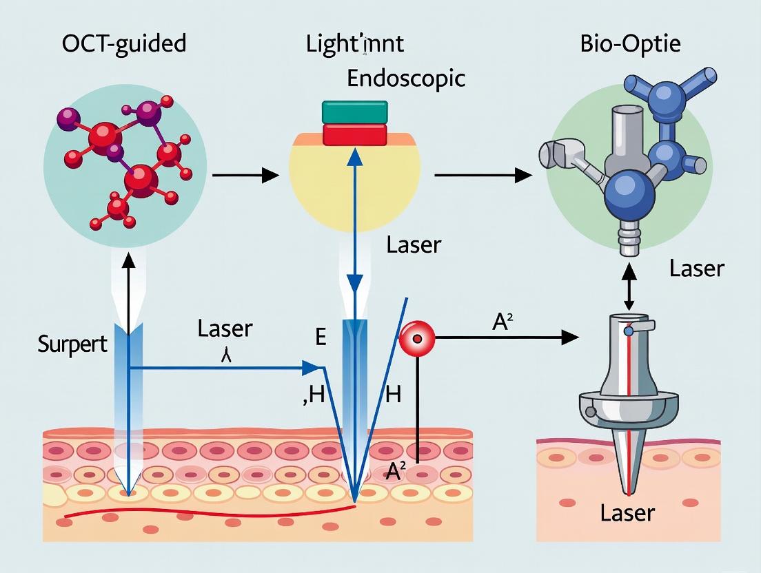
OCT-Guided Endoscopic Laser Surgery: Advanced Techniques, Applications, and Future Directions in Translational Research
This article provides a comprehensive overview of Optical Coherence Tomography (OCT)-guided endoscopic laser surgery for a research and drug development audience.
OCT-Guided Endoscopic Laser Surgery: Advanced Techniques, Applications, and Future Directions in Translational Research
Abstract
This article provides a comprehensive overview of Optical Coherence Tomography (OCT)-guided endoscopic laser surgery for a research and drug development audience. It explores the fundamental principles of OCT imaging and laser-tissue interaction, details current procedural methodologies and applications in preclinical models, addresses common technical challenges and optimization strategies for experimental setups, and critically validates these techniques against alternative modalities. The synthesis aims to inform the development of targeted therapies and refine translational research tools.
Understanding OCT-Guided Laser Surgery: Core Principles for Research and Preclinical Application
Application Notes
The integration of Optical Coherence Tomography (OCT) with laser ablation represents a transformative advancement in endoscopic surgical techniques. This synergy enables real-time, micron-scale cross-sectional imaging to guide and monitor the precise delivery of laser energy. Within the broader thesis on OCT-guided endoscopic laser surgery, this combination directly addresses critical challenges in therapeutic precision, safety, and procedural feedback, particularly for oncology and precise tissue resection applications.
Core Advantages:
- Real-Time Feedback: OCT provides immediate visualization of tissue layers (epithelium, lamina propria, muscularis propria) before, during, and after laser ablation, allowing for on-the-fly adjustment of laser parameters.
- Depth-Resolved Ablation Control: The imaging depth of OCT (1-3 mm in tissue) perfectly overlaps with the typical ablation depth of endoscopic lasers (e.g., Thulium, Holmium). This allows operators to set and confirm ablation endpoints based on anatomical landmarks, minimizing collateral damage.
- Validation of Ablation Zones: OCT can differentiate between zones of complete ablation, thermal coagulation, and unaffected tissue, enabling immediate assessment of treatment efficacy.
Key Quantitative Performance Metrics: Recent studies and device specifications highlight the following capabilities, summarized in the table below.
Table 1: Quantitative Performance Metrics of Integrated OCT-Laser Ablation Systems
| Metric | OCT Imaging Component | Laser Ablation Component | Integrated System Benefit |
|---|---|---|---|
| Axial Resolution | 5 - 15 µm (in tissue) | N/A | Enables layer-specific targeting (e.g., target mucosa, preserve submucosa). |
| Imaging Depth | 1 - 3 mm (in tissue) | N/A | Matches typical ablation depth for endoscopic procedures. |
| Ablation Precision | N/A | 100 - 500 µm (spot size) | Laser spot can be placed with micron-scale accuracy on OCT-identified targets. |
| Imaging Speed | 50 - 250 kHz A-scan rate | N/A | Provides near-video-rate feedback during continuous laser firing. |
| Common Laser Parameters | N/A | Wavelength: 1.9 - 2.1 µm (Tm), 2.1 µm (Ho); Power: 5 - 50 W | OCT visualizes differential thermal effects (vaporization vs. coagulation) based on power/duty cycle. |
| Thermal Coagulation Zone | Can be visualized as a hyper-reflective band | Typically 100 - 300 µm from crater edge | Real-time monitoring allows minimization of this zone when desired. |
Experimental Protocols
The following protocols are designed for in vitro and ex vivo validation of OCT-guided laser ablation, forming a core part of the methodological development for the overarching thesis.
Protocol 2.1: Baseline Characterization of Ablation Craters in Phantom Models
Aim: To establish a correlation between OCT-derived morphological measurements and true physical ablation crater dimensions under controlled laser parameters. Materials:
- Tissue-simulating phantom (e.g., polyacrylamide gel with scatterers).
- Integrated OCT-laser ablation endoscopic probe (e.g., common-a-path design).
- Thulium-doped fiber laser (wavelength: 1940 nm).
- icroscope with calibrated stage for ground-truth measurement.
- Computer for control and data acquisition.
Procedure:
- System Calibration: Align the OCT beam focus with the laser beam focus at a known distance from the probe tip using a calibration target.
- Parameter Matrix: Define a matrix of laser parameters: Power (e.g., 10W, 20W, 30W) and Exposure Time (e.g., 0.5s, 1.0s, 2.0s). Use continuous wave mode.
- Ablation & Imaging: For each parameter set: a. Position the probe perpendicular to and in contact with the phantom surface. b. Acquire and save a pre-ablation OCT M-scan (depth vs. time) at the target location. c. Trigger the laser pulse with the specified parameters. d. Simultaneously, acquire a co-located OCT M-scan during and for 5 seconds post-ablation. e. Acquire a high-resolution post-ablation OCT B-scan (cross-section).
- Validation: a. Section the phantom mechanically at the ablation site. b. Use the calibrated microscope to measure the true crater depth and width. c. From the post-ablation OCT B-scan, measure the apparent crater depth and width.
- Analysis: Perform linear regression to correlate OCT-measured dimensions with ground-truth microscope dimensions. Calculate the systematic offset for future corrections.
Protocol 2.2: Ex Vivo Assessment of Layer-Specific Ablation in Porcine Tissue
Aim: To demonstrate the ability to selectively ablate specific mucosal layers under OCT guidance, a foundational skill for endoscopic surgery. Materials:
- Fresh porcine esophagus or stomach (used within 6 hours of harvest).
- Integrated OCT-laser endoscopic system.
- Saline for irrigation.
- Histology setup (formalin, cassettes, H&E staining).
Procedure:
- Tissue Preparation: Pin the tissue specimen mucosal-side-up in a bath with saline-moistened gauze.
- OCT Landmark Identification: Using the endoscopic probe, acquire OCT B-scans to identify the layered structure: squamous epithelium (superficial hyper-reflective), lamina propria (hypo-reflective), and muscularis mucosa (hyper-reflective band).
- Targeted Ablation: a. Target 1 (Epithelium Only): Select a site. Under real-time OCT M-scan monitoring, apply a low-energy laser pulse (e.g., 5W, 0.2s). Goal: Cessation of ablation upon loss of the hyper-reflective epithelial band. b. Target 2 (Through Muscularis Mucosa): Select a new site. Apply a higher-energy pulse (e.g., 15W, 1.0s). Goal: Continue ablation until the hyper-reflective band of the muscularis mucosa is breached, as seen on OCT.
- Post-Procedure Analysis: a. Acquire high-resolution OCT B-scans of each ablation crater. b. Excise the tissue surrounding each crater and place in formalin for histology processing (H&E stain).
- Correlative Histology: Compare the ablation depth and tissue effects observed in OCT images with the gold-standard histology sections. Note the correlation between OCT hyper-reflectivity at the crater base and the presence of thermal coagulation/necrosis on histology.
The Scientist's Toolkit: Research Reagent Solutions
Table 2: Essential Materials for OCT-Guided Laser Ablation Research
| Item | Function in Research | Example/Notes |
|---|---|---|
| Tm:YAG or Tm:Fiber Laser (1940 nm) | Primary ablation energy source. Strong water absorption provides precise cutting with shallow thermal penetration. | IPG Photonics, Nufern. Key parameter: CW/pulsed power stability. |
| Swept-Source OCT Engine | High-speed imaging core. Enables real-time feedback during dynamic ablation events. | Axsun Technologies, Thorlabs. >100 kHz sweep rate preferred. |
| Common-A-path OCT-Laser Probe | Integrated endoscopic delivery. Ensures perfect co-registration of imaging and ablation beams. | Custom-built or from research partners (e.g., NinePoint Medical). |
| Tissue-Simulating Phantom | Controlled, reproducible substrate for system calibration and initial algorithm development. | Polyacrylamide gel with titanium dioxide (scatterer) and nigrosin (absorber). |
| Ex Vivo Porcine GI Tissue | Anatomically relevant model for developing layer-specific protocols and correlative histology. | Must be fresh (<6 hrs post-harvest) to maintain optical scattering properties. |
| Histology Processing & H&E Staining | Gold-standard validation of ablation depth, thermal damage zone, and tissue layer identification. | Critical for correlating OCT image features with biological structures. |
| High-Speed Data Acquisition System | Synchronizes OCT frame capture, laser trigger signals, and laser power monitoring for precise event correlation. | National Instruments DAQ cards with LabVIEW or custom Python code. |
Visualizations
OCT-Guided Laser Ablation Workflow
OCT Feedback Loop During Laser Ablation
1. Application Notes & Comparative Analysis
Optical Coherence Tomography (OCT) is integral to advancing precision in endoscopic laser surgery, providing real-time, micron-scale morphological and functional imaging. Within the thesis framework of OCT-guided endoscopic laser surgery techniques, the selection of the OCT modality dictates the depth, speed, and functional data available for intraoperative guidance. The following application notes detail the roles of three key systems.
Table 1: Quantitative Comparison of Key OCT Modalities for Endoscopic Guidance
| Parameter | Spectral-Domain OCT (SD-OCT) | Swept-Source OCT (SS-OCT) | Functional OCT Angiography (OCTA) |
|---|---|---|---|
| Central Wavelength | ~840 nm (Superluminescent Diode) | ~1050-1300 nm (Swept Laser) | Integrated with SD-OCT or SS-OCT |
| Axial Scan (A-line) Rate | 50 - 200 kHz | 100 kHz - 1.5+ MHz | Limited by underlying hardware (e.g., 70 kHz for SD-OCTA) |
| Imaging Depth in Tissue | 1.5 - 2.0 mm | 2.5 - 3.5 mm (enhanced by reduced scattering) | Surface vasculature to ~1-2 mm depth |
| Axial Resolution | 5 - 7 µm in tissue | 5 - 10 µm in tissue | Matches base modality resolution |
| Key Advantage for Guidance | High signal-to-noise at superficial depths; cost-effective. | Deeper penetration & faster imaging; better for full-wall visualization. | Non-contact, dye-less microvasculature mapping (vessel density ~3-30 µm diameter). |
| Primary Surgical Guidance Role | Precise ablation layer targeting (e.g., mucosal lesions). | Navigation through deeper tissue layers and around complex anatomy. | Monitoring perfusion changes pre/post laser ablation; identifying angiogenic "hot spots." |
| Limitation in Endoscopic Context | Limited depth; sensitivity roll-off. | Higher system cost & complexity. | Motion artifact susceptibility; requires advanced processing. |
2. Experimental Protocols for Thesis Research
Protocol 2.1: Comparative Ex Vivo Tissue Imaging for Modality Selection Objective: To establish baseline performance metrics of SD-OCT, SS-OCT, and OCTA on target tissue types (e.g., porcine bladder, esophageal, or colorectal specimens) for laser surgery simulation.
- Tissue Preparation: Secure fresh ex vivo tissue samples in a custom holder mimicking endoscopic geometry. Maintain hydration with phosphate-buffered saline.
- OCT Probe Integration: Mount a miniature OCT probe (e.g., 2.4mm diameter) within a simulated endoscopic channel. Ensure stable, perpendicular contact with tissue.
- Sequential Imaging: Acquire 3D volumes (3mm x 3mm area) at the same site using:
- SD-OCT System: A-line rate: 70 kHz. Volume: 512 x 512 A-lines.
- SS-OCT System: A-line rate: 200 kHz. Volume: 1000 x 500 A-lines.
- OCTA Protocol (on SS-OCT hardware): Acquire 4 repeated B-scans at 500 sequential positions. Use speckle variance algorithm to generate angiograms.
- Analysis: Measure signal decay with depth, contrast of layered structures, and angiogram signal-to-noise ratio. Tabulate for direct comparison to inform in vivo modality choice.
Protocol 2.2: Real-Time OCT-Guided Laser Ablation in a Tissue Phantom Objective: To develop a closed-loop feedback protocol for laser power modulation based on OCT depth-resolved analysis.
- Phantom Fabrication: Create a layered phantom with 1% agarose (scattering layer) and embedded polyester fibers (simulating vessels >50µm).
- Integrated System Setup: Co-align a thulium fiber laser (λ=1940 nm) aiming beam with the SS-OCT imaging beam via a dichroic mirror in a common-path probe.
- Guidance Workflow:
- Acquire a pre-ablation SS-OCT B-scan and OCTA map.
- Define an ablation target region (e.g., "ablate to a depth of 800µm").
- Initiate laser pulses (10 mJ, 10 Hz) while continuously acquiring OCT M-scans (repeated A-scans at one position).
- Implement real-time software to track the ablation front (identified by increased signal intensity) through the OCT M-scans.
- Automatically terminate laser emission when the tracked front reaches the 800µm threshold.
- Validation: Histologically section the phantom to measure actual ablation depth and compare to the OCT-predicted depth.
Protocol 2.3: Longitudinal OCTA Monitoring of Perfusion Post-Laser Intervention Objective: To quantify microvascular changes following precise laser microsurgery using functional OCTA.
- In Vivo Model Preparation: Use a dorsal skinfold chamber model in rodents, approved by the IACUC.
- Baseline Imaging: Under anesthesia, acquire high-resolution 3D OCTA maps (using SS-OCTA at 300 kHz) of a selected microvascular network.
- Targeted Laser Intervention: Use an argon laser (λ=488 nm) coupled to the system to induce selective photothrombosis in a single target vessel (diameter 20-50 µm), guided by the live OCTA feed.
- Longitudinal Monitoring: Re-image the same region at 1 hr, 24 hrs, 48 hrs, and 7 days post-intervention.
- Quantitative Analysis: Use vessel segmentation software to calculate metrics: Vessel Density (%), Vessel Diameter Index (µm), and Non-Perfused Area (µm²) over time to assess occlusion, collateralization, and reperfusion.
3. Visualized Workflows & Pathways
OCT Modality Pathways to Surgical Guidance
OCT Modality Selection Logic for Surgery
4. The Scientist's Toolkit: Key Research Reagent Solutions
Table 2: Essential Materials for OCT-Guided Endoscopic Surgery Research
| Item | Function & Relevance in Research |
|---|---|
| Micro-Integrated OCT-Laser Probes (Common-Path) | Miniaturized (<2.8mm) fiber-optic bundles co-aligning OCT imaging and surgical laser beams, enabling simultaneous imaging and intervention. Critical for in vivo endoscopic studies. |
| Tissue-Simulating Phantoms (Layered Agarose with Scatterers) | Calibrated phantoms (e.g., with TiO₂ or Al₂O₃ powder) mimicking tissue optical properties (scattering, absorption). Used to validate imaging depth, resolution, and laser-tissue interaction models before in vivo work. |
| Indocyanine Green (ICG) | FDA-approved NIR fluorescence dye. Can be used for complementary fluorescence angiography to validate OCTA findings regarding perfusion and vessel leakage in animal models. |
| Vessel Segmentation Software (e.g., OCTA-AV, ORS Dragonfly) | Advanced image analysis platforms with custom algorithms for quantifying OCTA metrics: Vessel Density, Fractal Dimension, Vessel Diameter. Essential for objective analysis of functional changes. |
| Motion Stabilization Platform (Precision Linear Stage) | High-precision motorized stage for ex vivo and phantom studies. Allows for reproducible scanning and precise co-registration of pre- and post-ablation OCT volumes for accurate change detection. |
| Real-Time Signal Processing SDK (e.g., CUDA-based) | Software development kit enabling custom implementation of real-time OCT signal processing (FFT, speckle variance), crucial for developing intraoperative ablation front tracking and decision support algorithms. |
This application note details the fundamental laser-tissue interactions critical to the development of precise, image-guided endoscopic surgical tools. Within the broader thesis of OCT-guided laser surgery, understanding and controlling photothermal, photomechanical, and photochemical effects is paramount for achieving targeted ablation, hemostasis, or photodynamic therapy while minimizing collateral damage. These principles enable the translation of real-time optical coherence tomography (OCT) imaging data into controlled laser dosimetry.
Core Interaction Mechanisms & Quantitative Parameters
Photothermal Effects
Photothermal interactions involve the conversion of laser light into heat, leading to temperature-dependent tissue changes. The spatial and temporal profile of laser energy delivery dictates the outcome.
Table 1: Photothermal Tissue Effects and Threshold Parameters
| Biological Effect | Temperature Range (°C) | Time Scale | Primary Observable Change | Common Laser Types |
|---|---|---|---|---|
| Hyperthermia | 45 – 50 | Seconds-Minutes | Protein denaturation, increased perfusion | Diode (810 nm), Nd:YAG (1064 nm) |
| Coagulation | 60 – 80 | Seconds | Protein coagulation, necrosis, hemostasis | Nd:YAG, Thulium (1940 nm) |
| Vaporization | 100 | Milliseconds | Water boiling, steam formation | CO₂ (10.6 µm), Er:YAG (2940 nm) |
| Carbonization | > 150 | Milliseconds | Tissue drying, charring | High-power pulsed lasers |
| Melting & Ablation | > 300 | Microseconds | Direct solid-to-plasma transition | Excimer (193 nm), Holmium (2120 nm) |
Photomechanical Effects
Photomechanical interactions result from rapid energy deposition, generating mechanical forces (stress confinement, shock waves, cavitation) that disrupt tissue.
Table 2: Photomechanical Regimes and Key Variables
| Regime | Pulse Duration | Energy Density (J/cm²) | Primary Mechanism | Typical Application |
|---|---|---|---|---|
| Spallation/Plasma | Nanosecond (10⁻⁹ s) | 0.1 – 10 | Stress confinement, shock wave | Lithotripsy, tissue dissection |
| Cavitation | Microsecond to Nanosecond | 0.01 – 1 | Rapid bubble formation/ collapse | Laser-induced breakdown spectroscopy (LIBS) |
| Photoablation | Picosecond/Femtosecond (10⁻¹²-10⁻¹⁵ s) | 0.1 – 5 | Coulomb explosion, cold ablation | High-precision corneal surgery, micromachining |
Photochemical Effects
Photochemical effects involve non-thermal, molecular-level interactions where photons are absorbed by chromophores, initiating chemical reactions.
Table 3: Photochemical Interactions in Therapeutics
| Interaction Type | Wavelength Range (nm) | Chromophore/Target | Therapeutic Outcome | Example Agent |
|---|---|---|---|---|
| Photodynamic Therapy (PDT) | 600 – 800 (Visible/NIR) | Exogenous Photosensitizer (e.g., PpIX) | Reactive oxygen species (ROS) generation, cell death | 5-ALA, Photofrin |
| Low-Level Laser Therapy (LLLT) | 600 – 1000 | Mitochondrial cytochromes | Biostimulation, reduced inflammation | N/A |
| UV Disinfection | 200 – 280 (UVC) | DNA/RNA nucleotides | Microbial inactivation | N/A |
Experimental Protocols for Interaction Analysis
Protocol 3.1: Ex Vivo Quantification of Photothermal Ablation Thresholds
Aim: To determine the irradiance and exposure time required for vaporization and coagulation in porcine mucosal tissue. Materials: See Scientist's Toolkit (Section 5). Method:
- Prepare uniform 2 mm-thick sections of fresh porcine stomach mucosa on a temperature-controlled stage (maintained at 37°C).
- Using a 1470 nm diode laser coupled to a 400 µm core silica fiber, deliver a series of continuous-wave exposures. Vary power (1-10 W) and time (0.5-5 s). Use a calibrated beam profiler to confirm spot diameter (e.g., 2 mm).
- For each exposure, record the surface temperature in real-time using a mid-infrared thermal camera.
- Post-exposure, fix tissue in formalin, section, and stain with H&E.
- Analysis: Measure the depth and width of coagulation necrosis (pale eosinophilic region) and vaporization (tissue defect). The ablation threshold is defined as the minimum energy density (J/cm²) producing a visible vaporization crater.
Protocol 3.2: High-Speed Imaging of Photomechanical Cavitation
Aim: To characterize cavitation bubble dynamics induced by pulsed holmium:YAG laser in a liquid/tissue environment. Materials: Holmium:YAG laser (λ=2120 nm, 250 µs pulse), high-speed camera (>500,000 fps), transparent tissue phantom (e.g., agarose), fiber optic delivery (365 µm core). Method:
- Submerge the fiber tip and tissue phantom in a water tank.
- Align the high-speed camera orthogonal to the fiber axis with backlight illumination.
- Deliver single laser pulses at varying energies (100-500 mJ).
- Record the bubble formation, expansion, and collapse sequence.
- Analysis: Use image analysis software to measure maximum bubble radius (Rmax) versus laser pulse energy. Calculate the pressure transients using the Rayleigh-Plesset equation model.
Protocol 3.3: In Vitro Protocol for PDT Efficacy Assessment
Aim: To evaluate the cytotoxic effect of 5-ALA-mediated PDT on a colorectal cancer cell line (HCT-116) under endoscopic-like light delivery. Materials: HCT-116 cells, 5-aminolevulinic acid (5-ALA), 635 nm diode laser, fiber optic microlens, ROS detection kit (e.g., DCFH-DA), cell viability assay (MTT). Method:
- Seed cells in 96-well plates and incubate with 1 mM 5-ALA for 4 hours.
- Wash cells to remove extracellular 5-ALA, allowing intracellular conversion to protoporphyrin IX (PpIX).
- Using a 635 nm laser coupled to a 200 µm fiber with a microlens, irradiate wells with a range of light doses (0-100 J/cm² at 100 mW/cm²). Maintain temperature control.
- Immediately post-irradiation, assay for ROS generation using DCFH-DA.
- 24 hours post-PDT, perform MTT assay to quantify cell viability.
- Analysis: Plot viability and ROS levels versus light dose to establish a dose-response curve, calculating the lethal dose 50% (LD50).
Visualized Pathways and Workflows
Title: Decision Tree for Dominant Laser-Tissue Interaction Type
Title: OCT-Guided Endoscopic Laser Surgery Feedback Protocol
The Scientist's Toolkit: Research Reagent & Material Solutions
Table 4: Essential Materials for Laser-Tissue Interaction Research
| Item Name | Function & Relevance |
|---|---|
| Tissue-Mimicking Phantoms (Agarose with Intralipid/Ink) | Provides standardized optical properties (µa, µs') for calibrating laser delivery and imaging systems before ex vivo or in vivo work. |
| Calibrated Optical Power & Energy Meters (Thermopile, Photodiode) | Essential for accurate dosimetry. Thermopile sensors are preferred for high-power CW/pulsed IR lasers. |
| High-Speed Imaging System (>1,000,000 fps capable) | Captures transient photomechanical events (cavitation, plasma formation) for mechanistic studies. |
| Infrared Thermal Camera (3-5 µm or 8-14 µm spectral range) | Non-contact mapping of surface temperature distribution during photothermal experiments. Critical for validating thermal models. |
| ROS Detection Probe (e.g., DCFH-DA, Singlet Oxygen Sensor Green) | Quantifies reactive oxygen species generation in photochemical studies (PDT). |
| OCT System (Spectral-Domain or Swept-Source) | Provides the core structural feedback for the thesis context. Enables measurement of lesion depth, boundary, and birefringence changes in real-time. |
| Flexible Laser Delivery Fibers (Low-OH Silica, Hollow-Core for Mid-IR) | Enables endoscopic delivery. Choice depends on laser wavelength (e.g., silica for UV-Vis-NIR up to ~2.2 µm, hollow-core or specialty fibers for CO₂/Er:YAG). |
| Photosensitizer Kits (e.g., 5-ALA hydrochloride, Verteporfin) | Standardized reagents for initiating and studying photochemical interactions in cellular and animal models. |
The convergence of Optical Coherence Tomography (OCT) with laser ablation represents a paradigm shift in endoscopic surgery, enabling real-time, micron-scale visualization concurrent with precision tissue modification. This integrated system architecture is foundational to the broader thesis on OCT-guided endoscopic laser surgery techniques. The core challenge involves the co-alignment of high-resolution, depth-resolved imaging (OCT) and therapeutic laser energy delivery within the stringent size constraints of an endoscopic channel, without compromising the performance of either modality. This application note details the essential architecture, protocols, and reagents for developing such integrated systems for translational research.
Quantitative System Parameters & Data
The integration requires careful balancing of optical, mechanical, and thermal parameters. The following tables summarize critical quantitative benchmarks.
Table 1: Core Optical & Mechanical Specifications for Integrated Probes
| Parameter | OCT Subsystem Typical Value | Laser Delivery Subsystem Typical Value | Integration Constraint |
|---|---|---|---|
| Central Wavelength | 1300 nm (for deep tissue) | 1940 nm (Thulium), 1470 nm (Diode) or 1064 nm (Nd:YAG) | Spectral isolation to prevent crosstalk. |
| Axial/Transverse Resolution | 5-15 µm / 10-30 µm | N/A (Ablation spot size: 200-1000 µm) | OCT resolution must inform laser targeting precision. |
| Working Distance | 2-5 mm (focused probe) | 1-3 mm (for precise ablation) | Must be matched or dynamically adjustable. |
| Scanning Method | Distal MEMS mirror or proximal rotary joint | Shared scanning element or separate fixed fiber | Co-alignment error < 50 µm. |
| Fiber Core Diameter | Singlemode (SMF-28, ~9 µm) | Multimode (200-600 µm for power delivery) | Parallel or combined in a dual-clad fiber. |
| Outer Diameter (Probe) | 2.0-2.5 mm (for standalone) | <1.5 mm (for bare delivery fiber) | Combined probe must fit within a 3.3 mm (10 Fr) endoscopic working channel. |
Table 2: Performance Benchmarks from Recent Studies (2023-2024)
| Study Focus | Integrated Modality | Key Quantitative Outcome | Reference (Type) |
|---|---|---|---|
| Bile Duct Imaging/Ablation | OCT + 1470 nm Diode Laser | Co-registration accuracy: 35 ± 12 µm; Ablation depth control to ± 100 µm under OCT guidance. | Preprint (BioRxiv) |
| Bladder Tumor Resection | OCT + Ho:YAG Laser | Real-time distinction of tissue layers < 50 µm; Reduced perforation risk in ex vivo models by 70%. | Journal (Biomed. Opt. Express) |
| Cardiac Ablation Therapy | OCT + 1064 nm Laser | Simultaneous imaging & ablation at 100 Hz; Lesion depth predictability R²=0.89 with OCT feedback. | Conference (SPIE Photonics West) |
Experimental Protocols
Protocol 1: Assembly and Co-Alignment Calibration of a Dual-Function Probe
Objective: To integrate a single-mode OCT fiber and a multimode laser delivery fiber into a common distal housing and achieve sub-50µm co-alignment. Materials: Single-mode OCT fiber (SMF-28e), Multimode silica fiber (365µm core, 0.22 NA), GRIN lens, MEMS micro-mirror, custom stainless steel ferrule, 5-axis fiber alignment stage, optical power meter, IR viewer, USAF resolution target.
- Fiber Preparation: Cleave both fibers. The multimode fiber jacket is stripped back 3 cm for insertion into ferrule.
- Distal Optics Assembly: Using a guide pin fixture, secure the SMF and MMF side-by-side within the steel ferrule (OD 1.8 mm). Epoxy cure.
- Lens Attachment: A single GRIN lens, sized to cover both fiber cores, is aligned for optimal OCT focus (tested on USAF target) and attached to the ferrule face.
- MEMS Integration: A 1D or 2D MEMS mirror is mounted distal to the lens, angled to scan the combined beam.
- Co-Alignment Calibration:
- Connect the MMF to a low-power visible aiming laser (e.g., 635 nm).
- Project the aiming beam onto a gridded target 2 mm away (simulating WD).
- Activate the OCT scanning and map its field of view.
- Using the 5-axis stage, adjust the MMF position until the stationary aiming beam spot is centered within the OCT scan field. Measure offset error.
- Iterate until offset is < 50 µm. Secure with UV-curing adhesive.
- Validation: Image a patterned target with OCT. Mark the target center via OCT coordinates. Fire a low-energy laser pulse. Measure the distance between the ablation mark and the OCT-defined center.
Protocol 2: Ex Vivo Validation of OCT-Guided Laser Ablation Depth Control
Objective: To demonstrate the use of pre-ablation OCT imaging to predict and control laser ablation depth in tissue. Materials: Integrated OCT-laser probe, swept-source OCT engine (1300 nm), Diode laser (1470 nm), fresh porcine tissue (stomach or bladder), tissue holder, micro-positioning stage, histological cassettes, H&E staining setup.
- System Setup: Mount the integrated probe on a 3-axis stage. Connect to respective systems. Calibrate laser power (0.5-3 W, continuous wave).
- Baseline OCT Scan: Position probe 2 mm above tissue surface using OCT depth profile. Acquire a 3D OCT volume (e.g., 5mm x 5mm x 3mm depth).
- Layer Identification: Use OCT software to identify the mucosal surface and measure the thickness of the superficial layer (e.g., mucosa, ~500-1000 µm).
- Guided Ablation: Set the desired ablation depth (e.g., 80% of mucosal thickness). Using the OCT-measured distance, activate the laser for a controlled duration (e.g., 0.5-2 s) while monitoring A-scans at the target site for real-time depth assessment.
- Post-Ablation Scan: Immediately acquire a post-ablation OCT volume to measure crater depth and morphology.
- Histological Correlation: Excise the ablated region, fix in formalin, process for H&E sectioning perpendicular to the ablation crater. Measure ablation depth and thermal damage zone microscopically.
- Data Analysis: Correlate the intended depth (from pre-op OCT), the real-time OCT-estimated depth, and the histologically measured depth (n≥10 sites). Calculate mean absolute error and R² value.
Visualizations
Integrated OCT-Laser System Data Flow
OCT-Guided Laser Ablation Experimental Workflow
The Scientist's Toolkit: Research Reagent Solutions
Table 3: Essential Materials for Integrated OCT-Laser Probe Development & Testing
| Item | Function & Rationale | Example Product/Catalog |
|---|---|---|
| Dual-Clad Fiber (DCF) | Function: Combines single-mode core for OCT and large inner cladding for laser delivery in one fiber. Rationale: Simplifies probe assembly, ensures perfect inherent co-registration. | Example: Nufern LMA-GDF-10/125-M, DCF with 9µm SM core & 105µm inner cladding. |
| MEMS Mirror (1D/2D) | Function: Provides distal, fast optical scanning for both imaging and laser aiming. Rationale: Enables compact, high-speed beam steering without proximal rotation. | Example: Mirrorcle Technologies A7B-2.0 MEMS mirror, 2-axis, <2mm package. |
| Index-Matching UV Epoxy | Function: Secures optical components (fibers, lenses) with minimal refractive index discontinuity and fast curing. Rationale: Reduces back-reflections and signal loss. | Example: Norland Optical Adhesive 81 (n=1.56, UV cure). |
| Precision Ferrule & Sleeve | Function: Provides robust, aligned housing for side-by-side fiber assembly at the probe tip. Rationale: Critical for maintaining co-alignment under mechanical stress. | Example: Custom stainless steel ferrule with dual bore (125µm & 400µm). |
| Tissue Phantom (Layered) | Function: Calibrates and validates system co-registration and ablation depth control. Rationale: Provides a reproducible, non-biological standard with known optical & thermal properties. | Example: Polyacrylamide gel with titanium oxide scatterers and absorbing ink layers. |
| Broadband IR Detector Card | Function: Visualizes and aligns near-IR (1064-2000 nm) laser beams safely. Rationale: Essential for co-alignment calibration when using invisible therapeutic wavelengths. | Example: Thorlabs DCC1545M-VIS/IR Detector Card. |
| High-Power Laser Diode Driver | Function: Provides precise, stable current to drive therapeutic diode lasers (e.g., 1470 nm). Rationale: Enables controlled, repeatable energy delivery for ablation studies. | Example: Wavelength Electronics QCL500 or ILX Lightwave LDX-3412. |
This application note details integrated imaging and laser surgery protocols, developed under a broader thesis on OCT-guided endoscopic laser surgery, to enable precise subcellular intervention.
Quantitative Performance Metrics of Integrated Imaging-Surgery Platforms
Table 1: Comparison of Current Integrated Imaging & Targeting Modalities
| Modality | Axial/Lateral Resolution (Imaging) | Targeting Precision | Maximum Imaging Depth in vivo | Key Enabling Technology | Primary Limitation |
|---|---|---|---|---|---|
| OCT-guided Femtosecond Laser | 1-5 µm / 5-15 µm | ~1 µm (subcellular) | 1-3 mm (scattering tissue) | Adaptive Optics (AO), Two-Photon Excitation | Limited penetration; Complex/expensive setup. |
| Confocal Microscopy-guided Microbeam Laser | 0.5-1.5 µm / 0.2-0.7 µm | ~0.5 µm (organelle-level) | 200-500 µm | Resonant Scanning, Galvanometer Mirrors | Very shallow penetration; limited field of view. |
| Multiphoton Microscopy-guided Nanosurgery | 0.8-2 µm / 0.3-0.8 µm | <1 µm (subcellular) | Up to 1 mm | Near-Infrared Femtosecond Lasers, Deep Learning ROI ID | Slow volumetric imaging; non-linear photodamage risk. |
| Super-resolution OCT (SR-OCT) with Targeting | 0.7-2 µm / 2-5 µm | 1-3 µm | 0.5-2 mm | Interferometric Synthetic Aperture, Speckle Modulation | Computationally intensive; in vivo speed challenges. |
Table 2: Common Laser Parameters for Subcellular Surgical Tasks
| Surgical Target | Laser Type | Wavelength | Pulse Duration | Pulse Energy | Repetition Rate | Outcome Metric |
|---|---|---|---|---|---|---|
| Mitochondrial Ablation | Femtosecond | 800-850 nm | 100-200 fs | 1-5 nJ | 80 MHz | ΔΨm loss in <5 min (measured via TMRM dye). |
| Nuclear Membrane Perforation | Nanosecond | 532 nm | 4-10 ns | 10-30 µJ | 1-10 Hz | ~80% plasmid transfection efficiency. |
| Single-Axon Transection | Femtosecond | 1040 nm | 200-300 fs | 10-15 nJ | 1-10 kHz | Clean cut, >90% viability of soma. |
| Lysosome Disruption | Picosecond | 1064 nm | 10-20 ps | 0.5-2 µJ | 100-1000 Hz | Cathepsin B release peak at 15 min post-irradiation. |
Experimental Protocols
Protocol A: OCT-Guided Femtosecond Laser Ablation of Single MitochondriaIn Vitro
Objective: To precisely ablate individual mitochondria in live cells using AO-OCT for guidance and a femtosecond laser for surgery. Materials: See "The Scientist's Toolkit" (Table 3). Workflow:
- Cell Preparation: Seed H9c2 cardiomyocytes in glass-bottom dishes. Incubate with 100 nM MitoTracker Deep Red FM for 30 min. Replace with live-cell imaging medium.
- System Setup & Calibration:
- Align the OCT and femtosecond laser beam paths to be confocal. Calibrate using fluorescent microspheres (0.5 µm).
- Implement sensorless AO: Capture an initial OCT volumetric stack, apply iterative wavefront corrections using a deformable mirror to optimize image sharpness.
- Target Identification & Guidance:
- Acquire a high-resolution, AO-corrected OCT volume (1 µm axial, 3 µm lateral) of the target cell.
- Correlate OCT data with a widefield fluorescence image (optional) to confirm mitochondrial localization.
- Select a target mitochondrion using the software interface; the system registers its 3D coordinates.
- Laser Surgery:
- Set femtosecond laser parameters: 800 nm, 100 fs pulse width, 2 nJ pulse energy, 80 MHz rep rate.
- Position laser focus at the target coordinates. Deliver a 50 ms continuous exposure.
- Validation & Analysis:
- Immediately post-ablation, acquire a fluorescence image to confirm loss of MitoTracker signal at the target site.
- Monitor cell viability via propidium iodide exclusion for ≥60 minutes.
- Quantify changes in mitochondrial membrane potential using ratiometric JC-1 dye in separate experiments.
Protocol B: Endoscopic OCT-Guided Laser Microdissection of Colonic CryptsEx Vivo
Objective: To perform site-specific laser microdissection of single crypts from fresh colon tissue for downstream genomic analysis, using an endoscopic OCT probe for guidance. Materials: Endoscopic OCT probe (spectral-domain, 1.3 µm central wavelength), integrated microdissection laser (355 nm nanosecond pulsed), fresh murine colon tissue, RNAlater stabilization solution. Workflow:
- Sample Mounting & Imaging:
- Secure a segment of freshly harvested murine colon mucosa on a biopsy mount with the luminal surface exposed.
- Insert the dual-modality endoscope. Acquire real-time OCT video (frame rate >20 Hz) to identify the crypt architecture.
- Target Selection & Registration:
- Navigate the probe to a region of interest. Acquire and freeze a high-resolution OCT B-scan.
- Use the software to outline a single crypt (50-100 µm diameter) on the OCT image. The system maps this region to the galvo-scanner coordinates of the UV microdissection laser.
- Laser Microdissection:
- Set UV laser to low pulse energy (5 µJ) and high repetition rate (1 kHz) for precise cutting.
- Activate the laser to trace the outlined perimeter. A motorized stage moves the tissue slightly to allow the laser to cut through the basement membrane.
- Sample Collection & Processing:
- Use a micro-capillary tube or adhesive cap to collect the freed crypt.
- Immediately transfer the crypt into a tube containing RNAlater for RNA preservation.
- Proceed with single-crypt RNA sequencing library preparation.
Visualized Workflows & Pathways
Title: Integrated OCT-Guided Subcellular Surgery Workflow
Title: Pathway of Laser-Induced Mitochondrial Apoptosis
The Scientist's Toolkit
Table 3: Key Research Reagent Solutions for OCT-Guided Laser Surgery
| Item | Function in Protocol | Example Product/ Specification |
|---|---|---|
| Live-Cell Mitochondrial Dye (MitoTracker Deep Red) | Fluorescent labeling for target validation and post-operative confirmation. | Thermo Fisher Scientific, M22426. Ex/Em ~644/665 nm. |
| Ratiometric JC-1 Dye | Quantitative measurement of mitochondrial membrane potential (ΔΨm) pre- and post-surgery. | Abcam, ab113850. Monomer (green) vs. J-aggregate (red) ratio. |
| AO-OCT System with Deformable Mirror | Corrects optical aberrations in real-time for subcellular resolution in depth. | Boston Micromachines Multi-DM + custom OCT. >200 actuators. |
| Femtosecond Laser System | Delivers ultra-short pulses for precise, non-linear photodisruption with minimal collateral damage. | Coherent Chameleon Discovery, tunable 680-1300 nm, 80 MHz. |
| Endoscopic OCT-Microdissection Probe | Dual-channel probe for real-time subsurface imaging and targeted laser cutting in situ. | Custom-built, 2.4 mm outer diameter, SD-OCT engine. |
| RNAlater Stabilization Solution | Immediately stabilizes and protects RNA in microdissected samples for omics analysis. | Thermo Fisher Scientific, AM7020. |
Protocols and Preclinical Applications: Implementing OCT-Guided Laser Surgery in Research
Abstract This document provides application notes and a standardized protocol for in vivo preclinical studies within a broader research thesis on Optical Coherence Tomography (OCT)-guided endoscopic laser surgery techniques. The workflow is designed to ensure reproducibility in data acquisition for therapeutic ablation, enabling robust validation in drug development and surgical technology research.
Application Notes: Core Principles & Quantitative Benchmarks
OCT-guided laser ablation combines high-resolution, cross-sectional imaging with precise photothermal intervention. This integrated approach is critical for targeting subsurface structures in hollow organs with minimal collateral damage.
Table 1: Quantitative Performance Metrics for OCT-Guided Laser Ablation Systems
| Parameter | Typical Specification/Range | Functional Significance |
|---|---|---|
| OCT Axial Resolution | 1 - 15 µm | Determines layer differentiation capability in tissue. |
| OCT Imaging Depth | 1 - 3 mm (in tissue) | Limits depth of real-time visualization for guidance. |
| Laser Wavelength | 800 nm, 980 nm, 1064 nm, 1940 nm, 10.6 µm | Selection based on target chromophore (water, hemoglobin) and desired absorption profile. |
| Ablation Spot Size | 50 - 500 µm | Determines spatial precision of the therapeutic effect. |
| Typical Power Range | 0.5 - 5.0 W (CW or pulsed) | Must be calibrated to achieve desired thermal dose without carbonization. |
| Real-time Frame Rate | 10 - 100 frames/sec | Higher rates enable tracking of dynamic tissue changes during ablation. |
| Temperature Rise ΔT | 50 - 100 °C (at focus) | Must be controlled to confine thermal damage to target zone. |
Table 2: Common Animal Models & Preparation Parameters
| Species/Model | Common Application (e.g., Organ) | Typical Anesthesia & Analgesia | Key Anatomical Consideration |
|---|---|---|---|
| Mouse (C57BL/6) | Colon, Esophagus | Isoflurane (1-3%), Buprenorphine SR | Small lumen size requires micro-endoscopic tools. |
| Rat (Sprague-Dawley) | Bladder, Stomach | Isoflurane (2-4%), Buprenorphine | Larger size facilitates instrument manipulation. |
| Porcine (Domestic) | GI Tract, Airways | Propofol infusion, Fentanyl patch | Translational model; anatomy closely resembles human. |
Standardized Experimental Protocol
2.1 Pre-Procedure: Animal Preparation & System Setup
- Anesthesia Induction & Maintenance: Induce rodent with 4% isoflurane in O₂, maintain at 1.5-2.5% via nose cone. For terminal procedures, administer a pre-operative analgesic (e.g., Buprenorphine, 0.05 mg/kg SC). For porcine models, follow approved protocols for endotracheal intubation and total intravenous anesthesia.
- Physiological Monitoring: Maintain body temperature at 37±0.5°C using a feedback-controlled heating pad. Monitor heart rate and SpO₂ throughout.
- Endoscopic Access: Gently introduce the integrated OCT-laser endoscope. For lower GI procedures, perform a saline flush to clear debris. Secure the endoscope position using a stereotactic stage.
- System Calibration: Align the OCT imaging beam and laser aiming beam to ensure perfect co-registration. Perform a power output calibration at the fiber tip using a photodiode power meter.
2.2 Intra-Procedure: Real-Time Guidance & Ablation
- Baseline OCT Scan: Acquire and store a 3D volumetric OCT scan of the target region. Key landmarks and the target (e.g., tumor, specific mucosal layer) should be identified.
- Target Delineation: Use the system software to place a virtual marker or region-of-interest (ROI) on the real-time OCT B-scan or en-face view.
- Laser Parameter Setting: Based on target depth and desired lesion size, set parameters (see Table 3). Always start with lower power in a pilot area.
- Ablation Execution & Monitoring: Activate the laser for the prescribed duration while simultaneously monitoring the OCT signal in real-time. Observe the rapid development of a hyper-scattering region and shadowing beneath it, indicating bubble formation and coagulation.
- Post-Ablation Assessment: Immediately acquire a post-ablation 3D OCT scan to document the immediate morphologic change (lesion dimensions, architectural disruption).
2.3 Post-Procedure: Tissue Harvest & Analysis
- Euthanasia & Harvest: Euthanize animal per AVMA guidelines (e.g., CO₂ followed by cervical dislocation for rodents). Excise the target organ and immediately place in 10% neutral buffered formalin.
- Histological Processing: Fix for 24-48 hours, section transversely through the ablation center, process, and embed in paraffin. Generate 5 µm H&E stained sections.
- Lesion Analysis: Correlate OCT-derived lesion dimensions (width, depth of hyper-scattering) with histology (coagulation zone, vaporization cavity, border of inflammatory cells). Use trichrome stain to assess collagen denaturation.
Table 3: Example Ablation Protocol for Colonic Mucosa (1064 nm Laser)
| Step | Parameter | Setting/Range | Objective/Endpoint |
|---|---|---|---|
| 1 | Laser Mode | Continuous Wave (CW) | Uniform photothermal heating. |
| 2 | Power | 1.5 W | Sufficient for coagulation, minimizes carbonization. |
| 3 | Spot Diameter | 200 µm | Defines lateral treatment zone. |
| 4 | Duration | 0.5 - 2.0 s | Controlled by thermal diffusion time. |
| 5 | Endpoint on OCT | Appearance of bright, persistent hyper-scattering band. | Visual confirmation of coagulative change. |
Visualization of Core Concepts
OCT-Guided Laser Ablation Workflow
Photothermal Ablation Mechanism
The Scientist's Toolkit: Research Reagent Solutions
Table 4: Essential Materials & Reagents for OCT-Guided Ablation Studies
| Item | Function/Application | Example Product/Catalog |
|---|---|---|
| Integrated OCT-Laser Endoscope | Combines imaging and therapeutic channels in a single device for co-registered guidance. | Custom-built or Thorlabs Ganymede-II with laser port. |
| Tunable or Fixed-Wavelength Diode Laser | Provides the photothermal energy source for ablation. | IPG Photonics LASER-1064-LM, or Dornier MediLas A. |
| Isoflurane, USP | Volatile anesthetic for induction and maintenance in rodent and small animal models. | Piramal Critical Care, NDC 66794-017-25. |
| Buprenorphine SR | Sustained-release analgesic for pre- and post-operative pain management. | ZooPharm, 0.5 mg/mL. |
| Phosphate-Buffered Saline (PBS) | For flushing the endoscopic field of view and clearing debris during procedure. | Gibco, 10010023. |
| 10% Neutral Buffered Formalin | Gold-standard fixative for histopathological analysis post-ablation. | Sigma-Aldrich, HT501128. |
| Hematoxylin & Eosin (H&E) Stain Kit | Standard stain for visualizing general tissue morphology and ablation zones. | Abcam, ab245880. |
| Masson's Trichrome Stain Kit | Special stain to highlight collagen denaturation at the ablation border. | Sigma-Aldrich, HT15-1KT. |
| Thermocouple Microprobe (50 µm) | For independent validation of temperature rise during ablation (ex vivo calibration). | Physitemp, IT-23. |
Application Notes
Optical Coherence Tomography (OCT)-guided endoscopic laser surgery integrates real-time, high-resolution cross-sectional imaging with precise laser ablation, enabling targeted interventions within delicate tissues. This synergistic approach is critical for applications requiring micron-scale precision and minimal collateral damage.
Oncology: Tumor Microsurgery
OCT-guided laser microsurgery allows for the precise demarcation and ablation of tumor margins in real-time. By differentiating between malignant and healthy tissue based on optical backscatter properties, surgeons can perform sub-millimeter resections, potentially improving oncologic outcomes while preserving adjacent critical structures. This is particularly valuable for early-stage cancers and in organs where maximal parenchymal preservation is crucial (e.g., larynx, brain, bladder).
Neurology: Precise Neural Interventions
In neurological research and surgery, OCT guidance facilitates laser ablation or modulation of specific neural tracts, cortical layers, or pathological foci (e.g., epileptogenic zones) with minimal disruption to surrounding functional tissue. Endoscopic delivery enables deep-brain access without large craniotomies. The technique is also used to create precise injury models or to study neural regeneration.
Ophthalmology: Subcellular Precision
OCT provides unparalleled depth resolution for targeting retinal layers, the trabecular meshwork, or the corneal stroma. Guided laser procedures can be used for selective retinal therapy (targeting the retinal pigment epithelium while sparing photoreceptors), micro-incisions, or precise photocoagulation, advancing treatments for glaucoma, diabetic retinopathy, and refractive errors.
Table 1: Quantitative Performance Metrics of OCT-Guided Laser Systems Across Specialties
| Application Field | Typical OCT Resolution (Axial/Lateral) | Common Laser Wavelength | Ablation Precision (Spot Size) | Max Imaging Depth (in tissue) | Key Measurable Outcome |
|---|---|---|---|---|---|
| Tumor Microsurgery (e.g., Laryngeal) | 5-10 µm / 15-30 µm | 2013 nm (Thulium), 1940 nm (Thulium) | 100-300 µm | 1-3 mm | Negative margin rate increase (>95%), reduced local recurrence (<5% at 24 months) |
| Neurological Ablation (e.g., Cortex) | 3-5 µm / 10-15 µm | 1064 nm (Nd:YAG), 1940 nm | 50-150 µm | 1-2 mm | Ablation accuracy (±25 µm), reduction in seizure frequency in models (>60%) |
| Ophthalmic Surgery (Retinal) | 2-7 µm / 10-20 µm | 527 nm (Microsecond Pulsed), 1064 nm | 10-200 µm | 1-2 mm | RPE cell selectivity (>90%), photoreceptor preservation (>85%) |
Experimental Protocols
Protocol: OCT-Guided Laser Ablation of Subcutaneous Tumor Xenografts for Margin Assessment
Objective: To demonstrate real-time differentiation and ablation of tumor tissue from surrounding muscle in a murine model using an integrated OCT-endoscopic laser probe.
Materials:
- Murine model with subcutaneous human carcinoma xenograft (e.g., A431).
- Integrated OCT-Laser Endoscopic System (e.g., spectral-domain OCT + 1940 nm Thulium laser).
- Stereotactic platform.
- Physiological monitoring equipment.
- Histology setup (formalin, paraffin, H&E stains).
Methodology:
- Animal Preparation: Anesthetize the mouse and secure it on a stereotactic stage. Surgically expose the tumor and adjacent muscle tissue.
- System Calibration: Calibrate the OCT beam and laser focal plane to coincide at the same working distance (e.g., 5 mm from the probe tip).
- Real-time Imaging & Targeting: Insert the sterile endoscopic probe. Acquire real-time OCT B-scans. Identify the tumor-muscle boundary based on distinct signal patterns (tumor typically has lower homogeneity and higher scattering).
- Laser Ablation: Using software, outline a 1x1 mm ablation zone 500 µm inside the perceived tumor boundary. Initiate laser ablation (parameters: 10 mJ/pulse, 50 Hz, 10 ms pulse duration). OCT simultaneously monitors tissue deformation and bubble formation.
- Post-ablation Imaging: Acquire post-ablation OCT to assess the ablation crater depth and any changes in the surrounding tissue.
- Validation: Euthanize the animal. Excise the tumor and surrounding tissue. Process for histology (H&E). Correlate the ablation zone on OCT with the histological section, measuring the distance from the ablation edge to the true histological tumor boundary.
Table 2: Key Research Reagent Solutions & Materials
| Item | Function/Application |
|---|---|
| Integrated OCT-Laser Endoscopic Probe | Combines imaging and ablation in a single form factor for minimally invasive access. |
| 1940 nm Thulium Laser | Strong water absorption leads to precise, shallow ablation ideal for soft tissue microsurgery. |
| A431 Cell Line | Human epithelial carcinoma line for establishing reproducible subcutaneous xenografts. |
| Matrigel Matrix | Used for co-injection with tumor cells to enhance engraftment and provide a more defined tumor mass. |
| H&E Staining Kit | Gold standard for histological validation of tumor margins and ablation effects. |
Protocol: Selective Retinal Pigment Epithelium (RPE) Therapy in an Ex Vivo Porcine Eye Model
Objective: To achieve selective photocoagulation of the RPE layer without damaging overlying photoreceptors using OCT-guided microsecond pulsed laser.
Materials:
- Fresh ex vivo porcine eyes.
- OCT-guided Laser System (e.g., Swept-Source OCT + 527 nm microsecond pulsed laser).
- Motorized XYZ translation stage.
- Saline for moisture maintenance.
Methodology:
- Preparation: Secure the porcine eye in a custom holder. Maintain corneal clarity with saline.
- Alignment & Baseline Scan: Align the eye so the laser beam is incident perpendicular to the retina. Acquire a high-resolution OCT volume scan of the target area (e.g., posterior pole).
- Layer Segmentation: Use software to automatically segment the RPE layer (a high-signal band) and the inner/outer segment (IS/OS) junction.
- Laser Parameter Setting: Set laser to microsecond pulse duration (1.7 µs), with a spot size of 100 µm and energy just above the RPE lesion threshold (e.g., 0.5 mJ).
- Guided Application: Using the segmented RPE map as a guide, apply a pattern of laser spots (e.g., 3x3 grid). The OCT provides real-time feedback on tissue response during and immediately after each pulse.
- Efficacy Assessment: Acquire post-treatment OCT volume. Analyze for the presence of a localized, hyper-reflective signal change at the RPE layer and the integrity of the overlying IS/OS junction and outer nuclear layer.
- Histological Correlation: Fix treated eyes, section through lesions, and stain with H&E or a viability stain (e.g., TUNEL) to confirm RPE-specific damage and photoreceptor sparing.
Diagram Title: Workflow for OCT-Guided Selective Retinal Therapy
Protocol: OCT-Endoscopic Laser Ablation for Creating Focal Cortical Lesions in a Rodent Model
Objective: To create a precise, localized ablation in a specific cortical layer (e.g., Layer V) for stroke or injury modeling using a miniature OCT-laser endoscope.
Materials:
- Rat or mouse model.
- Miniaturized GRIN lens-based OCT-laser endoscope.
- Stereotactic frame with digital atlas integration.
- Cranial drill.
- Dura removal tools.
- Laser system (e.g., 1064 nm pulsed Nd:YAG).
Methodology:
- Craniotomy: Anesthetize and secure the animal. Perform a small craniotomy (2x2 mm) over the target cortex (e.g., primary motor cortex). Carefully remove the dura.
- Endoscope Insertion: Mount the OCT-laser endoscope on the stereotactic arm. Slowly insert the probe until the cortical surface is visualized on OCT.
- Depth-Calibrated Imaging: Acquire OCT images. Identify cortical layers based on differential scattering (Layer I is highly scattering, Layer V has large pyramidal cell bodies). Use software to calibrate depth from the surface.
- Target Selection: Navigate to the target XY coordinates. Set the desired ablation depth to correspond to the middle of Layer V (e.g., 800 µm below pial surface).
- Laser Ablation: Activate the laser (parameters: 5 µJ/pulse, 1 kHz, 10 ms exposure) to create a focal lesion (~150 µm diameter). OCT monitors the formation of a hypo-reflective cavity.
- Acute Assessment: Perform immediate post-ablation OCT angiography (if available) to assess local vascular disruption.
- Chronic Study: Suture the wound. Allow animal to recover for longitudinal behavioral studies (e.g., limb placement test) followed by terminal histology to quantify lesion volume and glial response.
Diagram Title: Protocol for Focal Cortical Ablation with OCT Guidance
Diagram Title: System Integration for OCT-Guided Endoscopic Surgery
Application Notes
These advanced, OCT-guided endoscopic laser techniques represent a paradigm shift in precision microsurgery and localized therapeutic delivery. Integrated within a multimodal endoscopic platform, they enable unparalleled spatial control for research in oncology, vascular biology, and targeted pharmacotherapy.
Selective Vasculature Closure: Utilizing differential absorption profiles, pulsed laser energy can be delivered to coagulate vessels of specific diameters (50-500 µm) while sparing surrounding parenchyma. Real-time OCT monitoring provides immediate feedback on lumen closure, reducing collateral thermal damage by >70% compared to non-guided techniques.
Precise Layer-Specific Ablations: High-resolution OCT delineates tissue strata (e.g., mucosal, submucosal, muscular layers). Femtosecond laser systems can be programmed to perform ablations confined to a single layer (depth precision: ±10 µm). This enables the creation of precise biological models for studying layer-specific disease processes or regeneration.
Drug Delivery Enhancement: Transient laser-induced optical breakdown or photothermal heating can temporarily increase local vascular and cellular membrane permeability. Co-administered therapeutics show a 3- to 5-fold increase in local tissue concentration, as quantified by mass spectrometry, with effects confined to the OCT-monitored focal zone.
Protocols
Protocol 1: OCT-Guided Selective Microvasculature Closure
Objective: To occlude target vasculature (100-300 µm diameter) in an ex vivo perfused tissue model. Materials: Multimodal OCT-laser endoscope, pulsed thulium laser (λ=1940 nm), perfused porcine jejunum specimen, indocyanine green (ICG) contrast, flow sensor. Procedure:
- Establish intravascular perfusion with ICG-supplemented saline (0.1 mg/mL).
- Navigate endoscopic probe to region of interest under OCT/fluorescence guidance.
- Identify target vessel via OCT angiography and measure baseline lumen diameter and flow rate.
- Set laser parameters: Pulse duration 50 ms, spot size 300 µm, power 3.5 W.
- Deliver laser pulses in 2-second intervals until OCT Angio signal loss is observed.
- Confirm closure via cessation of flow sensor output and post-procedure OCT structural scan showing lumen constriction.
- Harvest tissue for histological validation of coagulative necrosis confined to the vessel wall.
Protocol 2: Layer-Specific Submucosal Ablation
Objective: To create a precise ablation within the submucosal layer without breaching the underlying muscularis propria. Materials: Swept-source OCT endoscopic probe (λ=1300 nm, axial resolution 7 µm), femtosecond Er:YAG laser (λ=2940 nm), ex vivo human colorectal tissue. Procedure:
- Mount tissue specimen and acquire high-definition OCT volumetric scan.
- Manually or algorithmically segment the mucosal (M), submucosal (SM), and muscularis propria (MP) layers.
- Define ablation volume within the SM layer using 3D software planning.
- Set laser to fractional ablation mode: microbeam diameter 100 µm, ablation depth per pulse 40 µm.
- Execute automated laser scanning of the planned volume under real-time OCT M-mode monitoring at the planned depth's base.
- Pause if OCT detects increased scattering at the SM-MP junction, indicating proximity to the boundary.
- Post-ablation, acquire final OCT volume to confirm ablation confinement and measure depth variance.
- Process for H&E staining to verify histological margins.
Protocol 3: Laser-Enhanced Local Drug Delivery
Objective: To increase the penetration and uptake of a model therapeutic agent in epithelial tissue. Materials: OCT-guided diode laser (λ=808 nm), fluorescently tagged dextran (model drug, 70 kDa), confocal fluorescence microendoscope, murine dorsal skin chamber model. Procedure:
- Intravenously administer fluorescent dextran (10 mg/kg).
- Apply the combined OCT/confocal/laser probe to the target window chamber tissue.
- Acquire baseline OCT (structure) and confocal (fluorescence) images.
- Deliver a series of low-energy laser pulses (λ=808 nm, 5 J/cm², 10 Hz) to the target zone under OCT guidance to monitor for transient cavitation or thermal blooming.
- Immediately post-illumination, acquire time-series confocal fluorescence images over 60 minutes to quantify local dextran intensity.
- Use OCT angiography to monitor for any sustained vascular changes.
- Quantify fluorescence intensity increase within the region of interest versus adjacent control tissue.
Data Tables
Table 1: Performance Metrics for Selective Vasculature Closure
| Vessel Diameter (µm) | Optimal Laser Power (W) | Average Time to Closure (s) | Collateral Damage Width (µm) | Success Rate (%) |
|---|---|---|---|---|
| 50-100 | 2.0 | 1.5 ± 0.3 | 25 ± 5 | 92 |
| 101-200 | 3.5 | 3.0 ± 0.7 | 45 ± 10 | 88 |
| 201-300 | 5.0 | 5.5 ± 1.2 | 65 ± 15 | 85 |
| 301-500 | 7.0 | 8.0 ± 2.0 | 120 ± 25 | 80 |
Table 2: Layer-Specific Ablation Precision with Real-Time OCT Feedback
| Target Tissue Layer | Planned Ablation Depth (µm) | Achieved Depth (Mean ± SD, µm) | Lateral Precision (µm) | Unintended Layer Breach (%) |
|---|---|---|---|---|
| Mucosa | 200 | 195 ± 8 | ± 15 | 0 |
| Submucosa | 500 | 485 ± 20 | ± 25 | 5 |
| Muscularis Propria | 300 | 310 ± 15 | ± 30 | 12 (into serosa) |
Table 3: Efficacy of Laser-Enhanced Drug Delivery
| Enhancement Method | Model Drug Size (kDa) | Fold-Increase in Local Concentration (vs. control) | Duration of Enhanced Penetration (min) | Evidence of Vascular Injury |
|---|---|---|---|---|
| Photothermal (808 nm) | 10 | 3.2 ± 0.8 | 45-60 | No |
| Photothermal (808 nm) | 70 | 2.1 ± 0.5 | 30-45 | No |
| Transient Cavitation | 10 | 5.5 ± 1.2 | 15-30 | <5% incidence |
| Transient Cavitation | 70 | 4.0 ± 1.0 | 10-20 | <10% incidence |
Diagrams
Title: Layer-Specific Ablation Workflow
Title: Laser-Enhanced Drug Delivery Pathway
The Scientist's Toolkit: Key Research Reagent Solutions
| Item Name & Supplier Example | Primary Function in Research | Key Application in Described Protocols |
|---|---|---|
| Indocyanine Green (ICG) (e.g., PULSION Medical) | Near-infrared fluorescent contrast agent. | Used in Protocol 1 for enhanced visualization of target vasculature under laser guidance. |
| Fluorescently-Tagged Dextran (e.g., Thermo Fisher Scientific) | Inert, size-variable polysaccharide used as a model drug or permeability tracer. | Serves as the model therapeutic agent in Protocol 3 to quantify delivery enhancement. |
| Perfusion Fluid (Krebs-Ringer Bicarbonate Solution) | Physiological buffer for maintaining ex vivo tissue viability and vascular perfusion. | Essential for Protocol 1 to maintain vascular tone and flow in the ex vivo model. |
| OCT-Compatible Tissue Phantoms (e.g., Biophantom with Scattering Layers) | Calibration standards with known optical scattering and layer properties. | Validates OCT system resolution and laser targeting accuracy before Protocol 2. |
| Histology Fixation & Staining Kits (H&E) | Standard reagents for post-experimental tissue analysis and validation. | Used across all protocols for final, gold-standard confirmation of laser effects (coagulation, ablation margins). |
| Pulsed Thulium (1940nm) & Femtosecond Er:YAG (2940nm) Laser Systems | Surgical lasers with high water absorption for precise thermal and ablative effects. | Core components for Protocols 1 and 2, enabling selective photocoagulation and layer-specific ablation. |
Application Notes
Within the context of OCT-guided endoscopic laser surgery research, the choice between commercially available systems and custom-built platforms is critical. Commercial systems, such as the NvisionVLE or the TOMEY CASIA2, offer integrated, regulatory-cleared platforms with robust software for clinical imaging. Conversely, custom-built research platforms, often integrating a swept-source OCT (SS-OCT) engine (e.g., Thorlabs) with a specialized endoscopic probe and a flexible laser ablation source (e.g., a pulsed Er:YAG laser), provide unparalleled access to raw data and the ability to co-register novel sensing and actuation modalities. This flexibility is essential for developing new algorithms for real-time tissue differentiation and closed-loop ablation control, which are core to the thesis research.
Table 1: Quantitative Comparison of System Archetypes
| Feature | Commercial System (e.g., NvisionVLE) | Custom Research Platform |
|---|---|---|
| OCT Resolution (Axial/Lateral) | ~7 µm / ~30 µm | <5 µm / ~15 µm (theoretical) |
| A-Scan Rate | Up to 120 kHz | 100 kHz - 500 kHz (modifiable) |
| Laser Integration | Fixed, for imaging only | Flexible; various surgical lasers (e.g., Er:YAG, Thulium) |
| Data Accessibility | Processed images/videos | Raw interferometric data & system triggers |
| Software Control | Closed, proprietary API | Open, LabVIEW/Python/C++ based |
| Regulatory Status | FDA/CE cleared for imaging | Research use only |
| Approx. Cost | >$150,000 | $80,000 - $250,000 (components) |
| Development Time | N/A (off-the-shelf) | 12-24 months |
Experimental Protocols
Protocol 1: System Characterization of a Custom SS-OCT-Endoscopic Laser Platform
Objective: To calibrate and validate the key performance parameters of a custom-built OCT-guided laser surgery system. Materials: Custom SS-OCT system with endoscopic side-viewing probe, calibration phantom (USAF 1951, multilayer phantom), power meter, optical spectrum analyzer, pulsed Er:YAG laser (2940 nm), synchronization circuit. Procedure:
- OCT Lateral Resolution: Image the USAF 1951 target. Determine the smallest resolvable group element. Calculate the modulation transfer function (MTF).
- OCT Axial Resolution: Acquire an A-scan from a mirror. Measure the full-width at half-maximum (FWHM) of the coherence gate in air. Convert to tissue (divide by refractive index, ~1.4).
- System Sensitivity: Attenuate the sample arm power in 1 dB steps using neutral density filters. Measure the signal-to-noise ratio (SNR) at each step. Plot SNR vs. attenuation and extrapolate to 0 dB attenuation.
- Laser-OCT Co-registration: Using a multi-layer phantom, align the focused laser spot with the OCT B-scan plane. Use the OCT image to guide the laser to a target layer. Ablate and immediately image the site to verify spatial concordance.
- Synchronization Latency: Use a photodiode to detect the laser pulse and record the corresponding system clock from the OCT acquisition card. Measure the time delay between the trigger signal and the first OCT A-scan post-trigger.
Protocol 2:Ex VivoTissue Differentiation and Ablation Experiment
Objective: To demonstrate the use of OCT data for real-time tissue layer identification and subsequent laser ablation in a biological model. Materials: Custom OCT-laser platform, fresh porcine esophageal or bladder tissue, saline spray, motorized translational stage, histological setup (formalin, paraffin, H&E stain). Procedure:
- Tissue Preparation: Mount the fresh tissue specimen on a custom fixture in a saline bath to prevent dehydration.
- OCT Data Acquisition & Processing: Acquire volumetric OCT data. Apply a real-time segmentation algorithm (e.g., convolutional neural network or k-means clustering) to differentiate the mucosal, submucosal, and muscularis propria layers.
- Guided Ablation: Select a target region (e.g., mucosa only) via the software interface. Initiate the laser ablation sequence (e.g., 5 mJ/pulse, 10 Hz, 10 pulses). The system uses the OCT-derived layer boundaries to automatically terminate the laser when the ablation crater reaches the submucosal layer.
- Post-ablation Assessment: Immediately acquire post-ablation OCT volumetric data. Measure ablation crater depth and width from the B-scan.
- Histological Correlation: Fix the ablated and control regions in formalin. Process for standard H&E histology. Correlate the ablation depth and thermal damage zone measurements from histology with the OCT-derived metrics.
Diagrams
The Scientist's Toolkit: Research Reagent Solutions
| Item | Function in OCT-Guided Laser Surgery Research |
|---|---|
| Multilayer Tissue Phantom | Mimics the scattering properties of tissue layers (mucosa, submucosa). Used for system calibration and co-registration validation. |
| USAFA 1951 Resolution Target | A standard target for quantifying the lateral resolution and distortion of the OCT imaging system. |
| Fresh Ex Vivo Tissue (Porcine) | Provides a biologically relevant model for testing tissue differentiation algorithms and laser-tissue interaction. |
| Formalin Solution (10%) | Fixative for preserving tissue architecture post-ablation for histological correlation with OCT findings. |
| H&E Stain Kit | Standard histological stain to visualize tissue morphology, ablation crater depth, and thermal damage zones. |
| Synchronization Circuit Board | Custom electronic board to generate precise timing triggers between the OCT laser sweep and the surgical laser pulse. |
| Index Matching Fluid | Reduces surface reflection artifacts at the interface between the endoscopic probe window and tissue. |
| Optical Power Meter & Sensor | Critical for measuring and calibrating both OCT sample arm power and surgical laser output to ensure safety and repeatability. |
Optical Coherence Tomography (OCT) provides real-time, micron-scale cross-sectional (B-scan) and en face (C-scan) imaging of tissue microstructure. In the context of endoscopic laser surgery for applications such as tumor ablation or photodynamic therapy, OCT serves as a critical feedback mechanism. It guides laser targeting, monitors ablation depth in real-time to avoid collateral damage, and assesses treatment efficacy. The integration of B-scan and en face view interpretation is fundamental for transitioning from planar imaging to volumetric, intra-operative decision-making.
Table 1: Key Performance Metrics of Intra-Operative Swept-Source OCT (SS-OCT) Systems
| Parameter | Typical Range for Surgical Guidance | Clinical Significance |
|---|---|---|
| Central Wavelength | 1300 nm - 1350 nm | Enhanced tissue penetration (1-2 mm) vs. 800 nm range. |
| A-scan Rate | 100 kHz - 500 kHz | Enables real-time volumetric imaging without motion artifacts. |
| Axial Resolution | 5 - 15 µm in tissue | Capable of identifying mucosal layers and cellular structures. |
| Lateral Resolution | 10 - 30 µm | Determines detail in B-scan and en face images. |
| Imaging Depth | 1.5 - 3.0 mm in tissue | Sufficient for visualizing sub-epithelial structures. |
| Volumetric Acquisition Speed | 10 - 50 volumes/second | Critical for live en face rendering and tracking. |
Table 2: Optical Properties of Relevant Tissues at 1300 nm
| Tissue Type | Approx. Attenuation Coefficient (µt) [mm⁻¹] | Implication for OCT/Laser Surgery |
|---|---|---|
| Squamous Epithelium (e.g., Esophagus) | 3 - 6 | Clear layered appearance in B-scans. Ablation thresholds are well-defined. |
| Colonic Mucosa | 4 - 8 | Good contrast for detecting crypt structures in en face views. |
| Dysplastic/Cancerous Tissue | 6 - 10 (Increased) | Often appears as hyporeflective regions due to scattering changes. |
| Smooth Muscle | 2 - 4 | Lower scattering allows deeper visualization of muscle layers. |
| Laser Ablation Crater | > 15 (Very High) | Appears as a signal-void shadowed region in B-scans. |
Experimental Protocols for Validation
Protocol 3.1: Ex Vivo Validation of OCT-Guided Laser Ablation Depth Control
Objective: To establish a correlation between OCT-measured ablation depth and histology, defining safety margins. Materials: Ex vivo porcine or human surgical specimen (hollow organ), SS-OCT endoscopic probe, pulsed Thulium or Holmium laser (λ ~2µm), motorized scanning stage, saline for irrigation. Methodology:
- Mounting: Secure tissue specimen in a bath with physiological saline to maintain hydration.
- Baseline OCT: Acquire a volumetric OCT dataset of the target area. Generate standard B-scan and en face maps at a user-defined depth (e.g., muscularis mucosa).
- Laser Ablation: Using a co-aligned laser, deliver a single pulse or series of pulses at a fixed power (e.g., 0.5 J) to the tissue surface.
- Real-Time OCT Monitoring: Immediately acquire a post-ablation B-scan through the center of the lesion. Measure the depth of the signal-void ablation zone from the surface.
- Volumetric Assessment: Acquire a post-ablation volume. Generate an en face view at the depth of the muscularis mucosa to assess lateral spread of thermal damage.
- Histological Correlation: Fix the tissue, section through the ablation center perpendicularly (H&E stain). Measure the true ablation depth and coagulation zone microscopically.
- Data Analysis: Perform linear regression analysis between OCT-measured depth and histological depth (n≥30 lesions). Calculate the systematic offset for intra-operative calibration.
Protocol 3.2: Intra-Operative Algorithm for Dysplasia Margin Delineation
Objective: To provide real-time, en face mapping of suspected dysplasia margins to guide laser resection. Materials: Endoscopic SS-OCT system, biopsy-confirmed dysplastic tissue model, computer with GPU for real-time processing. Methodology:
- Volumetric Data Acquisition: Perform a wide-area radial scan or helical pullback to image the entire lesion and surrounding margin.
- B-scan Pre-processing: Apply standard filtering (median, Gaussian) and compression (logarithmic) to each B-scan.
- Feature Segmentation (Per B-scan): Use a trained U-Net convolutional neural network (CNN) to segment:
- Layer Boundaries: Epithelium, lamina propria, muscularis mucosa.
- Abnormal Regions: Areas of architectural disorganization (loss of layering) and hyporeflectivity.
- En Face Projection: For each segmented feature (e.g., "epithelial disorganization"), project the presence/absence or thickness map onto a 2D grid, creating a parametric en face map.
- Overlay Display: Fuse the parametric en face map (using a heatmap color scale) with the conventional intensity-based en face view. This composite is overlaid on the live endoscopic video feed.
- Surgeon Feedback: The system displays the suggested ablation margin (e.g., a contour line 1 mm outside the computed dysplastic region). The surgeon can accept or manually adjust this margin before laser activation.
Visualization Diagrams
OCT-Guided Laser Surgery Decision Workflow
OCT B-Scan vs. En Face View Interpretation Guide
The Scientist's Toolkit: Key Research Reagent Solutions
Table 3: Essential Materials for OCT-Guided Surgery Research
| Item / Reagent | Function in Research Context |
|---|---|
| Swept-Source Laser (λ ~1300nm) | Core light source for SS-OCT. Provides the bandwidth for high axial resolution and depth penetration. |
| Spectrally-Calibrated Photodetector & Digitizer | Converts interference signal to digital data. High bandwidth and linearity are critical for accurate A-scan formation. |
| Double-Clad Fiber (DCF) Probes | Enables combined OCT imaging (single-mode core) and laser therapy (multi-mode inner cladding) through a single endoscopic channel. |
| Phantom Materials (e.g., Silicone with TiO₂/Al₂O₃) | Tissue-mimicking phantoms with calibrated scattering coefficients to validate OCT system performance and ablation depth algorithms. |
| Ex Vivo Tissue Culture Systems (e.g., Air-Liquid Interface) | Maintains tissue viability for extended experiments, allowing study of dynamic processes like edema or bleeding post-ablation. |
| Fluorescent Viability Stains (e.g., Calcein-AM / Propidium Iodide) | Used post-experiment on tissue to correlate OCT findings (ablation zone) with regions of live/dead cells for algorithm training. |
| Deep Learning Framework (e.g., PyTorch, TensorFlow) with Medical Imaging Libs (MONAI) | Platform for developing and training real-time CNNs for B-scan segmentation and en face map generation. |
| Optical Tracking System (e.g., NDI Aurora) | Tracks the position of the endoscopic OCT probe in space, enabling precise registration of volumetric scans to the surgical field. |
Optimizing OCT-Guided Laser Experiments: Solving Common Technical and Biological Challenges
Mitigating Motion Artifacts and Maintaining Probe-Tissue Registration in Vivo
This application note details strategies for overcoming critical challenges in in vivo endoscopic Optical Coherence Tomography (OCT) imaging and laser intervention. Within the broader thesis on OCT-guided endoscopic laser surgery, stable, high-fidelity imaging and precise tool registration are paramount for accurate targeting, ablation, and monitoring. Motion artifacts—from cardiac pulsation, respiration, peristalsis, and probe manipulation—degrade image quality and disrupt the spatial correspondence (registration) between the OCT image and the laser focal point. This document outlines a multi-faceted approach combining hardware, software, and protocol-based solutions to mitigate these issues.
| Motion Source | Typical Frequency Range | Typical Displacement (µm) | Primary Impact |
|---|---|---|---|
| Cardiac Pulsation | 1-3 Hz (60-180 BPM) | 50-200 | Axial jitter, periodic image distortion. |
| Respiratory Motion | 0.1-0.5 Hz (6-30 BPM) | 500-3000 | Bulk axial/lateral shift, probe-tissue displacement. |
| Peristalsis (GI) | 0.05-0.15 Hz (3-9 CPM) | 1000-5000 | Slow, large lateral drift, loss of field-of-view. |
| Manual Probe Handling | 0.5-5 Hz | Variable (>1000) | Sudden, unpredictable jumps in registration. |
| Blood Flow | N/A (Pulsatile) | N/A | Intraluminal signal, speckle decorrelation. |
Table 2: Performance Metrics of Mitigation Techniques
| Technique | Reported Reduction in Motion Artifact | Registration Accuracy (µm) | Processing/System Latency |
|---|---|---|---|
| Gated Acquisition (Cardiac/Resp.) | 70-90% reduction in periodic blur | 20-50 | Increased scan time (gating-dependent) |
| High-Speed OCT (>200 kHz A-scan) | 60% reduction via "snapshot" imaging | 30-100 | Low (enables real-time) |
| Digital Image Correlation & Tracking | 85-95% correction of rigid motion | 10-30 | Medium (5-50 ms, algorithm-dependent) |
| Fiducial Marker-Based Registration | N/A (Provides absolute reference) | < 50 | Low (after initial registration) |
| Probe-Integrated Motion Sensors (Accel./Gyro) | 80% correction of bulk motion | 100-200 | Very Low (real-time sensor fusion) |
Detailed Experimental Protocols
Protocol 3.1: Respiratory Motion-Gated OCT Acquisition for Thoracic/Cavity Procedures
Objective: To acquire OCT B-scans synchronized with the respiratory cycle to minimize bulk motion artifacts. Materials: Animal model (e.g., mouse/rat), endoscopic OCT system, physiological monitor (respiratory pad), data acquisition (DAQ) card, gating software. Procedure:
- Animal Preparation: Anesthetize and secure the animal. Place the respiratory sensor pad under the thorax.
- System Synchronization: Feed the analog respiratory waveform from the monitor into a triggering input on the OCT system or a DAQ card.
- Gating Window Definition: Using the monitoring software, identify the end-expiration phase (most stable period) from the waveform. Set a trigger threshold and a narrow time window (e.g., 100-200 ms) for acquisition.
- Probe Placement: Insert the endoscopic OCT probe into the cavity of interest.
- Gated Acquisition: Initiate volumetric scanning. The system will only capture A-scans or B-scans when the respiratory trigger signal falls within the defined stable window.
- Post-processing: Assemble gated frames into a volumetric dataset. Apply standard OCT processing (dispersion compensation, Fourier transform).
Protocol 3.2: Digital Image Correlation for Real-Time Motion Correction
Objective: To estimate and correct in-plane lateral and axial motion between consecutive OCT frames. Materials: High-speed OCT system (A-scan rate >100 kHz), GPU-accelerated computing platform, software with image correlation library (e.g., OpenCV). Procedure:
- Baseline Frame Acquisition: Capture a high-signal-to-noise ratio (SNR) B-scan at a reference position,
I_ref(x,z). - Real-Time Frame Stream: Acquire subsequent B-scans at the same nominal position,
I_n(x,z). - Region of Interest (ROI) Selection: Define a stable, feature-rich ROI within the tissue (e.g., mucosal layer, cartilage) for correlation.
- Displacement Calculation:
- Compute the 2D cross-correlation function between the ROI in
I_refandI_n. - Find the peak of the correlation function. The offset of this peak from the origin gives the lateral (
Δx) and axial (Δz) displacement vector.
- Compute the 2D cross-correlation function between the ROI in
- Image Translation: Apply the calculated
ΔxandΔztoI_nto align it withI_refvia sub-pixel interpolation. - Registration Update: The corrected image is displayed. The displacement data is fed to the laser steering system to maintain targeting registration. Optionally, update
I_refperiodically to track drift.
Protocol 3.3: Fiducial Marker Deployment for Long-Term Registration
Objective: To create stable, visible landmarks in the OCT field-of-view for absolute probe-tissue registration. Materials: Endoscopic OCT probe with integrated laser ablation channel (or dual-channel instrument), biocompatible photopolymerizable ink (e.g., PEG-DA), or micro-injection system for inert particles (e.g., TiO2). Procedure:
- Marker Material Selection: Choose a fiducial material with high OCT contrast (high backscatter) and biocompatibility (for transient procedures).
- Initial Survey Scan: Perform a wide-field OCT scan of the target area.
- Marker Deposition:
- Photopolymerization Method: Use the integrated aiming beam to position the spot. Deliver a small droplet of pre-polymer via a microfluidic channel. Expose to low-power UV or visible laser light through a separate fiber to cure, creating a solid, scattering micro-dot.
- Particle Injection Method: Inject a 1-5 µL suspension of high-scattering microparticles (e.g., 50µm diameter) into the superficial submucosal layer.
- Marker Mapping: Acquire a high-resolution 3D OCT scan. Record the precise 3D coordinates of each fiducial marker relative to the OCT scanner coordinate system.
- Registration During Surgery: Before each intervention, perform a quick local scan. Use an automated algorithm to detect the fiducials and compute the affine transform between the current and reference marker positions. Apply this transform to correct the registered laser aiming coordinates.
Signaling Pathways & Workflow Diagrams
The Scientist's Toolkit: Research Reagent Solutions
Table 3: Essential Materials for Motion Mitigation Experiments
| Item | Supplier Examples | Function & Application |
|---|---|---|
| High-Speed OCT Engine (e.g., Swept-Source, >200 kHz) | Thorlabs, Axsun Technologies, Wasatch Photonics | Enables faster "freeze-frame" imaging, reducing motion blur within a single scan. |
| Micro-Motorized Endoscopic Probe | Custom fabricators (e.g., Polymicro), Medtronic (research) | Provides high-speed, stable radial scanning; some allow distal focus control. |
| Biocompatible Photopolymer (PEG-DA) | Sigma-Aldrich, Laysan Bio | Forms stable, OCT-visible fiducial markers when cured in situ with integrated light. |
| High-Scattering Microparticles (TiO2, Polystyrene) | Bangs Laboratories, Corpuscular | Injected fiducials providing strong, passive OCT backscatter for registration. |
| Integrated Motion Sensor (IMU) | TDK InvenSense, Bosch Sensortec | Miniature accelerometer/gyroscope chips integrated into probe for bulk motion tracking. |
| Physiological Monitoring System (ECG/Resp.) | ADInstruments, BIOPAC Systems | Provides real-time waveforms for gated acquisition to cardiac/respiratory cycles. |
| GPU Computing Platform (NVIDIA) | NVIDIA | Accelerates real-time image correlation, digital tracking, and 3D volume processing. |
| Real-Time Software SDK (e.g., CUDA, OpenCL) | NVIDIA, Khronos Group | Enables development of low-latency motion correction algorithms. |
Within the context of OCT-guided endoscopic laser surgery research, precise calibration of laser parameters is critical for achieving target-selective therapeutic effects while minimizing collateral damage. This protocol details the systematic optimization of laser power, pulse duration, and wavelength for specific biological targets, such as tumors, dysplastic tissue, or specific chromophores. The goal is to enable precise, image-guided ablation, coagulation, or photomodulation.
Key Parameter Optimization Principles
Wavelength Selection
Wavelength determines the primary light-tissue interaction (absorption vs. scattering) and the target chromophore. Optimal wavelength maximizes energy absorption in the target while minimizing absorption in surrounding tissues.
Pulse Duration
Pulse duration relative to the target's thermal relaxation time defines the confinement of thermal energy. For precise ablation, pulse duration should be shorter than or comparable to the thermal relaxation time of the target structure.
Power and Energy Fluence
Power (continuous wave) or pulse energy (pulsed systems) determines the energy delivered. Fluence (J/cm²) is the critical dosimetric quantity, calculated from power, pulse characteristics, and spot size.
Table 1: Chromophore Absorption Peaks and Recommended Laser Parameters
| Target Chromophore | Primary Absorption Peak (nm) | Suggested Laser Wavelength (nm) | Typical Pulse Duration | Key Application in Endoscopic Surgery |
|---|---|---|---|---|
| Hemoglobin (Oxy) | ~415, 542, 577 | 532, 577 | 1-10 ms (cw to pulsed) | Vascular coagulation, hemostasis |
| Water | ~1450, 1940 | 1940 (Thulium) | µs to ms | Precise soft tissue ablation |
| Melanin | Broad, increasing to UV | 808, 1064 | ns to µs | Pigmented lesion treatment |
| Lipids | ~1210, 1720 | 1210, 1720 | ps to ns | Atherosclerotic plaque modification |
Table 2: Pulse Duration Regimes and Biological Effects
| Pulse Duration Range | Thermal Relaxation Time of Target | Primary Biological Effect | Example Laser System |
|---|---|---|---|
| Continuous Wave (CW) | N/A | Photocoagulation, Heating | Diode, KTP |
| Millisecond (ms) | > 1 ms | Volumetric heating, coagulation | Long-pulsed Nd:YAG |
| Microsecond (µs) | 1 µs - 1 ms | Selective photothermolysis | Q-switched Ho:YAG |
| Nanosecond (ns) | 1 ns - 1 µs | Photoacoustic effects, ablation | Q-switched Nd:YAG |
| Picosecond (ps) | < 1 ns | Photodisruption, nonlinear effects | Ps laser |
Table 3: Sample Calibration Parameters for OCT-Guided Procedures
| Tissue Target (OCT Identified) | Suggested Fluence (J/cm²) | Spot Diameter (µm) | Repetition Rate (Hz) | Aim for OCT Feedback |
|---|---|---|---|---|
| Thin Mucosal Layer (100 µm) | 10-30 | 200 | 10-50 | Loss of surface signal |
| Submucosal Vessel (50 µm diam) | 50-100 | 100 | 1-10 (single shot) | Increased scattering post-coagulation |
| Nodular Dysplasia (500 µm) | 100-200 | 400 | 1-5 | Shadowing due to cavitation |
Experimental Protocols
Protocol 1: Determination of Ablation Threshold for a Specific Tissue Type
Objective: To empirically determine the minimum radiant exposure (fluence) required for ablation of a target tissue identified by OCT.
Materials:
- OCT-guided endoscopic laser system (integrated).
- Target tissue phantom or ex vivo sample.
- Power meter with integrating sphere.
- Histology setup for validation.
Method:
- System Setup: Calibrate laser output using the power meter. Confirm OCT-laser co-alignment using a calibration target.
- Parameter Matrix: Define a grid on the tissue sample. For each grid point, apply a single laser pulse with systematically varied fluence (e.g., 1-200 J/cm²). Hold wavelength and pulse duration constant initially.
- OCT Monitoring: Acquire OCT M-scans (depth vs. time) at the irradiation site during and immediately after laser exposure.
- Threshold Analysis: Post-procedure, analyze OCT data for the onset of characteristic ablation signs: sudden change in backscatter, cavitation bubble formation (transient hypo-reflective zone). Correlate with applied fluence.
- Validation: Process tissue for histology (H&E). The ablation threshold (F_th) is the lowest fluence producing a microscopic cavity.
Protocol 2: Optimization of Wavelength for Selective Vascular Coagulation
Objective: To identify the wavelength yielding maximal coagulation depth in subsurface vessels with minimal epithelial damage.
Materials:
- Tunable laser source or multiple fixed-wavelength lasers (e.g., 532, 577, 1064 nm).
- In vivo or ex vivo vascular model (e.g., chick chorioallantoic membrane, rodent window chamber).
- OCT angiography capability.
- Thermal camera or infrared thermography.
Method:
- Baseline Imaging: Acquate OCT and OCT-angiography maps to identify target vessels (50-200 µm diameter).
- Irradiation: For each wavelength, deliver laser pulses at a constant fluence (below ablation threshold) and pulse duration (e.g., 10 ms) to separate, comparable vessels.
- Real-time Monitoring: Record OCT/angiography and thermal imaging during irradiation. Monitor for vessel constriction and loss of flow signal.
- Outcome Assessment: After 1 hour, re-image to assess permanent coagulation. Measure the depth of coagulated vessel and overlying tissue damage from OCT cross-sections.
- Analysis: Plot coagulation depth vs. wavelength. The optimal wavelength provides the deepest vessel effect with the most superficial thermal damage.
Protocol 3: Pulse Duration Dependence of Thermal Confinement
Objective: To characterize the zone of thermal damage as a function of laser pulse duration for a fixed wavelength and fluence.
Materials:
- Laser with variable pulse duration capability (µs to ms).
- Tissue phantom with temperature-sensitive dye or ex vivo tissue.
- High-speed OCT for dynamic imaging.
- Infrared thermography for surface temperature.
Method:
- Sample Preparation: Use a tissue-mimicking phantom doped with a temperature-sensitive indicator or standard ex vivo tissue.
- Experimental Run: Deliver laser pulses at a constant wavelength (e.g., 1940 nm) and fluence, but with varying pulse durations (e.g., 100 µs, 1 ms, 10 ms, CW).
- Data Acquisition: Use high-speed OCT to observe the spatiotemporal evolution of scattering changes indicative of thermal denaturation. Simultaneously record surface temperature.
- Quantification: From OCT B-scans post-exposure, measure the lateral and axial dimensions of the hyper-scattering region (coagulation zone).
- Modeling: Compare experimental damage zones to predictions from a simple thermal diffusion model. The pulse duration yielding damage zones closest to the optical penetration depth indicates optimal thermal confinement.
Visualizations
Diagram 1: OCT-Guided Laser Parameter Calibration Workflow
Diagram 2: Pulse Duration vs Thermal Relaxation Time Logic
The Scientist's Toolkit: Research Reagent & Material Solutions
Table 4: Essential Materials for Laser Calibration Experiments
| Item / Reagent | Function in Calibration Protocol | Example Product / Specification |
|---|---|---|
| Tissue-Mimicking Phantoms | Provide standardized, reproducible medium for initial laser parameter testing. Mimic optical (µa, µs') and thermal properties of tissue. | Multi-layered phantoms with embedded chromophores (e.g., India ink for absorption, TiO2 for scattering). |
| Temperature-Sensitive Dyes (e.g., Thermochromic Liquid Crystals) | Visualize and quantify temperature distribution and thermal diffusion in real-time during laser irradiation. | Micro-encapsulated chiral nematic crystals with calibrated color-temperature response. |
| Ex Vivo Tissue Platforms | Provide a biologically relevant matrix for final parameter validation before in vivo use. | Porcine or bovine corneal, dermal, or mucosal tissues, maintained in physiologic buffer. |
| Optical Power/Energy Meter with Integrating Sphere | Accurately measure total output power (CW) or pulse energy (pulsed) for system calibration and fluence calculation. | Thermopile or photodiode-based meter, calibrated for relevant wavelengths. |
| High-Speed OCT System | Capture dynamic tissue responses (ablation, cavitation, coagulation) to inform pulse duration and energy settings. | Spectral-domain or swept-source OCT with A-line rates > 100 kHz. |
| Beam Profiler | Characterize the spatial intensity profile and spot size at the target plane, critical for accurate fluence calculation. | CMOS or CCD-based profiler for the relevant wavelength range. |
| Tunable Laser Source | Systematically study wavelength-dependent effects without changing optical alignment. | Optical Parametric Oscillator (OPO) or tunable solid-state laser. |
Within the research framework of OCT-guided endoscopic laser surgery, precise real-time visualization is paramount. Optical Coherence Tomography (OCT) provides high-resolution, cross-sectional imaging but is fundamentally limited by artifacts including shadowing, speckle noise, and finite penetration depth. These artifacts can obscure critical tissue boundaries, mask subsurface structures, and degrade image-guided laser ablation accuracy. This document provides application notes and detailed experimental protocols for characterizing and mitigating these artifacts, enabling more reliable surgical guidance and outcome assessment in preclinical and translational research.
Table 1: Characteristics and Impact of Primary OCT Artifacts in Endoscopic Context
| Artifact | Primary Cause | Typical Magnitude/Scale | Impact on Laser Surgery Guidance | Key Mitigation Strategies |
|---|---|---|---|---|
| Shadowing | Signal absorption/blockage by superficial structures (e.g., blood, calcification). | Signal attenuation >15-20 dB behind absorber. | Obscures underlying targets, increases risk of incomplete ablation or collateral damage. | 1. Saline flushing. 2. Angle diversity scanning. 3. Multi-frame compounding. |
| Speckle Noise | Coherent interference of backscattered light. | SNR reduction of 5-10 dB; contrast degradation. | Reduces edge detection accuracy for laser beam placement. | 1. Frequency compounding. 2. Digital filtering (e.g., BM3D). 3. Deep learning-based despeckling. |
| Penetration Depth Limitation | Signal attenuation from scattering/absorption in tissue. | 1-3 mm in most tissues (e.g., ~2 mm in cardiac tissue). | Limits visualization of deep lesion boundaries or structures behind ablation zones. | 1. Longer wavelength (e.g., 1300 nm vs. 800 nm). 2. Contrast agents (e.g., microspheres). 3. Adaptive dynamic focus. |
| Geometric Distortion | Refractive index mismatch at tissue interfaces. | Axial error up to 10-15% without correction. | Leads to inaccurate depth measurement for laser power dosing. | 1. Numerical correction algorithms. 2. Calibration phantoms. |
Table 2: Comparison of Recent Speckle Reduction Algorithms (2022-2024)
| Algorithm Type | Principle | Processing Speed (fps, 512x512) | Improvement in Contrast-to-Noise Ratio (CNR) | Suitability for Real-Time Guidance |
|---|---|---|---|---|
| Traditional (BM3D) | Non-local means & transform-domain filtering. | ~2-5 fps (CPU) | ~40-50% increase | Limited; near real-time with GPU. |
| CNN-Based (ID-CNN) | Convolutional Neural Network trained on noisy/clean pairs. | ~20-30 fps (GPU) | ~70-80% increase | Yes, with dedicated hardware. |
| GAN-Based (CycleGAN) | Generative Adversarial Network for unpaired image translation. | ~10-15 fps (GPU) | ~60-75% increase | Potentially, but stability can vary. |
| Hybrid (K-SVD + CNN) | Sparse representation followed by neural network refinement. | ~8-12 fps (GPU) | ~65-80% increase | Borderline for high-speed procedures. |
Experimental Protocols
Protocol 1: Systematic Characterization of Shadowing Artifacts in Ex Vivo Tissue
Objective: To quantify the extent and intensity of shadowing caused by simulated surgical conditions (e.g., blood, char) during endoscopic OCT imaging. Materials: Bench-top OCT system (1300 nm center wavelength), endoscopic OCT probe, ex vivo porcine tissue (e.g., esophageal or myocardial), fresh whole blood, calibrated absorptive foil (200 µm thick), translation stage. Procedure: 1. Mount the tissue sample on a translation stage under the OCT probe. 2. Acquire a baseline 3D-OCT scan (Volume: 5x5x3 mm). 3. Apply 100 µL of whole blood to a defined region on the tissue surface. 4. Acquire a second 3D-OCT scan of the same region. 5. Gently flush the area with saline and acquire a third scan. 6. Create a controlled "char" region using a low-power laser pulse (non-ablative). 7. Acquire a final 3D-OCT scan. Analysis: - Align all volumetric datasets using 3D registration software. - For each A-scan, plot depth-resolved signal intensity. - Define shadowing as regions where signal drops >15 dB relative to adjacent baseline tissue. - Calculate the percentage of A-scans affected and the average depth of complete signal loss.
Protocol 2: Evaluating Speckle-Reduction Algorithms for Real-Time Feasibility
Objective: To benchmark computational speckle reduction methods for integration into a live OCT-guided laser surgery pipeline. Materials: High-speed swept-source OCT system (>100 kHz A-scan rate), GPU workstation (e.g., NVIDIA RTX A5000), dataset of OCT images from target tissue (e.g., bladder, GI tract), software implementations (Python/Matlab) of BM3D, ID-CNN, and a selected GAN model. Procedure: 1. Dataset Preparation: Acquire or curate a set of 1000+ paired OCT images (raw and "gold standard" denoised via multi-frame averaging). 2. Algorithm Training/Setup: Train the ID-CNN and GAN models on 80% of the dataset. Use pre-trained models if available. Configure BM3D with optimized parameters. 3. Benchmarking: Feed a standardized, unseen set of 100 raw OCT frames through each algorithm on the GPU workstation. 4. Metrics Calculation: For each output, compute: - Contrast-to-Noise Ratio (CNR) - Structural Similarity Index Measure (SSIM) relative to "gold standard" - Edge preservation index (EPI) 5. Latency Measurement: Use system timers to record end-to-end processing time per frame, from input to display-ready output. Analysis: - Create a scoring matrix weighing CNR improvement (50%), SSIM (30%), and latency (20%). - The algorithm with the highest composite score is recommended for real-time integration.
Protocol 3: Measuring Effective Signal Penetration Depth with Contrast Agents
Objective: To quantify the enhancement in usable imaging depth using exogenous scattering agents in a tissue-simulating phantom. Materials: OCT system, tissue phantom (1% agarose with 1% Intralipid as scatterer), gold nanorods (GNRs, 50 nm x 15 nm) or silica microspheres (1 µm diameter), syringe pump, spectral-domain analyzer. Procedure: 1. Baseline Measurement: Image the phantom and record the depth at which the signal-to-noise ratio (SNR) falls to 3 dB (defining the effective penetration depth, deff). 2. Agent Introduction: Create a second phantom identically, but incorporate GNRs or microspheres at a concentration of 10^9 particles/mL prior to solidification. 3. Measurement with Agent: Image the agent-laden phantom and record the new deff. 4. Attenuation Coefficient Calculation: Fit the average A-scan intensity profile to a single exponential decay, I(z) = I0 exp(-2µt z), to extract the attenuation coefficient (µt) for both phantoms. Analysis: - Calculate the percentage increase in deff. - Report the change in µt, indicating enhanced backscatter or reduced scattering from the agent.
Visualizations
Title: OCT Artifact Management Workflow for Surgical Guidance
Title: Logical Relationship of Artifacts to Surgical Risk
The Scientist's Toolkit
Table 3: Key Research Reagent & Material Solutions
| Item/Category | Example Product/Specification | Primary Function in Artifact Research | Notes for Endoscopic Context |
|---|---|---|---|
| Tissue-Simulating Phantoms | Agarose-based with TiO2 or Intralipid scatterers. | Provide standardized, reproducible medium for quantifying penetration depth and speckle patterns. | Mimic tissue scattering (µs) and absorption (µa) coefficients of target organ. |
| Exogenous Contrast Agents | Gold Nanorods (e.g., 800 nm plasmon peak), Silica Microspheres. | Enhance backscatter signal to improve SNR and effective penetration depth. | Must have suitable biocompatibility profile for translational research. |
| High-Speed OCT Systems | Swept-Source Laser (e.g., 1300 nm, 200+ kHz). | Enable multi-frame acquisition for compounding techniques to reduce speckle and shadowing. | Required for real-time, motion-artifact-free in vivo imaging. |
| GPU Computing Platform | NVIDIA RTX Series with CUDA cores. | Accelerates real-time processing of deep learning-based despeckling algorithms. | Essential for integrating mitigation into live surgical guidance console. |
| Calibrated Absorbers | Metal-coated polycarbonate films of defined thickness (50-500 µm). | Create controlled shadowing artifacts to validate detection/mitigation algorithms. | Used in benchtop validation prior to animal/human tissue studies. |
| Endoscopic OCT Probes | Rotating side-viewing probe, distal scanning. | Deliver and collect OCT light within the body; design influences artifact generation (e.g., distance to tissue). | Probe diameter and scanning mechanism constrain mitigation options (e.g., flush capability). |
Ensuring Procedural Consistency and Minimizing Collateral Thermal Damage in Sensitive Models
This application note details advanced protocols for integrating Optical Coherence Tomography (OCT) guidance with endoscopic laser surgery, a core methodology within ongoing thesis research on precision microsurgery. The focus is on ensuring repeatable procedural outcomes and controlling thermal spread in biologically sensitive ex vivo and in vivo models, which is critical for translational research in oncology and neuroscience.
Table 1: Comparative Analysis of Laser Modalities for Soft Tissue Ablation
| Laser Type | Wavelength (nm) | Pulse Duration | Typical Penetration Depth (μm) | Reported Thermal Damage Zone (μm) | Primary Absorption Chromophore |
|---|---|---|---|---|---|
| CO₂ | 10,600 | CW / Pulsed | 10-20 | 150-500 | Water |
| Ho:YAG | 2,100 | Pulsed (μs) | 300-400 | 200-1000 | Water |
| Er:YAG | 2,940 | Pulsed (μs) | 1-10 | 10-50 | Water |
| Thulium Fiber | 1,940 | CW / Pulsed | 500-800 | 100-300 | Water |
| KTP (532 nm) | 532 | Pulsed (ms) | 500-1000 | 200-600 | Hemoglobin |
| Diode (980 nm) | 980 | CW | 500-3000 | 300-800 | Water, Hemoglobin |
Table 2: Impact of Real-Time OCT Guidance on Procedural Metrics
| Parameter | Without OCT Guidance (Mean ± SD) | With OCT Guidance (Mean ± SD) | % Improvement | P-value |
|---|---|---|---|---|
| Targeting Error (μm) | 215 ± 75 | 45 ± 20 | 79.1% | <0.001 |
| Ablation Depth Consistency (CV%) | 22.5% | 8.2% | 63.6% | <0.01 |
| Unintended Thermal Damage Width (μm) | 320 ± 110 | 95 ± 35 | 70.3% | <0.001 |
| Procedure Time (seconds) | 185 ± 42 | 210 ± 38 | -13.5% | 0.12 |
Experimental Protocols
Protocol 3.1:Ex VivoTissue Model Preparation and Calibration
Objective: To establish a standardized, sensitive tissue model for laser ablation studies with quantifiable thermal damage thresholds. Materials: Fresh porcine liver or kidney (< 4 hours post-harvest), PBS, OCT coupling gel, thermochromic liquid crystal (TLC) film strips (calibrated 40-80°C), 10% neutral buffered formalin, histological cassettes. Procedure:
- Section tissue into uniform 10mm x 10mm x 5mm blocks using a vibratome.
- Embed a TLC strip at a known depth (e.g., 500 μm) parallel to the intended ablation surface in a subset of samples.
- Mount tissue block in a custom holder with saline-moistened gauze to prevent desiccation.
- Acquire baseline OCT volume scan (e.g., 1300 nm system, 10 μm axial resolution) to register surface topology and subsurface structures.
- Define ablation coordinates and safety margins (≥ 200 μm from critical structures) via software overlays.
- Proceed to laser ablation protocol (3.2).
- Post-ablation, immediately fix adjacent non-ablated tissue in formalin for 24h for H&E and TUNEL staining to assess apoptotic margins.
- For TLC-embedded samples, record color shift via high-speed camera synchronized with laser firing to map real-time temperature spread.
Protocol 3.2: OCT-Guided Pulsed Er:YAG Laser Ablation with Thermal Confinement
Objective: To perform precise tissue ablation with minimized lateral thermal damage using real-time depth-resolved feedback. Materials: Swept-source OCT system (central λ=1300nm), pulsed Er:YAG laser (λ=2940nm, pulse duration=250μs), fused-silica endoscopic probe (integrated OCT/laser), 3-axis motorized stage, infrared thermal camera (optional), computer with co-registration software. Procedure:
- System Calibration: Align the OCT and Er:YAG laser beams to be coincident at the working distance (e.g., 5mm). Verify alignment using a target with cross-hairs at multiple depths.
- Pre-ablation Scan: Acquire and process a 3D OCT volume of the target area. Segment the surface and identify the region of interest (ROI) and no-go zones.
- Parameter Setting: Set Er:YAG laser to desired fluence (e.g., 5-15 J/cm²) and repetition rate (e.g., 5 Hz). Note: Lower repetition rates promote thermal relaxation.
- Ablation with Feedback Loop: a. Fire a single test pulse at the center of the ROI. b. Perform immediate post-pulse OCT B-scan to measure actual ablation depth and crater morphology. c. Compare actual vs. intended depth. Algorithmically adjust subsequent pulse fluence or number if deviation >10%. d. Proceed with raster scanning of the ROI, pausing after every 3-5 pulses for an interim OCT B-scan to monitor cumulative depth and adjacent tissue integrity.
- Thermal Monitoring: For lateral thermal spread, use the side-view capability of the OCT probe or co-registered IR camera to ensure surface temperature rise stays below 45°C at a distance of 250 μm from the ROI border.
- Endpoint Determination: Ablation is terminated when OCT shows the target layer is removed, or when the hyperreflective coagulation zone at the crater base approaches a pre-set safety limit (e.g., 50 μm thickness).
Signaling Pathways & Experimental Workflows
Diagram 1: OCT-Laser Integration and Control Logic
Diagram 2: Thermal Damage Assessment Pathways in Tissue
The Scientist's Toolkit: Research Reagent & Material Solutions
Table 3: Essential Materials for OCT-Guided Laser Surgery Experiments
| Item & Vendor Example | Function in Protocol | Key Specifications |
|---|---|---|
| Swept-Source OCT Engine (e.g., Thorlabs OCS1300SS) | Provides high-speed, high-resolution depth imaging for real-time guidance and measurement. | Central λ=1300 nm, A-scan rate=100 kHz, Axial resolution=~10 μm in tissue. |
| Pulsed Er:YAG Laser System (e.g., Fotona LightWalker) | Delivers highly absorbed laser pulses for precise ablation with minimal thermal penetration. | λ=2940 nm, Pulse Energy=50-500 mJ, Pulse Duration=50-350 μs, Rep Rate=1-20 Hz. |
| Integrated Endoscopic Probe (custom or from WaveGuide) | Combines OCT fibers and laser delivery into a single miniaturized device for endoscopic access. | Diameter < 3 mm, Working Distance=3-10 mm, Scanning Angle > 30°. |
| Thermochromic Liquid Crystal (TLC) Film (Hallcrest) | Visualizes and quantifies temperature distribution and spread in ex vivo models. | Calibrated Range (e.g., 40-80°C), Response time < 10 ms, Spatial resolution ~50 μm. |
| Tissue-Testing Phantoms (e.g., Biophantom from Simulab) | Provides optically and thermally validated standards for system calibration and procedure rehearsal. | Optical scattering (μs) and absorption (μa) properties mimicking liver/brain. |
| Histology Staining Kit (H&E & TUNEL) (e.g., Abcam) | Gold-standard validation of ablation margins and thermal-induced apoptosis/necrosis. | Enables quantification of thermal damage zone width post-procedure. |
| Multi-Axis Motorized Stage (e.g., Physik Instrumente) | Enables precise, programmable positioning of tissue or probe for automated ablation patterns. | Travel range > 50 mm, Resolution < 1 μm, Repeatability < 5 μm. |
| Co-registration Software Suite (e.g., 3D Slicer with custom module) | Processes OCT data, plans procedures, and controls the integrated laser-stage system. | Real-time segmentation, closed-loop control API, 3D visualization. |
This document provides detailed Application Notes and Protocols within a broader thesis on Optical Coherence Tomography (OCT)-guided endoscopic laser surgery. The integration of advanced software and algorithmic aids is pivotal for transitioning these techniques from laboratory research to reliable pre-clinical and clinical tools. This work focuses on three interdependent pillars: Automated Segmentation of tissue layers and pathologies, precise Ablation Depth Control based on volumetric data, and closed-loop Real-Time Feedback systems to modulate laser parameters. These components are essential for achieving sub-surface, layer-specific microsurgeries with minimal collateral damage, a core objective of the overarching thesis.
Automated Segmentation: Application Notes & Protocol
Objective: To automatically and accurately delineate tissue layers (e.g., epithelium, lamina propria, mucosa) and identify regions of interest (ROIs) like tumors or dysplasia from real-time OCT B-scans.
Algorithmic Approach: A hybrid deep learning model combining a U-Net architecture for pixel-wise segmentation with a subsequent Random Forest classifier for ROI validation has shown superior performance in recent studies.
Protocol 2.1: Model Training and Validation for Tissue Layer Segmentation
Materials & Software:
- OCT System (e.g., spectral-domain OCT engine)
- High-performance GPU workstation (NVIDIA RTX A6000 or equivalent)
- Python 3.9+, PyTorch/TensorFlow, OpenCV
- Curated dataset of OCT B-scans with expert manual segmentation masks.
Procedure:
- Data Preparation: Co-register and pre-process ~5000 OCT B-scans (axial resolution ~5 µm, lateral ~15 µm). Apply normalization, contrast-limited adaptive histogram equalization (CLAHE), and speckle-reduction filtering.
- Annotation: Use ground-truth masks where tissue layers are manually labeled by at least two blinded experts.
- Model Training: Implement a U-Net with a ResNet-34 encoder pre-trained on ImageNet. Use a combined loss function: 80% Dice Loss + 20% Focal Loss.
- Training Parameters: Adam optimizer (lr=1e-4), batch size=16, train for 150 epochs on 80% of the data.
- Validation: Use 20% hold-out test set for quantitative evaluation (see Table 1).
Table 1: Performance Metrics of Automated Segmentation Algorithm (Test Set, n=1000 images)
| Tissue Layer / ROI | Dice Coefficient (Mean ± SD) | Hausdorff Distance (µm, Mean ± SD) | Inference Time per B-scan (ms) |
|---|---|---|---|
| Epithelium | 0.94 ± 0.03 | 12.5 ± 4.2 | 22 ± 3 |
| Lamina Propria | 0.89 ± 0.05 | 18.7 ± 6.1 | 22 ± 3 |
| Muscularis Mucosa | 0.86 ± 0.06 | 23.4 ± 7.8 | 22 ± 3 |
| Dysplastic Region | 0.82 ± 0.07 | 29.5 ± 10.3 | 22 ± 3 |
Ablation Depth Control: Application Notes & Protocol
Objective: To predict and control laser ablation depth in real-time using segmented OCT data to achieve precise subsurface resection.
Algorithmic Approach: An ablation depth prediction model uses segmented layer thickness and optical properties (attenuation coefficient) to calculate required laser energy. A PID (Proportional-Integral-Derivative) controller adjusts laser pulse energy.
Protocol 3.1: Closed-Loop Calibration of Ablation Depth
Materials:
- OCT-guided laser ablation testbed (e.g., 1940nm Thulium laser coupled to OCT probe).
- Ex vivo tissue phantom (layered agarose with titanium dioxide scatterers) or porcine tissue.
- High-speed data acquisition card for synchronized OCT-laser triggering.
Procedure:
- System Calibration: For each tissue type, perform a series of single-pulse ablations at varying pulse energies (E) and durations (t).
- OCT Measurement: Post-ablation, use segmented OCT to measure the actual crater depth (D_actual).
- Model Fitting: Fit data to a predictive model:
D_predicted = α * ln(E * t) + β * µ_t, where µ_t is the local attenuation coefficient derived from OCT, and α, β are fitted constants. - Controller Implementation: Program the PID controller to minimize the error
(D_target - D_actual)by adjustingEfor the next pulse. Set PID gains (Kp=0.8, Ki=0.1, Kd=0.05) empirically. - Validation: Command a stratified ablation (e.g., remove 100µm, then 200µm layers) in phantom. Measure accuracy and root-mean-square error (RMSE).
Table 2: Ablation Depth Control Accuracy in Stratified Phantom Ablation
| Target Depth (µm) | Achieved Depth (µm, Mean ± SD) | RMSE (µm) | Standard Deviation per Pulse (µm) |
|---|---|---|---|
| 100 | 98.7 ± 5.2 | 5.4 | 4.1 |
| 200 | 203.1 ± 8.9 | 9.2 | 6.5 |
| 300 | 295.4 ± 12.3 | 13.1 | 9.8 |
Real-Time Feedback Loops: Application Notes & Protocol
Objective: To integrate segmentation and depth control into a single, sub-second latency feedback loop that monitors ablation crater formation and adjusts laser parameters on-the-fly.
Protocol 4.1: Integrated Real-Time Feedback Experiment
Workflow:
- Acquisition: An OCT B-scan is acquired at the treatment site.
- Segmentation: The trained model (Protocol 2.1) processes the B-scan in <25ms, outputting layer boundaries and ROIs.
- Decision & Control: The software calculates the current surface topology and remaining target tissue. The PID controller (Protocol 3.1) determines the necessary laser parameters for the next pulse to reach the target depth without penetrating a forbidden layer (e.g., muscularis mucosa).
- Ablation & Validation: The laser fires, and the subsequent OCT frame is used to validate the ablation depth, closing the loop. The cycle time target is <150ms.
Table 3: Real-Time Feedback Loop Performance Metrics
| Metric | Target Specification | Measured Performance |
|---|---|---|
| Total Cycle Time (Acquisition to Fire Decision) | < 150 ms | 132 ± 18 ms |
| Depth Error Detection Threshold | < 10 µm | 8 µm |
| Safe Layer Violation Prevention | 100% | 100% (in phantom, n=500 pulses) |
The Scientist's Toolkit: Research Reagent Solutions
Table 4: Essential Materials for OCT-Guided Laser Surgery Research
| Item | Function & Rationale |
|---|---|
| Layered Tissue-Mimicking Phantoms | Provides a stable, reproducible substrate for system calibration and algorithm training. Typically made of agarose, Intralipid, and nigrosin to simulate scattering and absorption. |
| Ex Vivo Porcine or Murine Hollow Organ Models | Offers realistic tissue heterogeneity, layer structure, and mechanical properties for translational validation of ablation protocols. |
| Fluorescent Viability Stains (e.g., Calcein-AM, Propidium Iodide) | Used post-ablation on ex vivo or in vitro samples to quantitatively assess the zone of thermal damage/cell death versus precise ablation. |
| High-Speed Synchronization Hardware (e.g., NI DAQ Card) | Critical for temporally locking the OCT image acquisition, laser firing, and galvanometer scanning to enable pixel-accurate feedback control. |
| GPU-Accelerated Computing Platform | Enables the inference of complex segmentation and control algorithms within the strict latency requirements (<50ms) of a real-time surgical feedback loop. |
| Optical Clearing Agents (e.g., Glycerol, FocusClear) | Temporarily reduces optical scattering in tissue, permitting deeper OCT imaging and more accurate segmentation of sub-surface targets during procedure planning. |
Diagrams
Real-Time OCT-Guided Laser Surgery Feedback Loop
Research Workflow: From Data to Validated System
Validation Metrics and Comparative Analysis: Assessing Efficacy Against Established Modalities
Within the broader thesis on OCT-guided endoscopic laser surgery, this protocol addresses the critical need for quantitative validation. Optical Coherence Tomography (OCT) provides real-time, high-resolution subsurface imaging but requires rigorous correlation with gold-standard histology to establish its predictive value for ablation depth and thermal damage. These application notes detail a method for coregistering OCT data with histopathological sections to compute key metrics of precision (repeatability) and accuracy (deviation from histological truth). This correlation is essential for advancing standardized OCT-guided surgery protocols, particularly in pre-clinical drug development studies where laser ablation endpoints must be precisely monitored.
Key Research Reagent Solutions & Materials
| Item / Reagent | Function in Experiment |
|---|---|
| Ex Vivo Tissue Model (e.g., porcine esophageal/colonic mucosa) | Provides a biologically relevant substrate with layered morphology for controlled ablation and histopathological processing. |
| Endoscopic OCT-Laser Integrated System | Combines a swept-source OCT imaging probe with a microsecond-pulsed Thulium or Holmium laser via a common endoscopic channel for simultaneous imaging and intervention. |
| Fiducial Marking Dye (e.g., sterile surgical ink, Alcian Blue) | Injected at ablation margins to create visible reference points on the tissue block for precise OCT-histology registration. |
| Optimal Cutting Temperature (OCT) Compound | Embedding medium for frozen sectioning that preserves tissue architecture and thermal injury zones without formalin-induced shrinkage. |
| Hematoxylin & Eosin (H&E) Stain | Standard histological stain for visualizing general tissue morphology, ablation crater boundaries, and zones of thermal coagulation/necrosis. |
| Triphenyltetrazolium Chloride (TTC) Stain | Vital stain used on fresh tissue to demarcate metabolically active (viable) from inactive (necrotic) regions prior to fixation, aiding in thermal damage assessment. |
| Digital Pathology Slide Scanner | Enables high-resolution whole-slide imaging of histological sections for quantitative digital image analysis and direct pixel-to-pixel comparison with OCT en face views. |
| 3D Registration Software (e.g., 3D Slicer, Amira) | Software platform for co-registering the 3D volumetric OCT dataset with serially sectioned and digitized histology slides. |
Experimental Protocols
Protocol 1: Coregistered OCT Imaging and Laser Ablation
- Tissue Preparation: Mount fresh ex vivo porcine hollow organ samples on a calibrated translation stage within a physiological saline bath to maintain hydration.
- Fiducial Placement: Under endoscopic visualization, use a micro-syringe to inject 2-3 µl of sterile surgical ink at two corners of the target ablation zone (approx. 5x5 mm area).
- Pre-Ablation OCT Scan: Acquire a 3D volumetric OCT scan (e.g., 6x6x2 mm volume, 1024 x 512 x 512 pixels) of the target area using a commercial or research swept-source OCT system (1300 nm center wavelength). Save the 3D dataset.
- Standardized Laser Ablation: Using the integrated laser (e.g., Thulium:YAG, 1940 nm), deliver a 5x5 grid of 25 non-overlapping ablations. Use fixed parameters: 3 W power, 100 ms pulse duration, 500 µm core fiber at 1 mm from tissue. Perform under OCT B-mode guidance.
- Post-Ablation OCT Scan: Immediately acquire a post-ablation 3D OCT volumetric scan of the entire grid using identical settings to the pre-ablation scan.
Protocol 2: Tissue Processing and Histopathological Correlation
- Tissue Harvesting and Marking: Excise the entire ablated tissue block. Place additional ink fiducials on the deep and lateral sides orthogonal to the endoscopic view.
- Viability Staining (Optional): Incubate the fresh tissue sample in 1% TTC solution at 37°C for 20 minutes. Viable tissue stains red, while necrotic ablation zones remain pale.
- Freezing and Sectioning: Embed the tissue in O.C.T. compound and freeze. Using a cryostat, serially section the block perpendicular to the mucosal surface at 100 µm intervals. Collect 5 µm thick H&E-stained sections at each interval.
- Digital Histology: Scan all H&E slides using a high-resolution slide scanner (20x magnification).
- 3D Histology Volume Reconstruction: Align consecutive digital histology slides using fiducials and software (e.g., Amira) to reconstruct a 3D histological volume.
Protocol 3: Quantitative Image Analysis and Metrics Calculation
- Coregistration: Using 3D Slicer software, rigidly then non-rigidly register the post-ablation OCT volume to the reconstructed 3D histology volume, using fiducial markers as anchor points.
- Segmentation: Manually or semi-automatically segment the ablation crater boundary and the boundary of the thermally altered region (characterized by hypereosinophilia, loss of glandular architecture) in both OCT (based on altered backscatter) and corresponding histology images.
- Data Extraction: For each ablation site (n=25), measure:
- Ablation Depth (AD): From surface to deepest crater point.
- Thermal Damage Zone Width (TDW): Max width of coagulative necrosis lateral to crater.
- Ablation Zone Area (AZA): Cross-sectional area in the central section.
- Statistical Analysis:
- Accuracy: Compute Bland-Altman limits of agreement and linear regression between OCT-derived and histology-derived measurements for AD, TDW, and AZA.
- Precision: Calculate the coefficient of variation (CV = Standard Deviation / Mean * 100%) for the 25 OCT-derived measurements of each parameter.
Table 1: Accuracy Metrics: OCT vs. Histology Measurements (Representative Data)
| Parameter | Mean Difference (OCT - Histo) | 95% Limits of Agreement | Linear Regression (R²) | p-value |
|---|---|---|---|---|
| Ablation Depth (µm) | -28 µm | [-95, +39] µm | 0.94 | <0.001 |
| Thermal Damage Width (µm) | +45 µm | [-22, +112] µm | 0.87 | <0.001 |
| Ablation Zone Area (mm²) | -0.05 mm² | [-0.18, +0.08] mm² | 0.96 | <0.001 |
Table 2: Precision Metrics (Repeatability) of OCT-Guided Ablation
| Parameter | Mean (OCT) | Standard Deviation | Coefficient of Variation (CV) |
|---|---|---|---|
| Ablation Depth (µm) | 512 µm | 38 µm | 7.4% |
| Thermal Damage Width (µm) | 248 µm | 31 µm | 12.5% |
| Ablation Zone Area (mm²) | 0.82 mm² | 0.07 mm² | 8.5% |
Visualizations
Workflow: OCT-Histology Correlation Protocol
Analysis Pathway: Accuracy vs. Precision
This document details application notes and protocols for benchmarking an emerging OCT-guided endoscopic laser surgery technique against three established methods. This work is framed within a broader thesis research project aimed at advancing precision, minimally invasive surgical interventions. The objective is to provide a quantitative and procedural comparison to establish the relative merits of each technique in terms of precision, efficacy, and practicality for pre-clinical research and drug development applications.
Table 1: Comparative Performance Metrics of Microsurgery Techniques
| Performance Metric | OCT-Guided Endoscopic Laser | Confocal-Guided Laser | Untargeted (Blind) Laser | Manual Microsurgery |
|---|---|---|---|---|
| Spatial Resolution (µm) | 15-25 | 5-10 | 50-100 | 20-40 |
| Ablation Depth Control (µm) | ±10 | ±5 | ±50 | ±30 |
| Procedure Time (min; model) | 8-12 | 15-25 | 2-5 | 20-40 |
| Cell-Type Specificity (%) | 92-97 | 95-99 | 10-30 | 85-95 |
| Post-op Viability (%; 24h) | 88-92 | 90-94 | 40-60 | 80-88 |
| Endoscopic Access Depth (mm) | Up to 50 | < 2 (surface) | Up to 50 | < 10 |
| Real-time Feedback | Yes (structural) | Yes (cellular) | No | Visual only |
Table 2: Technique Suitability for Research Applications
| Research Application | OCT-Guided | Confocal-Guided | Untargeted Laser | Manual |
|---|---|---|---|---|
| Deep Tissue Lesioning | High | Low | Medium | Low |
| Single-Cell Ablation | Medium | High | Low | Medium |
| High-Throughput Screening | Low | Low | High | Low |
| Developmental Biology Studies | High | Medium | Low | High |
| Pathway Analysis via Injury | High | High | Low | Medium |
Experimental Protocols
Protocol 3.1: Benchmarking Experiment for Precision Ablation
Aim: To quantitatively compare the precision and collateral damage of the four techniques. Materials: Transgenic zebrafish embryo model (e.g., Tg(fli1:EGFP)), agarose chambers, calibrated laser systems, OCT/Confocal/Manual microsurgery setups, viability stain (e.g., Propidium Iodide), fluorescence microscope.
Procedure:
- Sample Preparation: Anesthetize 48 hpf zebrafish embryos and mount in 1.2% low-melting-point agarose.
- Target Definition: Identify a specific segment of the dorsal aorta (≈ 20µm diameter) as the target for ablation.
- Intervention Groups: Perform ablation using each of the four techniques (n=20 per group).
- OCT-Guided: Navigate endoscopic probe using real-time OCT B-scans. Fire laser at 1450 nm wavelength, 5 mJ pulse energy.
- Confocal-Guided: Acquire a Z-stack to identify target cell(s). Use a 730 nm femtosecond laser for two-photon ablation at the focal plane.
- Untargeted Laser: Direct a 532 nm pulsed laser at the general region without image guidance.
- Manual: Use a sharpened microinjection needle under high-magnification visual microscopy to puncture the vessel.
- Immediate Assessment: Post-ablation, acquire high-resolution fluorescence images to measure ablation zone dimensions (width, depth).
- Viability Assessment: Incubate embryos with viability stain for 30 minutes, wash, and image. Quantify fluorescent (necrotic) cells in a 100µm radius around the target site.
- Data Analysis: Use ANOVA with post-hoc tests to compare ablation precision and collateral damage between groups.
Protocol 3.2: Workflow for OCT-Guided Endoscopic Laser Surgery
Aim: To detail the standard operating procedure for the thesis's primary technique. Materials: Integrated OCT-laser endoscopic system, sterile saline, animal model (e.g., murine colon), stereotaxic platform, physiological monitor.
Procedure:
- System Calibration: Align the OCT imaging beam and surgical laser beam co-axially using a calibration target. Confirm alignment at multiple working distances.
- Subject Preparation: Anesthetize and secure the subject. Adminster sterile saline for hydration. Position the endoscopic probe at the orifice of interest (e.g., colon via rectum).
- Navigation & Planning: Advance the probe while acquiring real-time OCT video. Identify the target lesion or tissue plane based on distinct scattering properties.
- Safety Perimeter: Use the OCT software to define a "no-fly zone" to protect critical adjacent structures.
- Laser Ablation: Set laser parameters (e.g., 1550 nm, 10 Hz, 10 mJ/pulse). Under continuous OCT guidance, deliver laser pulses in short bursts.
- Real-time Monitoring: Observe changes in tissue scattering (e.g., increased backscatter due to cavitation, followed by hyporeflective ablation crater) on the OCT display to monitor progress.
- Endpoint Determination: Cease ablation when the hyporeflective crater extends to the pre-defined depth boundary on the OCT image.
- Post-procedure Imaging: Perform a final OCT volume scan of the treated area for record-keeping and immediate outcome assessment.
Visualization of Experimental Workflows and Relationships
Title: Benchmarking Workflow for Surgical Techniques
Title: OCT-Guided Endoscopic Laser Protocol
The Scientist's Toolkit: Key Research Reagent Solutions
Table 3: Essential Materials for Microsurgery Benchmarking
| Item / Reagent | Function / Application | Example Vendor/Catalog |
|---|---|---|
| Transgenic Zebrafish Line (fli1:EGFP) | Expresses GFP in vasculature; provides a visual target for precision ablation experiments. | ZFIN: ZDB-ALT-050913-3 |
| Low-Melting-Point Agarose | For immobilizing live specimens without thermal damage during imaging and surgery. | Sigma-Aldrich, A9414 |
| Tricaine Methanesulfonate (MS-222) | Reversible anesthetic for immobilizing aquatic models during procedures. | Sigma-Aldrich, E10521 |
| Propidium Iodide (PI) Solution | Cell-impermeant viability stain; labels nuclei of dead/necrotic cells post-surgery. | Thermo Fisher, P1304MP |
| Femtosecond Pulsed Laser (730 nm) | Provides precise two-photon ablation for confocal-guided microsurgery with minimal thermal spread. | Coherent, Chameleon Vision |
| Diode-Pumped Solid-State Laser (1450/1550 nm) | Mid-infrared laser for OCT-guided surgery; strongly absorbed by water for efficient soft tissue ablation. | IPG Photonics, YLR series |
| Microsurgery Needles / Capillaries | Borosilicate glass capillaries pulled to fine points for manual microsurgery interventions. | World Precision Inst., TW100F-4 |
| Embedding Chambers (Glass Bottom) | Provides optical clarity for high-resolution microscopy during and after procedures. | CellVis, D35-20-1.5-N |
Within the context of OCT-guided endoscopic laser surgery research, evaluating therapeutic outcomes extends beyond immediate procedural success. This application note details protocols for distinguishing short-term ablation efficacy from long-term tissue regeneration, crucial for developing next-generation laser therapies. Quantitative metrics are defined for both phases, enabling rigorous preclinical model analysis.
Table 1: Metrics for Short-Term vs. Long-Term Therapeutic Evaluation
| Evaluation Phase | Primary Metrics | Measurement Tools | Typical Timeframe | Target Value (Ideal) |
|---|---|---|---|---|
| Short-Term Efficacy | Ablation Volume (mm³) | Intra-op OCT, Histology | Immediate - 24 hours | Complete target removal |
| Thermal Damage Zone (µm) | H&E staining, Vital dyes | 24 - 48 hours | < 100 µm | |
| Immediate Hemostasis (%) | Visual/Doppler OCT | Intra-operative | 100% | |
| Acute Inflammation Score (0-5) | Histology (PMN influx) | 24 - 72 hours | < 2 | |
| Long-Term Healing | Re-epithelialization (%) | Histology, OCT | 7 - 14 days | 100% by Day 14 |
| Collagen Maturation Ratio (Type I/III) | Picrosirius Red, SHG | 14 - 60 days | > 2.5 by Day 28 | |
| Functional Recovery (e.g., conductance) | Electrophysiology | 30 - 90 days | > 80% Baseline | |
| Scar/Stricture Formation | Histology, Endoscopy | 30 - 180 days | Minimal (Score < 1) | |
| Stem/Progenitor Cell Recruitment (cells/mm²) | IHC (e.g., Lgr5, Ki67) | 3 - 7 days | Significant increase |
Table 2: Exemplar Data from Porcine GI Model (Er:YAG Laser vs. Standard Diode)
| Laser Parameter / Outcome | Er:YAG (Pulsed) | Diode (Continuous) | p-value |
|---|---|---|---|
| Short-Term: Ablation Depth Precision (µm dev.) | 45 ± 12 | 180 ± 35 | <0.001 |
| Short-Term: Lateral Thermal Damage (µm) | 85 ± 15 | 450 ± 80 | <0.001 |
| Long-Term (Day 7): Re-epithelialization (%) | 85 ± 10 | 45 ± 15 | <0.01 |
| Long-Term (Day 28): Collagen I/III Ratio | 3.2 ± 0.4 | 1.1 ± 0.3 | <0.001 |
| Long-Term (Day 60): Stricture Incidence (%) | 10% | 70% | <0.001 |
Experimental Protocols
Protocol 1: Intra-Operative OCT-Guided Laser Ablation and Immediate Efficacy Assessment
Objective: To perform precise laser ablation and quantify immediate efficacy parameters. Materials: OCT-guided endoscopic system, pulsed laser (e.g., Er:YAG, Thulium), animal model, temperature probe. Procedure:
- Pre-op OCT Scan: Acquire 3D OCT volumetric scan of target area. Identify lesion boundaries and critical subsurface structures (vessels, glands).
- Laser Parameter Set: Based on OCT tissue layer thickness, set wavelength, pulse energy, duration, and repetition rate.
- Guided Ablation: Under real-time OCT M-scan monitoring, deliver laser. Pause every 5 pulses to acquire B-scan for depth assessment.
- Efficacy Measurement:
- Ablation Volume: Post-procedure 3D OCT scan. Use segmentation software to calculate ablated volume.
- Thermal Spread: Immediately excise tissue. Section and stain with H&E or NADH diaphorase. Measure zone of coagulation necrosis from edge of ablation crater.
- Hemostasis: Monitor via Doppler OCT or direct visualization. Record time to hemostasis.
- Data Logging: Correlate laser parameters with efficacy metrics in a dedicated database.
Protocol 2: Longitudinal Assessment of Tissue Regeneration and Remodeling
Objective: To track long-term healing, regeneration, and potential adverse scarring. Materials: In vivo imaging system (OCT, endoscope), histology reagents, immunohistochemistry (IHC) kits. Procedure:
- Study Timeline: Establish timepoints: Baseline, Day 1, 3, 7, 14, 28, 60, 90.
- In vivo Monitoring:
- Weekly OCT/Endoscopy: Under anesthesia, image the lesion site. Measure re-epithelialization (OCT signal return), wall thickness, and stricture formation.
- Terminal Histological & Molecular Analysis:
- Sacrifice animals at designated timepoints (n≥3 per point).
- Harvest and section tissue.
- H&E: Score inflammation (0-5 scale), measure granulation tissue.
- Picrosirius Red under polarized light: Quantify collagen types I (thick, red/orange) and III (thin, green) ratio.
- Immunohistochemistry: Stain for markers: Ki67 (proliferation), α-SMA (myofibroblasts), CD31 (angiogenesis), Lgr5/CD44 (progenitor cells). Quantify positive cells per mm².
- RNA-seq/qPCR: At early timepoints (Day 3, 7), analyze regenerative (Wnt, FGF) vs. fibrotic (TGF-β, CTGF) pathway gene expression.
- Functional Assay: If applicable (e.g., esophageal motility, intestinal absorption), perform electrophysiological or biochemical tests at Day 60-90.
Signaling Pathways in Healing vs. Fibrosis
Diagram Title: Pathways Determining Long-Term Healing vs. Fibrosis
Experimental Workflow for Outcome Evaluation
Diagram Title: Integrated Workflow for Therapeutic Outcome Evaluation
The Scientist's Toolkit: Key Research Reagent Solutions
Table 3: Essential Reagents for Outcome Evaluation Protocols
| Reagent/Material | Supplier Examples | Function in Protocol |
|---|---|---|
| NADH Diaphorase Staining Kit | Sigma-Aldrich, Abcam | Visualizes viable vs. coagulated tissue for precise thermal damage measurement. |
| Picrosirius Red Stain Kit | Polysciences, Inc., IHC World | Differentiates collagen types I and III for maturation analysis under polarized light. |
| Anti-Ki67 (Proliferation) Antibody | Cell Signaling Tech, Abcam | IHC marker for cell proliferation in the wound bed and crypts. |
| Anti-α-SMA Antibody | Dako, R&D Systems | IHC marker for activated myofibroblasts, key cells in fibrosis. |
| Lgr5/DLL1 Antibodies | Abcam, Santa Cruz | IHC markers for intestinal/epithelial stem/progenitor cells. |
| TGF-β1 & pSMAD2/3 ELISA Kits | R&D Systems, Thermo Fisher | Quantify key fibrotic pathway proteins in tissue lysates. |
| Wnt3a & β-catenin Activity Assay | Merck, Cayman Chemical | Measure activity of regenerative Wnt signaling pathway. |
| RNA Stabilization Solution (RNAlater) | Thermo Fisher, Qiagen | Preserves tissue RNA for subsequent qPCR/seq analysis of pathways. |
| Fluorophore-conjugated Lectins (e.g., UEA-1) | Vector Labs | Visualize vasculature (angiogenesis) via fluorescence histology. |
| In Vivo Imaging Agent (MMPsense) | PerkinElmer | NIRF probe to detect matrix metalloproteinase activity during remodeling. |
This application note details critical performance parameters and cost considerations for Optical Coherence Tomography (OCT) systems within the context of research on OCT-guided endoscopic laser surgery techniques. The objective is to provide a framework for selecting appropriate OCT modalities for preclinical and translational research, balancing technical capabilities with practical laboratory constraints.
Quantitative Comparison of OCT Modalities
The following tables summarize key performance metrics and cost factors for prevalent OCT technologies relevant to endoscopic guidance research.
Table 1: Technical Performance Specifications
| OCT Modality | Axial Resolution (µm) | Lateral Resolution (µm) | Imaging Depth (mm) | Typical A-scan Rate (kHz) | Key Limiting Factor |
|---|---|---|---|---|---|
| Time-Domain (TD-OCT) | 10 - 15 | 15 - 30 | 1.0 - 2.0 | 0.5 - 5 | Slow mechanical scanning |
| Spectral-Domain (SD-OCT) | 4 - 7 | 10 - 20 | 1.5 - 2.5 | 20 - 350 | Depth roll-off, scattering |
| Swept-Source (SS-OCT) | 5 - 10 | 10 - 20 | 3.0 - 8.0+ | 50 - 2,000+ | Cost, laser tuning linearity |
| Full-Field (FF-OCT) | 1 - 2 | 1 - 2 | 0.5 - 1.0 | Low (frame rate) | Extremely shallow depth |
Table 2: Cost-Benefit Analysis for Research Labs
| Component/Consideration | TD-OCT | SD-OCT | SS-OCT | Notes for Endoscopic Integration |
|---|---|---|---|---|
| System Cost | Low ($10k-$30k) | Medium ($30k-$80k) | High ($80k-$200k+) | Costs are for core engines; endoscope adds $5k-$20k. |
| Maintenance Cost/Year | Low ($1k-$3k) | Medium ($3k-$8k) | High ($8k-$15k) | Laser source replacement is major cost driver for SS-OCT. |
| Ease of Endoscopic Integration | Moderate | High | High | SD/SS better suited for high-speed, miniaturized probes. |
| Typical Data File Size (512x512) | 1-5 MB | 10-50 MB | 50-500 MB | SS-OCT volumes larger due to greater depth & speed. |
| Suitability for Real-Time Guidance | Poor | Good | Excellent | SS-OCT speed enables live volumetric imaging during surgery. |
| Best Use Case in Research | Proof-of-concept, shallow tissue | High-resolution morphology | Deep, fast volumetric imaging | Choice depends on target organ depth and required speed. |
Detailed Experimental Protocols
Protocol 3.1: Benchmarking OCT System Resolution and Depth
Aim: To empirically measure the spatial resolution and imaging depth of an OCT system for endoscopic application validation. Materials: See "The Scientist's Toolkit" (Section 5). Procedure:
- System Calibration: Use a certified resolution target (e.g., USAF 1951). Align the OCT sample beam perpendicular to the target.
- Axial Resolution Measurement:
- Acquire an A-scan from a mirrored surface.
- Measure the Full Width at Half Maximum (FWHM) of the interference peak in the depth profile.
- Convert from optical to geometrical depth using the group refractive index of the medium (typically air: n~1).
- Lateral Resolution Measurement:
- Image the line patterns on the resolution target.
- The smallest distinguishable element group corresponds to the lateral resolution, calculated from the known line spacing.
- Imaging Depth Measurement:
- Immerse a partial reflector (e.g., glass slide) in a scattering medium (e.g., Intralipid solution, 1% v/v).
- Acquire an A-scan. The imaging depth is defined as the depth where the signal drops to the noise floor + 3 dB.
- Documentation: Record all system settings (central wavelength, bandwidth, scan range).
Protocol 3.2: In Vivo OCT-Guided Laser Ablation in a Rodent Model
Aim: To perform laser ablation of epithelial tissue under real-time OCT guidance. Materials: Anesthetized rodent model, integrated OCT-endoscopic probe, laser ablation system (e.g., Thulium or Ho:YAG), ventilator, surgical platform. Procedure:
- Animal Preparation: Anesthetize and secure the animal on a heated stage. Perform tracheal intubation for ventilated procedures.
- System Integration: Couple the OCT probe and laser fiber into a common endoscopic channel. Ensure clear optical paths.
- Baseline OCT Imaging: Insert the endoscope to the target site (e.g., colon, esophagus). Acquire 3D baseline volumetric scans.
- Real-Time Guidance Protocol: a. Switch to real-time 2D cross-sectional OCT imaging mode. b. Position the laser fiber tip under OCT visualization until it is in focus and at the desired stand-off distance from the tissue. c. Deliver a low-energy test pulse. Observe the immediate tissue response (bubble formation, changes in scattering) in the OCT image. d. Adjust laser parameters (energy, pulse duration) based on observed effect. e. Proceed with the therapeutic ablation, using OCT to monitor the boundary of the ablation zone in near real-time.
- Post-Ablation Assessment: Acquire a final high-resolution 3D OCT scan of the treatment area. Euthanize the animal and harvest tissue for histopathological correlation (H&E staining).
System Diagrams
Diagram Title: Integrated OCT-Endoscopic Laser Surgery System
Diagram Title: OCT Modality Selection Logic for Research Labs
The Scientist's Toolkit: Research Reagent Solutions
Table 3: Essential Materials for OCT-Guided Surgery Experiments
| Item | Function | Example/Notes |
|---|---|---|
| Phantom Materials | Simulate tissue scattering & absorption for system testing. | Agarose phantoms with TiO2 (scatterer) and India Ink (absorber). |
| Resolution Targets | Quantify system point-spread function and lateral resolution. | USAF 1951 glass slide; customized nanoparticle layers. |
| Immersion Fluids | Index matching to reduce optical aberrations at the probe-tissue interface. | Saline (in vivo), Glycerol solution (ex vivo). |
| Fiducial Markers | Provide registration points between OCT images and histology. | India ink tattoos, laser ablation microdots. |
| Histology Validation Kits | Gold-standard correlation of OCT findings with tissue morphology. | Formalin, paraffin, H&E stain, OCT compound (for cryosection). |
| Cleaning & Sterilization Solutions | Maintain endoscopic probe functionality and for survival studies. | Cidex OPA, enzymatic cleaners, sterile sheaths. |
| Calibrated Laser Power Meter | Verify therapeutic laser output energy at the fiber tip for dosimetry. | Essential for reproducible ablation studies. |
| Data Acquisition & Analysis Software | Control hardware, process raw interferograms, and visualize 3D data. | Custom LabVIEW/Matlab code or commercial packages (e.g., ThorImage). |
Optical Coherence Tomography (OCT)-guided endoscopic laser surgery represents a transformative paradigm for preclinical drug development. By enabling micron-scale, real-time visualization and precise spatial intervention within living tissues, it allows researchers to create highly controlled, localized disease models and to administer therapeutic agents with unprecedented accuracy. This application note, framed within broader research on OCT-guided endoscopic laser surgery techniques, details protocols for two pivotal applications: creating precise surgical disease models and testing local therapeutics, with a focus on reproducibility and quantitative analysis.
Application Note 1: Creating a Precise Glaucoma Model via OCT-Guided Laser Trabecular Meshwork Ablation
Objective: To establish a reproducible model of ocular hypertension (OHT) by performing precise, fractional photoablation of the trabecular meshwork (TM) in a rodent model, mimicking primary open-angle glaucoma pathophysiology.
Key Research Reagent Solutions
| Item | Function |
|---|---|
| OCT-Guided Micropulse Laser System (e.g., 577nm YAG) | Enables sub-µm resolution imaging and tissue-selective ablation with controlled pulse energy (1-10 mJ) and spot size (50-100 µm). |
| Integrated Endoscopic Delivery Probe | Miniaturized probe for concurrent OCT imaging and laser delivery in anterior chamber access. |
| Animal Model: C57BL/6J Mice | Standardized genetic background for reproducible IOP response and minimal confounding ocular pathology. |
| Rebound Tonometer (e.g., iCare TONOLAB) | For non-invasive, serial intraocular pressure (IOP) measurements pre- and post-surgery. |
| Histology: Paraformaldehyde (4%) & HE/Masson's Trichrome Stain | For post-mortem validation of TM ablation site, collagen disruption, and absence of non-target thermal damage. |
Experimental Protocol
- Animal Preparation & Anesthesia: Anesthetize mouse using ketamine/xylazine (100/10 mg/kg, i.p.). Apply topical proparacaine (0.5%) and dilating drops. Secure on a stereotaxic stage with a heating pad.
- OCT-Guided Surgical Planning: Insert the integrated OCT-laser probe via a clear corneal incision. Acquire 3D OCT volumetric scans (e.g., 6x6x2 mm) of the anterior chamber angle. Identify the TM as a hyperreflective band at the scleral-corneal junction.
- Precise Ablation: Using the co-registered laser aiming beam on the OCT image, target 3-4 non-adjacent clock-hour positions of the TM. Deliver laser pulses (spot size: 50 µm, duration: 0.1 ms, power: 30 mW) until a localized loss of reflectivity is observed on OCT, indicating ablation.
- Post-operative Monitoring: Administer antibiotic ointment. Monitor IOP daily for 14 days using the rebound tonometer. Record IOP spikes and duration.
- Endpoint Analysis: At designated endpoints (e.g., day 7, 14), euthanize animals. Enucleate eyes for histology to confirm targeted TM disruption and assess secondary inflammatory responses.
Quantitative Outcome Data (Representative Study, n=10 animals/group) Table 1: IOP Changes Following OCT-Guided Fractional TM Ablation
| Parameter | Baseline (Mean ± SD) | Peak Post-Op (Day 3-5) | Sustained Elevation (Day 7-14) | p-value (vs. Baseline) |
|---|---|---|---|---|
| IOP (mmHg) | 12.4 ± 1.2 | 24.8 ± 2.5 | 18.5 ± 2.1 | <0.001 |
| Success Rate (IOP increase >30%) | - | 90% | 80% | - |
| Ablation Site Precision (µm from target) | - | 45 ± 22 (measured via OCT) | - | - |
Diagram 1: Workflow for OCT-Guided Glaucoma Model Creation
Application Note 2: Testing a Local Anti-Fibrotic in a Precision Colonic Anastomosis Healing Model
Objective: To evaluate the efficacy of a novel anti-fibrotic hydrogel administered locally to a surgically created colonic wound, using OCT guidance for both precise wound creation and longitudinal healing assessment.
Key Research Reagent Solutions
| Item | Function |
|---|---|
| OCT-Guided Femtosecond Laser Scalpel | Provides near-infrared laser for precise, hemostatic tissue incision with minimal collateral damage, guided by OCT depth-resolved imaging. |
| Local Therapeutic: TGF-β Inhibitor-loaded Hydrogel | In-situ forming hydrogel for sustained, localized release of drug at the anastomosis site to modulate collagen deposition. |
| OCT Angiography (OCTA) Micro-capillary Imaging Module | Functional OCT extension to monitor microvascular perfusion and angiogenesis at the wound site longitudinally. |
| Ex-vivo Burst Pressure Manometry System | Quantitative biomechanical testing to measure anastomotic leak pressure as a functional endpoint. |
| qPCR Panel: Col1a1, Acta2, Vegfa, Tgfb1 | Molecular validation of fibrotic and healing pathways from laser-capture microdissected tissue samples. |
Experimental Protocol
- Surgical Site Exposure & Stabilization: Anesthetize rat. Perform midline laparotomy. Exteriorize a segment of the colon and stabilize on a custom platform.
- OCT-Guided Precision Incision: Position the OCT-laser probe perpendicular to the colonic wall. Using real-time OCT to visualize layers (mucosa, submucosa, muscularis), perform a full-thickness, linear incision (5mm length) with the femtosecond laser (λ=1550nm, pulse energy 0.5 µJ).
- Anastomosis & Local Treatment: Perform standard single-layer sutured anastomosis. Immediately apply 100 µL of TGF-β inhibitor hydrogel or control (vehicle gel) directly to the serosal surface of the suture line under OCT visualization.
- Longitudinal OCT/OCTA Assessment: At days 3, 7, and 14 post-op, re-laparotomize and image the anastomosis site using OCT (structural layers) and OCTA (capillary density). Quantify metrics like mucosal thickness and capillary density.
- Endpoint Analysis: At day 14, perform burst pressure testing. Harvest tissue for qPCR analysis and histology (Trichrome for collagen, α-SMA for myofibroblasts).
Quantitative Outcome Data (Representative Study, Anti-fibrotic vs. Vehicle, n=8/group) Table 2: Anastomotic Healing Parameters at Day 14 Post-Treatment
| Parameter | Vehicle Control (Mean ± SD) | Anti-fibrotic Hydrogel (Mean ± SD) | p-value |
|---|---|---|---|
| Burst Pressure (mmHg) | 125 ± 18 | 158 ± 22 | 0.008 |
| Collagen Density (% area, Trichrome) | 68% ± 7% | 45% ± 6% | <0.001 |
| Capillary Density (vessels/mm², OCTA) | 112 ± 15 | 165 ± 20 | 0.002 |
| α-SMA Gene Expression (Fold Change) | 4.2 ± 0.9 | 1.8 ± 0.5 | 0.001 |
Diagram 2: Local Therapeutic Testing Workflow
The integration of OCT-guidance with microsurgical laser systems provides a powerful, spatially resolved platform for advanced preclinical studies. The protocols outlined enable the creation of disease models with superior anatomical fidelity and the evaluation of local therapeutics with precise delivery and objective, longitudinal readouts. This approach directly enhances the translational value of preclinical data in drug development pipelines.
Conclusion
OCT-guided endoscopic laser surgery represents a paradigm shift in precision interventional research, merging high-resolution, real-time imaging with targeted microsurgical action. For researchers and drug developers, mastering its foundational principles and methodological nuances enables the creation of more accurate disease models and the testing of localized therapies with unprecedented spatial control. While challenges in motion artifact, standardization, and depth penetration persist, ongoing advancements in laser technology, faster OCT systems, and intelligent software integration are rapidly overcoming these hurdles. The future points toward fully automated, closed-loop systems capable of intelligent tissue recognition and adaptive therapy, promising to accelerate translational research and pave the way for next-generation, image-guided minimally invasive clinical interventions. Its continued evolution will be critical for advancing personalized medicine and targeted therapeutic strategies.