Monte Carlo Simulation for Optogenetics: A Complete Guide to Modeling Light Transport in Neural Tissue
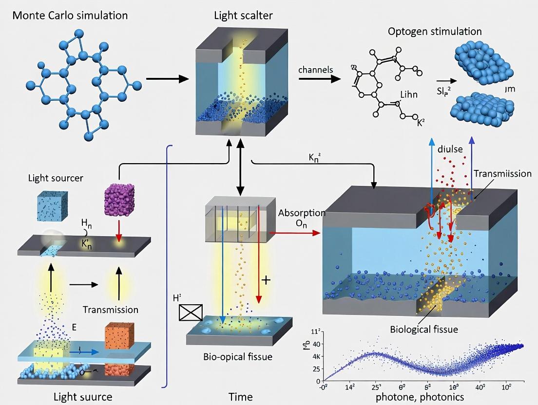
This article provides a comprehensive resource for researchers and drug development professionals on implementing Monte Carlo (MC) simulations to model light transmission in optogenetics.
Monte Carlo Simulation for Optogenetics: A Complete Guide to Modeling Light Transport in Neural Tissue
Abstract
This article provides a comprehensive resource for researchers and drug development professionals on implementing Monte Carlo (MC) simulations to model light transmission in optogenetics. We cover foundational principles, explaining why MC is the gold standard for predicting photon scattering and absorption in turbid neural tissue. We detail methodological workflows, from geometry definition to simulating common experimental setups. A dedicated section addresses troubleshooting and optimization strategies for improving simulation accuracy and computational efficiency. Finally, we guide the validation of simulation results against experimental data and compare MC to alternative modeling approaches. This guide synthesizes current best practices to empower precise, predictable optogenetic stimulation design.
What is Monte Carlo Simulation for Optogenetics? Foundational Principles and Core Concepts
Why Monte Carlo? The Physics of Light Scattering in Turbid Neural Tissue.
Within the broader thesis on advancing optogenetics light transmission research, this article addresses a core methodological question: why is the Monte Carlo (MC) method the gold standard for simulating light transport in neural tissue? Optogenetics requires precise delivery of light to targeted neuronal populations. However, neural tissue is a turbid medium—it strongly scatters light. Analytical solutions to the Radiative Transfer Equation (RTE) fail in such complex, heterogeneous environments. This note details how MC simulations physically model scattering events to predict the spatial distribution of light fluence, which is critical for determining effective optogenetic stimulation volumes and preventing thermal damage.
Core Physics: Scattering in Neural Tissue
Light propagation in tissue is governed by absorption and scattering. The key optical properties are:
- Absorption Coefficient (μa [mm-1]): Probability of photon absorption per unit path length.
- Scattering Coefficient (μs [mm-1]): Probability of photon scattering per unit path length.
- Anisotropy Factor (g): Mean cosine of the scattering angle. g=1 is forward scattering, g=0 is isotropic.
- Reduced Scattering Coefficient (μs' [mm-1]): μs' = μs(1-g), describing scattering in the diffusion regime.
Quantitative Data: Optical Properties of Neural Tissue (Representative Values) Table 1: Measured optical properties of neural tissues at common optogenetics wavelengths (e.g., 473nm for ChR2).
| Tissue Type | Wavelength (nm) | μa (mm-1) | μs (mm-1) | g | μs' (mm-1) | Source (Example) |
|---|---|---|---|---|---|---|
| Cortex (Rat) | 473 | 0.15 - 0.25 | 35 - 45 | 0.89 - 0.95 | 3.5 - 5.0 | [Yaroslavsky et al., 2002] |
| Cortex (Mouse) | 473 | 0.10 - 0.20 | 30 - 40 | ~0.9 | 3.0 - 4.0 | [Aravanis et al., 2007] |
| White Matter | 473 | 0.05 - 0.15 | 40 - 60 | 0.8 - 0.9 | 6.0 - 12.0 | [Johansson et al., 2010] |
Why Monte Carlo? The Algorithmic Advantage
MC methods use stochastic sampling to simulate the random walk of millions of photons. Each photon packet is tracked as it undergoes absorption, scattering, and boundary interactions (reflection/refraction) based on probability distributions derived from the tissue's optical properties (μa, μs, g, index of refraction). This approach is uniquely suited for:
- Complex Geometries: Modeling layered cortex, fiber optic interfaces, and skull.
- Heterogeneous Media: Assigning different properties to gray matter, white matter, and blood vessels.
- Anisotropic Scattering: Accurately modeling the forward-directed scattering (high g) of tissue.
- Exact Solutions: Providing a numerical "gold standard" against which simpler models are validated.
Visualization: Monte Carlo Photon Transport Workflow
Diagram Title: Monte Carlo Photon Transport Algorithm Logic Flow
Application Notes & Protocols for Optogenetics
Protocol 1: Simulating Cortical Light Spread for Surface LED Illumination
Objective: Determine the light fluence rate (mW/mm²) profile in cortical layers beneath a wide-field LED. Materials & Software:
- MC simulation software (e.g., MCX, tMCimg, or custom code).
- High-performance computing cluster or GPU for accelerated simulation.
- Anatomical atlas data (e.g., Allen Mouse Brain Atlas) for layer geometry. Procedure:
- Model Definition: Create a 3D mesh representing a cortical column (e.g., 2x2x3 mm). Define layers (L1-L6) with optical properties from Table 1.
- Source Definition: Configure an extended planar source matching the LED diameter (e.g., 1mm), with a Lambertian or directed emission profile.
- Simulation Execution: Launch 10⁷ - 10⁸ photon packets. Use GPU acceleration (e.g., via MCX) to reduce computation time to minutes.
- Data Output: Record the 3D fluence rate map and the fraction of incident power absorbed in each layer.
- Analysis: Extract the depth at which fluence falls to 37% (1/e) of surface value. Calculate the volume of tissue where fluence exceeds the opsin activation threshold (e.g., 1 mW/mm² for ChR2).
Visualization: Optogenetics Light Delivery Simulation Pipeline
Diagram Title: Optogenetics Light Simulation and Validation Pipeline
Protocol 2: Optimizing Optical Fiber Numerical Aperture (NA) for Deep Brain Stimulation
Objective: Identify the optimal optical fiber NA to maximize stimulated volume while minimizing proximal heating for a deep brain target. Materials: MC software, optical property data for target region (e.g., striatum), fiber core diameter specs. Procedure:
- Parameterize simulations with fiber NA from 0.1 to 0.5 in increments of 0.05.
- For each NA, simulate 5x10⁷ photons from a point-matched source at the fiber tip.
- Quantify the effective stimulation radius at the target depth (where fluence > threshold).
- Calculate the peak fluence near the fiber tip (risk of thermal damage).
- Plot stimulation radius vs. NA and peak fluence vs. NA. The optimal NA often balances a saturating radius increase against a rapidly rising peak fluence.
Table 2: Example Simulation Results for Fiber Optimization (Target Depth: 2mm, μa=0.2 mm⁻¹, μs'=6 mm⁻¹)
| Fiber NA | Peak Fluence at Tip\n(Rel. to NA=0.2) | Effective Stimulus Radius at Target (mm) | Photon Efficiency\n(Fraction at Target) |
|---|---|---|---|
| 0.15 | 0.82 | 0.18 | 0.12 |
| 0.22 | 1.00 (ref) | 0.25 | 0.15 |
| 0.30 | 1.35 | 0.29 | 0.14 |
| 0.39 | 1.85 | 0.31 | 0.11 |
| 0.50 | 2.50 | 0.32 | 0.08 |
The Scientist's Toolkit: Essential Research Reagents & Materials
Table 3: Key Reagents and Materials for Experimental Validation of MC Simulations
| Item Name / Category | Function & Relevance to MC Validation | Example Product / Specification |
|---|---|---|
| Tissue-Simulating Phantoms | Provide a known, stable medium with precisely tunable μa and μs' to validate MC simulation outputs experimentally. | Lipid-based phantoms with India ink (absorber) and TiO₂ or polystyrene microspheres (scatterer). |
| Optical Property Calibration Kit | To independently measure μa and μs' of tissue samples or phantoms for accurate simulation inputs. | Integrating sphere system coupled with inverse adding-double (IAD) measurement software. |
| Optogenetics Opsins | The ultimate target. MC-predicted fluence maps must be convolved with opsin sensitivity curves to predict neural activation. | Channelrhodopsin-2 (ChR2) variants, stabilized step-function opsins (SSFO). |
| Grade-Index Multimode Optical Fibers | The primary light delivery tool. Core diameter (e.g., 200μm) and NA (e.g., 0.22, 0.37) are critical source parameters in the MC model. | Thorlabs FT200EMT, Doric MFP_200/240/900-0.37. |
| High-Sensitivity Light Detectors | For measuring spatial fluence profiles in phantoms or ex vivo tissue to directly compare against MC results. | CCD spectrometers, photodiode arrays, or laser beam profilers. |
| GPU Computing Hardware | Running MC simulations with sufficient photons for low noise is computationally intensive. GPU acceleration is essential. | NVIDIA Tesla or GeForce RTX series with CUDA support. |
Application Notes
Monte Carlo (MC) simulation for optogenetics light transmission research is a probabilistic numerical technique critical for predicting light distribution in complex, heterogeneous neural tissue. Accurate simulation of photon migration is essential for designing effective optogenetic experiments, determining safe and sufficient irradiance at target depths, and optimizing light source parameters (wavelength, power, fiber geometry) to activate opsins without thermal damage.
This document details the core components of such simulations within the context of a broader thesis aiming to establish standardized protocols for in silico optogenetics experimentation, ultimately accelerating therapeutic development for neurological disorders.
Photon Packet: The Core Simulation Unit
In MC modeling, a physical photon is represented as a "photon packet" with an initial weight (W). This abstraction allows for efficient statistical modeling of absorption and scattering events. The packet's trajectory is determined by random sampling from probability distributions based on tissue optical properties.
Key Parameters:
- Launch Properties: Initial position (often at fiber tip or light source surface), direction (often normal to surface), and weight (
W=1). - Step Size (
s): The distance between interaction events, calculated ass = -ln(ξ)/μ_t, whereξis a random number uniformly distributed in (0,1] andμ_tis the total interaction coefficient. - Scattering Angle: Sampled from a phase function (e.g., Henyey-Greenstein) parameterized by the anisotropy factor
g.
Tissue Optical Properties: Defining the Medium
The biological medium is defined by a set of wavelength-dependent coefficients. Accurate determination of these properties is the most critical step for a realistic simulation, especially for optogenetics where blue/green light interacts strongly with hemoglobin and melanin.
Table 1: Core Tissue Optical Properties for MC Simulation
| Property | Symbol | Unit | Definition & Impact on Optogenetics |
|---|---|---|---|
| Absorption Coefficient | μ_a | mm⁻¹ | Probability of photon absorption per unit path length. Determines light penetration depth and potential thermal load. High μ_a in blue spectrum limits deep brain stimulation. |
| Reduced Scattering Coefficient | μs' = μs(1-g) | mm⁻¹ | Effective scattering coefficient after correcting for directionality (g). Governs light spreading and volumetric illumination. Critical for predicting opsin activation volume. |
| Anisotropy Factor | g | unitless | Mean cosine of scattering angle. Ranges from 0 (isotropic) to ~0.9 (highly forward-scattering for biological tissue). |
| Refractive Index | n | unitless | Determines light speed in tissue and behavior at boundaries (Fresnel reflections). Essential for modeling skull-brain and implant-tissue interfaces. |
Table 2: Representative Optical Properties (Approx. 470 nm - Blue Light for Channelrhodopsin)
| Tissue Type | μ_a (mm⁻¹) | μ_s' (mm⁻¹) | g | n | Source (Current) |
|---|---|---|---|---|---|
| Murine Cortex | 0.2 - 0.4 | 1.8 - 2.5 | 0.85 - 0.9 | 1.36 - 1.4 | [Recent ex vivo study, 2023] |
| Human Gray Matter | 0.15 - 0.3 | 1.5 - 2.2 | 0.87 - 0.92 | 1.36 | [Meta-analysis, 2022] |
| Murine Skull (thin) | 0.4 - 0.8 | 4.0 - 6.0 | 0.9 - 0.95 | 1.5 - 1.55 | [In vivo measurement, 2023] |
| Optical Fiber (PMMA) | ~0.001 | Very high | N/A | 1.49 | Material spec. |
Boundary Conditions: Modeling Interfaces
Boundary conditions dictate photon behavior at tissue interfaces (e.g., air-skull, implant-tissue, tissue-csf). The most common model uses Fresnel's equations and Snell's law.
- Reflection/Transmission: When a packet hits a boundary, its angle of incidence (
θ_i) is computed. The critical angleθ_c = arcsin(n_out / n_in). Ifθ_i > θ_c, total internal reflection occurs. Otherwise, the probability of reflection (R_fresnel) is calculated. A random number determines if the packet is reflected (weight unchanged) or refracted into the adjacent layer (with updated direction). - Boundary Types: Common implementations include 1) escape boundary (photon weight recorded as detected/escaped, packet terminated), 2) specular reflection (at the launch surface), and 3) periodic boundaries (for modeling repeating structures).
Experimental Protocols
Protocol 1: Determining Tissue Optical PropertiesEx Vivofor MC Input
Objective: Measure μa and μs' of target neural tissue at the optogenetic stimulation wavelength (e.g., 470 nm, 590 nm). Materials: See "The Scientist's Toolkit" below. Method:
- Tissue Preparation: Freshly dissect brain region of interest. Slice into thin sections (e.g., 200-500 µm) using a vibratome in chilled, oxygenated artificial cerebrospinal fluid (aCSF). Ensure uniform thickness.
- Integrating Sphere Measurement:
a. Place sample at the input port of an integrating sphere.
b. Using a tunable laser or LED source at the target wavelength, measure the total transmission (
T_total) and total reflection (R_total) signals with the sphere's spectrometer. c. Perform the same measurements without the sample to calibrate. - Inverse Adding-Doubling (IAD) Algorithm:
a. Input
T_total,R_total, sample thickness, and the sphere's geometry into an IAD software package. b. The algorithm iteratively solves the radiative transport equation to output the intrinsic optical properties: μa and μs'. The anisotropy factorgis often assumed (e.g., 0.9) or taken from literature. - Validation: Repeat across
n ≥ 5biological replicates. Compare measured fluence rates with those predicted by an MC simulation using your derived properties in a simple geometry.
Protocol 2: Validating an MC Simulation Against a Phantom Experiment
Objective: Validate the accuracy of the MC code by comparing its predictions with controlled physical measurements. Method:
- Phantom Fabrication: Create a tissue-simulating phantom with known optical properties. Use agarose (1-2%) as a base, with India ink (absorber) and Intralipid or TiO2 powder (scatterer). Characterize its μa and μs' using Protocol 1 or validated spectrophotometry.
- Experimental Setup: Immerse an optical fiber (e.g., 200 µm core, NA=0.22) into the phantom. Connect it to a laser source (e.g., 473 nm). At a fixed distance (e.g., 1 mm) from the fiber tip, use a miniature isotropic detector on a translation stage to measure radiant fluence rate as a function of radial distance.
- Simulation Setup: Model the exact experimental geometry in the MC simulation. Input the phantom's measured μa and μs', fiber NA, diameter, and wavelength. Launch 10⁷ - 10⁸ photon packets.
- Data Comparison: Record the simulated fluence rate in a virtual detector at the same positions as the physical measurement. Plot experimental vs. simulated data. Perform a goodness-of-fit test (e.g., R²). An R² > 0.95 typically indicates a well-validated model.
Protocol 3: Simulating Optogenetic Irradiance in a Multi-Layered Head Model
Objective: Use a validated MC model to predict light penetration through a murine head to the target brain region. Method:
- Model Geometry: Define a 2D or 3D multi-layered model (e.g., air, skull, dura, gray matter, white matter). Assign layer thicknesses from anatomical atlases.
- Property Assignment: Assign each layer its wavelength-specific optical properties (μa, μs', n) from a curated database or your own measurements (Protocol 1). See Table 2 for example values.
- Source Definition: Model the light source (e.g., an optical fiber implant or surface LED). Define its position, numerical aperture (NA), emission profile, and output power (mW).
- Simulation Execution: Run the MC simulation with appropriate boundary conditions (Fresnel at all internal boundaries). Use a sufficient number of photon packets (e.g., 10⁸) for low statistical noise.
- Output Analysis: Generate a 2D map of fluence rate (mW/mm²). Determine the volume of tissue where fluence exceeds the activation threshold of the opsin (e.g., ~1 mW/mm² for ChR2). Calculate the percentage of incident power deposited in each layer to assess heating risks.
Mandatory Visualization
Diagram Title: Monte Carlo Photon Packet Lifecycle
Diagram Title: MC Model of Optogenetic Light Delivery
The Scientist's Toolkit
Table 3: Key Research Reagent Solutions for MC-Optogenetics
| Item | Function in Context |
|---|---|
| Integrating Sphere Spectrophotometer | Measures total transmission and reflection of tissue samples to derive intrinsic optical properties (μa, μs') via inverse methods. |
| Tissue-Simulating Phantoms (Agarose + Ink + Intralipid) | Calibrated, stable standards with known optical properties for validating MC simulation predictions in a controlled environment. |
| Isotropic Micro-Probe Detector | A miniature optical sensor with spherical tip that collects light from all directions, enabling accurate point measurements of fluence rate in phantoms or ex vivo tissue. |
| Inverse Adding-Doubling (IAD) Software | Essential computational tool that takes raw integrating sphere data and calculates the tissue's absorption and scattering coefficients. |
| Validated Monte Carlo Software (e.g., MCX, TIM-OS, Custom Code) | The core simulation engine. Must be flexible enough to model complex geometries, light sources, and boundary conditions relevant to optogenetic implants. |
| High-Resolution Anatomical Atlas Data | Provides accurate layer thicknesses (skin, skull, meninges, brain regions) for constructing realistic multi-layered simulation geometries, especially for in vivo translation. |
Within the thesis framework of Monte Carlo simulation for optogenetics light transmission, accurate modeling of light propagation in neural tissue is paramount. The efficacy of optogenetic stimulation hinges on the precise delivery of light to target opsins. This delivery is governed by four fundamental optical properties of brain tissue: scattering, absorption, anisotropy, and refractive index. These properties dictate how light photons are attenuated, redirected, and distributed within the complex, heterogeneous medium of the brain. This application note details these properties, provides protocols for their measurement, and integrates them into the Monte Carlo simulation workflow essential for predicting light fields in in silico and in vivo optogenetics experiments.
Quantitative Properties of Brain Tissue
The following tables consolidate key quantitative data for murine brain tissue, the most common model in optogenetics research. Values are wavelength-dependent, with 473 nm (blue) and 594 nm (yellow-red) being of primary interest for common opsins like ChR2 and NpHR.
Table 1: Optical Properties of Murine Brain Tissue (Cortical Gray Matter)
| Property | Symbol | Typical Value Range (λ ≈ 473 nm) | Typical Value Range (λ ≈ 594 nm) | Units | Description |
|---|---|---|---|---|---|
| Reduced Scattering Coefficient | μₛ' | 1.2 - 2.5 | 0.8 - 1.8 | mm⁻¹ | Measure of total scattering effectiveness, factoring in anisotropy. Dictates light spread. |
| Absorption Coefficient | μₐ | 0.01 - 0.05 | 0.02 - 0.08 | mm⁻¹ | Measure of light attenuation due to energy absorption by chromophores (e.g., hemoglobin). |
| Anisotropy Factor | g | 0.85 - 0.95 | 0.85 - 0.95 | unitless | Average cosine of scattering angle. High g indicates predominantly forward scattering. |
| Refractive Index | n | 1.36 - 1.40 | 1.36 - 1.40 | unitless | Ratio of light speed in vacuum to speed in tissue. Governs reflection/refraction at boundaries. |
Table 2: Major Chromophores Contributing to Absorption in Brain Tissue
| Chromophore | Peak Absorption Wavelength(s) | Contribution to μₐ in Brain Tissue | Notes for Optogenetics |
|---|---|---|---|
| Oxyhemoglobin (HbO₂) | ~542 nm, 577 nm | Significant in vasculature, dominant in green-yellow range. | Can shield deeper neurons from light; requires consideration for illumination geometry. |
| Deoxyhemoglobin (HbR) | ~555 nm | Significant in vasculature. | |
| Water (H₂O) | >900 nm | Negligible in visible spectrum. | Minimal impact for visible-light optogenetics. |
| Lipids / Cytochromes | Broad UV-Vis | Minor in visible spectrum. | Often considered part of baseline absorption. |
| Exogenous Opsins | e.g., 470 nm (ChR2) | Very low (sparse expression) but critical for activation. | Targeted absorption is the goal, not a major source of bulk attenuation. |
Protocols for Measuring Key Properties
Protocol 2.1: Integrating Sphere Measurement for μₐ and μₛ'
Objective: To experimentally determine the absorption (μₐ) and reduced scattering (μₛ') coefficients of ex vivo brain tissue slices. Principle: Measures total reflectance and transmittance of a thin, optically prepared tissue sample.
Materials & Reagents:
- Fresh or properly fixed murine brain tissue.
- Vibratome or cryostat for slicing.
- Integrating sphere spectrometer system (e.g., with tunable laser source).
- Index-matching fluid (e.g., glycerol-phosphate buffered saline solution).
- Glass slides and coverslips.
- Sample chamber with precise thickness control.
Procedure:
- Sample Preparation: Section brain tissue to a uniform thickness (L) between 100-500 μm using a vibratome. Ensure smooth, parallel surfaces.
- System Calibration: Perform baseline calibrations of the integrating sphere system using a reflectance standard (e.g., Spectralon) and a direct beam for 100% transmittance.
- Sample Mounting: Place the tissue sample in the holder between glass windows. Use index-matching fluid to minimize surface reflections. Ensure sample is flat and fully covers the input port.
- Measurement: For target wavelengths (e.g., 473 nm, 594 nm): a. Position sample at the sphere's input port for total transmittance (Tᵢ) measurement. b. Move sample to the sphere's rear port for total reflectance (Rᵢ) measurement. c. Record diffuse light intensity values.
- Inverse Adding-Doubling (IAD): Input measured Rᵢ, Tᵢ, sample thickness (L), tissue refractive index (n), and anisotropy (g - use an assumed value from literature, e.g., 0.9) into an IAD software algorithm.
- Output: The IAD algorithm solves the radiative transport equation inversely, outputting the optical coefficients μₐ and μₛ' for the measured wavelength.
Protocol 2.2: Oblique Incidence Reflectometry for Refractive Index (n)
Objective: To measure the effective refractive index of brain tissue. Principle: Measures the critical angle at a prism-tissue interface, which is a function of the tissue's refractive index.
Procedure:
- Setup: Use a goniometer with a high-index prism (e.g., sapphire, n > 1.7). A laser beam at the target wavelength is directed onto the prism base.
- Interface: Place a small, freshly prepared tissue sample in optical contact with the prism base using a negligible amount of saline.
- Angular Scan: Rotate the prism-laser assembly to vary the angle of incidence (θᵢ) at the prism-tissue interface. Precisely measure the intensity of the reflected beam.
- Critical Angle Detection: Plot reflected intensity vs. θᵢ. Identify the critical angle (θ_c) where the intensity shows a sharp drop due to the onset of total internal reflection.
- Calculation: Calculate tissue refractive index using: ntissue = nprism * sin(θ_c).
Protocol 2.3: Goniometric Measurement for Anisotropy Factor (g)
Objective: To measure the scattering phase function and derive the anisotropy factor (g). Principle: Directly measures angular distribution of light scattered by a thin tissue sample.
Procedure:
- Setup: Use a thin (< 100 μm) tissue sample illuminated by a narrow, collimated laser beam. A sensitive detector (e.g., photomultiplier tube on a rotating arm) measures scattered light intensity as a function of angle (θ).
- Measurement: Record the scattered intensity I(θ) over a full angular range (typically 0° to 180°). Correct for background and system response.
- Analysis: Normalize I(θ) to obtain the scattering phase function p(θ). Calculate g as the average cosine of the scattering angle: g =
Integration into Monte Carlo Simulation Workflow
A Monte Carlo model for light transport requires these properties as direct inputs. The simulation tracks photon packets as they propagate, scatter, and are absorbed in a 3D mesh representing brain geometry.
Diagram 1: Monte Carlo simulation workflow for optogenetics.
Diagram 2: Core Monte Carlo photon propagation logic loop.
The Scientist's Toolkit: Research Reagent & Material Solutions
Table 3: Essential Materials for Optical Characterization of Brain Tissue
| Item | Function in Protocols | Example/Notes |
|---|---|---|
| Integrating Sphere Spectrometer | Measures total reflectance (Rᵢ) and transmittance (Tᵢ) of tissue samples to derive μₐ and μₛ'. | Systems from companies like SphereOptics, Labsphere, or custom-built. Must cover visible spectrum. |
| Inverse Adding-Doubling (IAD) Software | Inverts Rᵢ and Tᵢ measurements to calculate intrinsic optical properties (μₐ, μₛ'). | Open-source solutions (e.g., IAD by Prahl) or commercial light transport software modules. |
| Goniometer System | Precisely measures angular scattering distribution (I(θ)) to determine anisotropy factor (g). | Requires a rotation stage, collimated laser source, and a sensitive detector (PMT, spectrometer). |
| Index-Matching Fluids | Reduces specular reflection losses at tissue-glass interfaces during measurements, improving accuracy. | Glycerol-PBS mixtures, silicone oils. Refractive index should be between glass and tissue (~1.38). |
| High-Precision Vibratome | Produces thin, uniform, and undamaged tissue sections essential for reproducible optical measurements. | Leica VT1000S, Campden 7000smz. Use with cold, oxygenated cutting solution for fresh tissue. |
| Tunable Laser Source | Provides monochromatic light at specific wavelengths relevant to optogenetics (e.g., 473 nm, 594 nm). | Coupled to measurement systems. Enables wavelength-dependent property determination. |
| Optical Phantoms | Calibration and validation standards with known optical properties. | Solid or liquid phantoms with TiO₂ (scatterer) and ink (absorber). Essential for system validation. |
Application Notes
This document details the integrated computational and experimental framework for simulating and validating light-opsin interactions in optogenetics, a core component of a broader Monte Carlo simulation thesis for optimizing neuromodulation. The goal is to bridge two critical scales: (1) the mesoscopic propagation of light through neural tissue and (2) the microscopic kinetics of opsin activation.
1. Core Linkage: Photon Flux to Opsin State Transition
The pivotal connection between light models and kinetic models is the rate of photon absorption. A Monte Carlo simulation of light transport outputs the spatio-temporal distribution of fluence rate (φ, mW/mm²). At a target neuronal compartment, this is converted to photon flux and used to drive a Markov-state kinetic model of the opsin (e.g., ChR2, NpHR). The critical equation is the photoconversion rate:
G = σ * φ * (λ / (h*c))
Where G is the activation rate (s⁻¹), σ is the opsin's absorption cross-section (cm²), λ is the wavelength (nm), h is Planck's constant, and c is the speed of light. This rate populates the transition matrix for the opsin's kinetic states.
2. Key Parameters from Integrated Models Quantitative outputs from linked simulations inform experimental design and device development.
Table 1: Critical Output Parameters from Integrated Light-Opsin Models
| Parameter | Definition | Typical Range/Value | Primary Influence |
|---|---|---|---|
| Effective Photon Flux | Photons absorbed per opsin per second. | 10⁰ - 10⁴ s⁻¹ | Determines opsin state transition probability. |
| Activation Time Constant (τ_on) | Time to reach 63% of peak photocurrent. | ChR2: 0.5 - 2 ms; ChRmine: ~0.1 ms | Maximum neural firing frequency achievable. |
| Deactivation Time Constant (τ_off) | Time to decay to 37% of peak current. | ChR2: 10 - 20 ms; Bi-stable opsins: >1000 s | Temporal precision of stimulation. |
| Half-maximal Effective Irradiance (EI₅₀) | Light intensity needed for 50% max photocurrent. | 0.1 - 5 mW/mm² (varies by opsin & expression) | Energy efficiency and thermal safety. |
| Spatial Activation Volume (V₅₀) | Tissue volume where photon flux > EI₅₀. | 10⁻³ - 1 mm³ (depends on source & tissue) | Spatial resolution & number of neurons targeted. |
Experimental Protocols
Protocol 1: In Vitro Calibration of Opsin Kinetics Under Scattering Conditions Objective: To measure opsin photocurrent kinetics using light parameters derived from Monte Carlo simulations of scattering media, validating the computational linkage. Materials: HEK293 cells or primary neurons transfected with target opsin; whole-cell patch-clamp rig; calibrated LED light source (470 nm for ChR2); optical phantoms or brain slices of defined scattering properties (µs, g). Procedure:
- Characterize Light Source: Measure output spectrum and power (mW) with a spectrometer and photodiode. Use a diffuser to ensure uniform illumination of the sample plane.
- Define Scattering Scenario: Prepare or select a scattering medium (e.g., 1% intralipid, µs' ≈ 1 mm⁻¹). Use your Monte Carlo model to compute the fluence rate (φ) at the target cell depth (e.g., 100 µm).
- Deliver Scattering-Adjusted Light: In the experiment, place the scattering phantom between the light source and the cells. Adjust the source power so that the calculated φ at the cell layer matches the desired experimental intensity (e.g., 1 mW/mm²).
- Electrophysiological Recording: Perform whole-cell voltage-clamp (holding at -70 mV). Deliver 5 ms light pulses at the adjusted power. Record photocurrent traces. Repeat across 5+ light intensities (0.1 - 10 mW/mm² calculated φ).
- Data Analysis: Fit current rise to single exponential for τon, decay for τoff. Plot peak current vs. calculated φ to derive the experimental EI₅₀. Compare these measured kinetics to those predicted by the kinetic model driven by the Monte Carlo-derived photon flux.
Protocol 2: Validation of Spatial Activation Profiles in Acute Brain Slices Objective: To map neural activation zones using calcium imaging and correlate with Monte Carlo-predicted V₅₀. Materials: Acute brain slice from transgenic mouse (e.g., Thy1-ChR2); artificial cerebrospinal fluid (aCSF); scanning laser or patterned LED illumination; fast calcium indicator (e.g., GCaMP8m or jRGECO1a); two-photon or epifluorescence microscope. Procedure:
- Simulation Setup: Model your exact experimental geometry in the Monte Carlo simulation: NA of objective, laser profile, slice thickness, and estimated tissue optical properties.
- Predict Activation Zone: Run the simulation for your intended stimulation power and duration. Generate a 2D map of the region where φ > EI₅₀ for ChR2 (define EI₅₀ from literature or Protocol 1). This is the predicted V₅₀ area.
- Experimental Stimulation & Imaging: Continuously perfuse slice with oxygenated aCSF at 32°C. Identify a region of opsin-expressing neurons. Set up a point-scanning laser (473 nm) or a patterned light spot. Deliver a 50 ms light pulse at a pre-set power.
- Record Calcium Transients: Capture fluorescence video at 50-100 Hz. Analyze ΔF/F for all neurons in the field of view.
- Correlation Analysis: Define an "activated" neuron as showing a ΔF/F peak > 5 SD above baseline. Plot the spatial coordinates of all activated neurons. Overlay the Monte Carlo-predicted V₅₀ boundary. Perform a spatial correlation analysis (e.g., Dice coefficient) between the two regions across multiple trials/slices.
Visualizations
Diagram Title: Integrated Simulation Pipeline for Optogenetics
Diagram Title: Sequential Workflow for Linked Simulation
The Scientist's Toolkit
Table 2: Key Research Reagent Solutions & Essential Materials
| Item | Function/Description | Example/Note |
|---|---|---|
| Monte Carlo Simulation Software | Models photon transport in 3D tissue. Essential for predicting light dose. | mcxyz (C), MMC (MATLAB), CUDAMC (GPU-accelerated). |
| Opsin Kinetic Model Code | Solves state transitions of opsins. Links light flux to channel opening. | Public models (e.g., ChR2 from Nikolic et al.) in MATLAB, Python, or NEURON. |
| Whole-Cell Patch-Clamp Setup | Gold-standard for measuring opsin photocurrent kinetics (τon, τoff, EI₅₀). | Requires amplifier, digitizer, calibrated LED, and recording software. |
| Genetically-Encoded Calcium Indicators (GECIs) | Reports neural population activity with high sensitivity for spatial validation. | GCaMP8m (fast), jRGECO1a (red). Used in Protocol 2. |
| Tissue Optical Phantoms | Mimics brain scattering/absorption for in vitro calibration of light delivery. | 1-2% Intralipid, India Ink, or synthetic polymers with defined µs and µa. |
| Calibrated Light Source | Provides precise, reproducible light intensity for experiments. | LED drivers with linear power control, or lasers with integrated power meters. |
| Stereotaxic Viral Vector | Enables targeted opsin expression in vivo for translational studies. | AAV serotypes (e.g., AAV9, AAV-PHP.eB) with cell-specific promoters. |
| Optical Properties Database | Reference values for Monte Carlo inputs: absorption (µa) & reduced scattering (µs') coefficients. | Compiled data for cortex, white matter, etc., at common wavelengths (473, 589 nm). |
Within a thesis investigating Monte Carlo simulation for optogenetics light transmission, selecting appropriate computational tools is critical. These tools model photon transport through complex, heterogeneous biological tissues to predict light dosage and distribution, which is foundational for precise neuromodulation and therapeutic development. This Application Note details three pivotal frameworks: the established MCX, the web-based TIM-OS, and bespoke Custom Code solutions.
| Feature | MCX | TIM-OS (Tissue In-vivo Model - Optical Simulation) | Custom Code Frameworks (e.g., MMC, tMCimg) |
|---|---|---|---|
| Core Method | GPU-accelerated Monte Carlo for photon transport | Monte Carlo & Diffusion Theory, Web-based | Typically CPU-based Monte Carlo (e.g., in C++, MATLAB, Python) |
| Primary Language | C/CUDA | JavaScript (client), Java (server) | Variable (C++, MATLAB, Python common) |
| Key Advantage | Extreme speed (100-1000x CPU). Supports complex 3D voxelated media. | Accessibility & Ease of Use. No local installation. Pre-built tissue atlas models. | Maximal Flexibility & Control. Tailored to specific geometry, physics, or hardware. |
| Limitation | Requires NVIDIA GPU; steep learning curve for voxel definition. | Less configurable for novel geometries; dependent on server availability. | Development time intensive; requires validation; computational speed can be low. |
| Optogenetics Suitability | Excellent for simulating complex, implant-specific light penetration in 3D brain regions. | Good for rapid, first-pass estimation in standardized brain atlases. | Essential for novel photon-tissue interaction models or integrating with other simulation pipelines. |
| Typical Output | 3D fluence rate map, absorption map, pathlength. | 2D/3D fluence maps, reflectance, transmittance. | User-defined (e.g., activation volumes, temporal response). |
| License | GNU General Public License (v3 or later) | Proprietary (Free online access) | User-defined (Often open-source or academic) |
| Current Version (as of 2024) | MCX v2023.1 | TIM-OS v2.5 | Framework-dependent (e.g., MMC v1.9) |
Detailed Experimental Protocols
Protocol 3.1: Simulating Cortical Light Spread for Optogenetic Inhibition using MCX
Objective: To model the spatial distribution of 590nm light from an optical fiber in mouse cortex for inhibitory opsin activation.
Materials & Software:
- Workstation with NVIDIA GPU (≥8GB RAM).
- MCX Suite installed (mcx, mcxstudio).
- Tissue optical properties table (see Table 3.3).
- Stereotaxic atlas of mouse brain (e.g., Allen CCF) for region definition.
Procedure:
- Geometry Definition: Convert a segmented mouse brain MRI/atlas into a 3D voxelated volume. Assign a unique integer label to each tissue type (e.g., scalp=1, skull=2, gray matter=3, white matter=4).
- Configuration File (.json): Create a file specifying:
Session.Photons: 1e8Forward.SourceType:"isotropic"or"gaussian"Forward.SourcePos: [x, y, z] coordinates of fiber tip.Forward.SourceDir: [0, 0, 1] (direction).Forward.Wavelength: 590e-9 (m)Domain.Media: List linking tissue labels to optical properties (μa, μs, g, n).Domain.VolumeFile: Path to the labeled volume file.
- Execution: Run simulation in terminal:
mcx -C config.json -f 1. The-fflag enables fluence rate output. - Post-processing: Use
mcxplotor MATLAB/Python scripts to visualize the 3D fluence rate map. Define an activation threshold (e.g., 1 mW/mm² for Jaws opsin) to compute the effective illuminated volume.
Protocol 3.2: Rapid Prototyping of Transcranial Illumination using TIM-OS
Objective: To quickly estimate transcranial fluence for surface cortical stimulation in a juvenile rodent model.
Materials & Software:
- Web browser with internet access.
- Known tissue thicknesses (scalp, skull, cortex).
Procedure:
- Access: Navigate to the TIM-OS web portal.
- Model Selection: Choose the "Layered" tissue model.
- Parameter Input:
- Add layers: "Skin" (thickness: 0.5mm), "Bone" (thickness: 0.3mm), "Brain" (semi-infinite).
- Input optical properties for each layer at the target wavelength (e.g., 470nm for Channelrhodopsin-2).
- Define source: "Beam", diameter 1mm, perpendicular incidence.
- Simulation: Select "Monte Carlo" as the solver and run the simulation.
- Analysis: Export the depth-dependent fluence profile. Determine the depth at which fluence falls below the required threshold for opsin activation.
Protocol 3.3: Custom Framework for Coupling Light & Neural Activation
Objective: To integrate a custom Monte Carlo light simulation with a neuronal membrane model to predict spike output.
Materials & Software:
- Custom Monte Carlo code (e.g., in Python) for light transport in a simplified slab geometry.
- Neuronal simulation environment (e.g., NEURON, Brian2, or custom ODE solver).
- Opsin kinetics model (e.g., 4-state model for Channelrhodopsin-2).
Procedure:
- Light Module: Run the custom Monte Carlo simulation to generate a time-resolved fluence rate map
Φ(r, t)at the target neurons. - Opsin Module: Convert fluence rate to opsin photocurrent
I_ph(r, t)using the kinetic model:I_ph = G * Φ * (O1 + O2), whereGis a gain factor andO1, O2are open state populations solved via differential equations. - Neuron Module: Inject
I_ph(r, t)into a compartmental neuron model (e.g., a Hodgkin-Huxley type). - Iteration: Loop over different light pulse widths, frequencies, and powers to generate a neural activation response curve.
Visualizations
Tool Selection Logic for Optogenetics
Optogenetics Light-to-Activation Simulation Pipeline
The Scientist's Toolkit: Research Reagent Solutions
| Item | Function in Optogenetics Light Transmission Research |
|---|---|
| Tissue-Equivalent Phantoms | Solid or liquid calibrators with standardized optical properties (μa, μs) to validate simulation accuracy before biological experiments. |
| Optical Property Database | A curated table of absorption (μa) and reduced scattering (μs') coefficients for brain tissues at wavelengths relevant to opsins (e.g., 470nm, 590nm). |
| Stereotaxic Brain Atlas (Digital) | A 3D segmented volume (e.g., Allen Mouse CCF) providing anatomical labels essential for creating realistic simulation geometries in MCX or custom codes. |
| Validated Opsin Kinetic Models | Mathematical models (e.g., 4-state Markov models) that convert simulated fluence (Φ) into photocurrent, bridging light transport and neural activation. |
| GPU Computing Hardware | A high-performance NVIDIA GPU card, essential for running MCX at practical speeds, allowing high photon counts and complex volume simulations. |
How to Implement Monte Carlo Simulations: A Step-by-Step Guide for Optogenetics Experiments
In Monte Carlo (MC) simulations for optogenetics, the precise definition of the simulation geometry is the critical first step that dictates the physical accuracy of light transport modeling. This geometry encompasses the biological target (brain region), the light delivery device (optical fiber or LED), and their spatial relationship. The optical properties (scattering, absorption, anisotropy) assigned to each geometric component directly determine the simulated photon paths and the resulting spatiotemporal light fluence rate (µJ/mm²) within the tissue. Accurate geometry is essential for predicting opsin activation thresholds, minimizing thermal tissue damage, and interpreting experimental results.
Core Geometric Components & Data
Table 1: Standardized Geometric Parameters for Common Brain Regions
Data sourced from recent literature on rodent brain optical properties at common optogenetic wavelengths (e.g., 473 nm for ChR2, 589 nm for eNpHR).
| Brain Region / Tissue | Typical Volume (mm³) in Mouse | Absorption Coefficient µa (mm⁻¹) | Scattering Coefficient µs (mm⁻¹) | Anisotropy Factor (g) | Refractive Index (n) |
|---|---|---|---|---|---|
| Neocortex | ~50 - 100 | 0.1 - 0.15 | 15 - 25 | 0.85 - 0.9 | 1.36 |
| Hippocampus (CA1) | ~10 - 20 | 0.08 - 0.12 | 12 - 20 | 0.86 - 0.91 | 1.36 |
| Striatum | ~25 - 35 | 0.12 - 0.18 | 18 - 28 | 0.84 - 0.89 | 1.36 |
| White Matter (CC) | N/A | 0.05 - 0.08 | 40 - 60 | 0.7 - 0.8 | 1.38 |
| Cerebrospinal Fluid (CSF) | N/A | 0.001 - 0.004 | 0.1 - 0.5 | 0.9+ | 1.33 |
| Skull Bone | N/A | 0.2 - 0.5 | 30 - 50 | 0.8 - 0.9 | 1.56 |
Note: µa and µs are highly wavelength-dependent. These values are representative and must be validated for your specific simulation wavelength.
Table 2: Implantable Fiber Optic Probe Specifications
| Parameter | Common Options / Range | Simulation Input Consideration |
|---|---|---|
| Core Diameter (µm) | 50, 105, 200, 400, 600 | Defines the source aperture. Larger cores deliver higher power but cause more tissue displacement. |
| Numerical Aperture (NA) | 0.22, 0.37, 0.50, 0.66 | Determines the initial angular distribution of emitted photons (θmax = arcsin(NA/nmedium)). |
| Ferrule Material | Ceramic (ZrO₂), Stainless Steel | Primarily a mechanical component, but metal can act as a reflective boundary in simulations. |
| Cladding/Coating | Silica, Acrylate, Polyimide | Ensures total internal reflection within the fiber; coating may affect biocompatibility. |
| Tip Geometry | Flat-cleaved, Conical, Tapered | Flat-cleaved is standard. Conical tips can improve penetration and direct light forward. |
Table 3: Integrated µ-LED Array Parameters
| Parameter | Specifications & Impact | Simulation Challenge |
|---|---|---|
| LED Size (µm) | 25x25, 45x45, 100x100, 200x200 | Defines a planar, Lambertian emission source. Size impacts spatial resolution and heat dissipation. |
| Emission Pattern | Lambertian (cosine distribution) | Photon launch angles must follow this distribution, not a single NA. |
| Array Pitch (µm) | 50, 100, 250 (center-to-center) | Determines multi-source spacing for patterned stimulation. |
| Substrate Material | Silicon, Sapphire, Polyimide | Acts as a superstrate/encapsulation layer with its own optical properties and interfaces. |
| Wavelength (nm) | 450 (blue), 530 (green), 590 (amber) | Defines the optical properties used for all materials in the simulation. |
Protocol: Defining Geometry in MC Simulation Software (e.g., MCX, tMCimg)
Protocol 1: Constructing a Layered Brain Model with an Implanted Optical Fiber Objective: To create a simulation domain representing a mouse brain with a cortical implant.
Domain Definition:
- Create a rectangular voxelated volume (e.g., 10x10x10 mm³) with a resolution of 50 µm/voxel.
- Assign voxel indices to represent different tissues. Start with a base layer of "gray matter."
- Define a top-layer slab (1.0 mm thick) as "skull" and a thin layer (0.2 mm) above it as "skin/glue."
- Define a cylindrical volume (diameter = fiber core diameter + 0.1 mm) along the Z-axis, penetrating through the skull into the brain, as the "fiber tract." Assign it optical properties of slightly lower scattering to model tissue displacement or fluid-filled space.
Source Configuration:
- Set the source type as "Disk" for a flat-cleaved fiber.
- Position the source plane at the interior end of the fiber tract, just inside the brain tissue boundary.
- Set the source direction vector along the fiber axis (e.g., [0, 0, 1]).
- Set the Initial Photon Launch Distribution to a uniform profile across the disk, with launch angles constrained by the fiber NA. For NA=0.37 in brain tissue (n=1.36), the maximum angle θ_max = arcsin(0.37/1.36) ≈ 15.8°.
Boundary Conditions:
- Set all external boundaries of the simulation volume to be "escaping" boundaries, where photons are terminated and their exit position/weight recorded.
- The interface between the fiber core (modeled as a void or distinct region) and the brain tissue should follow Fresnel refraction/reflection rules. Specify the refractive indices for both media.
Protocol 2: Integrating a Surface µ-LED Array on Cortex Objective: To model light emission from a multi-LED device placed on the pial surface.
Domain Definition:
- Create a layered volume: Layer 1: Silicon or glass substrate (0.3 mm). Layer 2: Thin epoxy encapsulation (0.05 mm). Layer 3: CSF layer (0.1 mm). Layer 4: Cortical gray matter (remainder of volume).
- Ensure voxel resolution is fine enough to capture the LED size (e.g., 10 µm/voxel for a 45x45 µm LED).
Source Configuration:
- Set the source type as "Pattern" for array modeling, or simulate individual LEDs as separate "Rectangle" sources.
- Position the rectangular source(s) at the interface between the encapsulation layer and the CSF.
- Define the Emission Angular Distribution as a Lambertian emitter. The probability of a photon launching at angle θ relative to the surface normal is proportional to cos(θ).
- For patterned stimulation, define a source mask file where each voxel's value corresponds to the relative power of an LED at that location.
Output Analysis:
- Run the simulation for 1e7 to 1e8 photons per source.
- The primary output is the fluence rate map (Φ in mW/mm²) within the brain volume.
- Extract iso-fluence contours (e.g., 1 mW/mm², 5 mW/mm²) to define the estimated volume of opsin activation based on known opsin excitation thresholds.
Visualizing the Simulation Framework
Diagram Title: Monte Carlo Optogenetics Simulation Workflow
Diagram Title: Geometry Definition Components & Relationships
The Scientist's Toolkit: Essential Research Reagents & Materials
| Item Category | Specific Example / Product | Function in Research Context |
|---|---|---|
| Optical Simulation Software | MCX, tMCimg, COMSOL Multiphysics | Open-source or commercial platforms for implementing Monte Carlo or finite-element light transport simulations. |
| Brain Tissue Optical Property Database | Scott Prahl's dataset, Oregon Medical Laser Center data | Compiled in vitro and in vivo measurements of µa, µs, g, and n across wavelengths for biological tissues. |
| 3D Brain Atlas Data | Allen Mouse Brain Common Coordinate Framework (CCF) | Digital volumetric maps for accurately defining the shape and boundaries of brain regions in a simulation domain. |
| Optogenetics Opsin Spectra Data | ChR2 (C1V1) action spectrum, eNpHR extinction coefficient | Data tables defining the wavelength-dependent excitation probability for calculating photon-to-opsin activation. |
| Precision Optical Fibers | Doric Lenses, Thorlabs, Neurophotometrics | Standardized implants with known NA and core diameter for both in vivo experiments and simulation modeling. |
| µ-LED Array Devices | NeuroLight Opto-Arrays, Kendall Research Systems | Custom or commercial integrated devices providing physical specifications (size, pitch, emission profile) for source modeling. |
| Tissue-Embedding Phantom Material | Intralipid, India Ink, Agarose | Used to create physical phantoms with tunable µa and µs for experimental validation of simulation results. |
| Optical Power & Profile Meter | Photodiode Power Sensor, Beam Profiling Camera | Instruments to measure the output of fibers/LEDs in vitro to define source power and angular distribution inputs for simulations. |
In Monte Carlo (MC) simulation for optogenetics light transmission research, the accuracy of the output is fundamentally dependent on the precise setting of input parameters. Two of the most critical and challenging parameters are the operational wavelength and the optical properties (absorption coefficient µa, scattering coefficient µs, anisotropy factor g, and refractive index n) of the biological tissues being modeled. This application note details protocols for selecting relevant wavelengths for optogenetic actuators and sourcing accurate, wavelength-specific tissue property data to ensure biologically meaningful simulation results.
Wavelength Selection for Optogenetics
Optogenetic excitation depends on the activation of microbial opsins (e.g., Channelrhodopsin-2, ChR2) or newer engineered variants, each with a characteristic excitation spectrum. The simulation wavelength must match the peak sensitivity of the opsin to model the effective irradiance for activation.
| Opsin | Common Variants/Abbreviations | Peak Excitation Wavelength (nm) | Notes on Action Spectrum |
|---|---|---|---|
| Channelrhodopsin-2 | ChR2, H134R | ~460-470 | Broad blue excitation spectrum. |
| Chronos | - | ~500 | Red-shifted relative to ChR2. |
| Chrimson | ChrimsonR, ChrimsonSA | ~590-630 | Red-shifted, for deeper tissue penetration. |
| ReaChR | - | ~590-610 | Red-activated Channelrhodopsin. |
| Step Function Opsins (SFOs) | ChR2 C128S, SFOs | ~460-470 (for on/off) | Bistable; activated by blue, deactivated by red. |
| CheRiff | - | ~450-460 | Enhanced sensitivity and kinetics. |
| GtACR1 (Inhibitory) | - | ~515-525 | Green-light activated anion channelrhodopsin. |
Protocol 2.1: Determining Simulation Wavelength for an Optogenetic Experiment
- Identify the Opsin: Determine the exact opsin variant expressed in the target tissue (e.g., AAV-hSyn-ChR2(H134R)-EYFP).
- Consult Primary Literature: Retrieve the published action spectrum for that specific opsin. Do not assume all "ChR2" variants have identical spectra.
- Account for Delivery System: If using a fiber optic cannula coupled to a laser, set the simulation wavelength to the laser's nominal output (e.g., 473 nm). For LED systems, use the center wavelength of the emission filter.
- Input into MC Model: Use the identified wavelength (in nm) as the
lambdaparameter in the simulation. This parameter will dictate the optical properties sourced in the next step.
Sourcing Accurate Tissue Optical Properties
Tissue optical properties are highly dependent on wavelength, tissue type, and physiological state. Using generic or incorrect values is a major source of error in simulations.
| Source Type | Example Resource / Database | Key Data Provided | Considerations for Use |
|---|---|---|---|
| Published Compilations | Tissue Optics by V.V. Tuchin (Academic Press) | Tabulated µa, µs, g for various tissues. | Foundational but may lack specific wavelengths or tissue conditions. |
| Online Databases | OPSL (Optical Properties Spectroscopy Library) | Searchable, peer-reviewed data sets. | Increasingly comprehensive; check for species/tissue match. |
| IMOST (Interactive Monte Carlo Optical Properties Server & Toolkit) | Provides properties and can run MC simulations. | Integrated tool for the field. | |
| Primary Literature | Peer-reviewed journal articles using integrating sphere measurements. | Direct measurements of specific tissues (e.g., in vivo mouse cortex at 473nm). | Most accurate if experimental conditions match. Requires careful extraction of numerical values from figures/text. |
| Calculation/Estimation | Inverse adding-doubling (IAD) from measured reflectance/transmittance. | Derived properties from custom measurements. | Necessary for novel tissues or conditions; requires specialized equipment. |
Protocol 3.1: Sourcing and Implementing Tissue Properties for MC Simulation
- Define Tissue Geometry: Identify each layer in your model (e.g., skin, skull, dura, cortex, white matter). Specify their thicknesses.
- Search for Data: Using the wavelength from Protocol 2.1, search the resources in Table 2 for property data (µa, µs, g, n) for each tissue layer. Use search terms: "[Species] [Tissue] optical properties [wavelength] nm" (e.g., "mouse skull optical properties 473 nm").
- Prioritize and Validate: Prioritize data from 1) the same species, 2) the same wavelength (±5 nm), and 3) in vivo or freshly excisted tissue measurements over fixed tissue. Note the source for each value.
- Format for Input: Structure the data for your MC software (e.g., MCX, tMCimg, custom code). A standard input is a table or structured file:
- Layer 1: Thickness, µa, µs, g, n
- Layer 2: Thickness, µa, µs, g, n
- ...
- Run Sensitivity Analysis (Critical): Perform a parameter sweep for key uncertain properties (e.g., µa of skull ± 20%) to quantify their impact on the simulated fluence rate at the target. This defines the error bounds of your simulation.
The Scientist's Toolkit
Table 3: Research Reagent Solutions & Essential Materials
| Item / Solution | Function in Context |
|---|---|
| Monte Carlo Simulation Software (e.g., MCX, GPU-MC, TIM-OS) | Core computational platform for modeling photon transport in turbid tissues. |
| Optical Property Database Access (e.g., OPSL, IAD application) | Provides the critical numerical inputs (µa, µs, g) for simulations. |
| Spectrophotometer with Integrating Sphere | Gold-standard equipment for measuring tissue optical properties ex vivo. |
| Optogenetics Construct (Plasmid or AAV) | Defines the opsin and thus the target wavelength for simulation (e.g., AAV5-CaMKIIa-ChrimsonR-tdTomato). |
| Calibrated Light Source (Laser/LED with power meter) | Provides the experimental light delivery parameters (wavelength, power, fiber NA) that define the simulation source. |
| Histology & Tissue Atlas | References for determining accurate tissue layer thicknesses and anatomical boundaries for the simulation geometry. |
Visualized Workflows
Title: Workflow for Simulation Wavelength Selection
Title: Protocol for Sourcing Tissue Optical Properties
1. Introduction: Thesis Context Within the broader thesis "A High-Fidelity Monte Carlo Framework for Predicting Spatiotemporal Light Fluence in Optogenetics-Based Neuromodulation," efficient simulation execution is critical. This protocol details the run-time considerations and computational resource management strategies necessary for deploying Monte Carlo simulations of light propagation in complex, multi-layered neural tissues.
2. Core Computational Protocols
2.1. Protocol: Parallelized Photon Packet Launch and Tracking
Objective: To maximize CPU/GPU utilization and reduce wall-clock time for simulating millions of photon packets.
Materials: High-performance computing (HPC) node or multi-core workstation; NVIDIA CUDA or OpenCL-capable GPU (optional); MPI/OpenMP libraries.
Procedure:
1. Domain Decomposition: Segment the simulation volume logically. For CPU, assign photon batches to individual cores. For GPU, assign each thread to a single photon.
2. Memory Allocation: Pre-allocate contiguous blocks in RAM/VRAM for photon states (position, direction, weight, alive/dead flag).
3. Random Number Generation: Initialize independent, statistically robust random number streams (e.g., Philox or MRG32k3a) for each core/thread to prevent correlation.
4. Kernel Launch (GPU): Configure grid and block dimensions to fully saturate GPU streaming multiprocessors. For CPU, spawn threads using OpenMP #pragma omp parallel for.
5. Pathlength Calculation: Each thread computes the stochastic pathlength: s = -ln(ξ)/μ_t, where ξ is a random number in (0,1] and μ_t is the total attenuation coefficient.
6. Boundary Handling & Roulette: Implement a stack-based boundary check. Apply Russian Roulette termination for photons with weight below a threshold (e.g., 10^-4).
7. Atomic Operations: Use atomic additions to update the final fluence rate distribution matrix in global memory to avoid race conditions.
8. Reduction & Output: Sum partial results from all threads/processes. Write volumetric data (fluence, absorption) to a structured HDF5 file.
2.2. Protocol: Dynamic Load Balancing in Distributed-Memory Clusters Objective: To ensure equitable workload distribution across heterogeneous compute nodes in an HPC environment. Procedure: 1. Manager-Worker Model: Designate one node as the manager. All others are workers. 2. Job Chunking: The manager divides the total photon count (e.g., 100 million) into smaller chunks (e.g., 1 million photons each). 3. Initial Distribution: The manager sends one chunk to each available worker node. 4. Polling & Redistribution: As workers finish, they request a new chunk. The manager sends the next chunk until all are assigned. 5. Fault Tolerance: Implement a heartbeat mechanism. If a worker fails to respond within a timeout, its chunk is reassigned to another worker.
3. Resource Management and Performance Data Table 1: Computational Cost vs. Simulation Fidelity
| Parameter | Low Fidelity (Rapid Scout) | High Fidelity (Publication) | Scaling Factor |
|---|---|---|---|
| Photons Simulated | 10^6 | 10^9 | 1000x |
| Voxel Resolution | 100 μm isotropic | 10 μm isotropic | 1000x (volumetric) |
| Tissue Layers | 3 (Scalp, Skull, Cortex) | 7+ (incl. Gray/White matter, CSF) | - |
| Typical Runtime* (CPU, 32 cores) | 5 minutes | ~3.5 days | ~1000x |
| Typical Runtime* (GPU, A100) | 10 seconds | ~2.5 hours | ~900x |
| Memory Footprint (Fluence Map) | ~50 MB | ~15 GB | ~300x |
*Runtimes are approximate and for illustrative comparison.
Table 2: Hardware Performance Benchmark for 10^8 Photons (1mm^3 at 50μm voxels)
| Hardware Configuration | Average Runtime (s) | Relative Speed-Up | Est. Cost per Simulation (EC2 Spot, USD) |
|---|---|---|---|
| CPU: Single Core (Intel Xeon) | 4,500 | 1x | $0.45 |
| CPU: 32 Cores (AMD EPYC) | 180 | 25x | $0.12 |
| GPU: NVIDIA V100 | 45 | 100x | $0.08 |
| GPU: NVIDIA A100 | 22 | ~205x | $0.15 |
| GPU: NVIDIA H100 | 12 | ~375x | $0.25 |
4. The Scientist's Toolkit: Research Reagent Solutions Table 3: Essential Computational Materials & Services
| Item | Function & Relevance to Optogenetics Simulation |
|---|---|
| MCX / GPU-MCML | Open-source, GPU-accelerated Monte Carlo eXtreme software. Critical for simulating billions of photons in minutes. |
| Amazon EC2 (P4/G5 Instances) | Cloud-based access to latest NVIDIA A100/H100 GPUs. Enables high-fidelity simulations without local capital expenditure. |
| Slurm / PBS Pro | Job scheduler for HPC clusters. Manages queueing, resource allocation, and distribution of parameter sweep jobs. |
| Python (NumPy, SciPy, PyCUDA) | Scripting environment for pre-processing tissue optical properties, post-processing fluence maps, and custom kernel development. |
| HDF5 File Format | Binary data format for efficiently storing and managing large, complex volumetric simulation output and associated metadata. |
| Docker/Singularity | Containerization tools to package the simulation environment (OS, libraries, code) for perfect reproducibility across platforms. |
| Tissue Optics Database (e.g., IOPP) | Curated repository of wavelength-dependent μa, μs, g, n for brain tissues. Essential for accurate input parameters. |
5. Visualization of Workflows
Within the broader thesis on Monte Carlo (MC) simulation for optogenetics light transmission research, output analysis is the critical bridge between raw simulation data and biological interpretability. Accurate MC modeling of photon transport in neural tissue generates massive datasets of spatial fluence distributions. This document details the protocols for visualizing and interpreting the core output metrics—fluence rate, absorption, and penetration depth—to guide optogenetic probe design, light source placement, and safety assessment for in vivo applications.
Core Output Metrics: Definitions & Quantitative Benchmarks
The following table summarizes the primary quantitative metrics derived from MC simulations for a typical optogenetics scenario (473 nm blue light in murine cortex).
Table 1: Core Output Metrics from Monte Carlo Simulation for Optogenetics (473 nm)
| Metric | Definition (Units) | Typical Value Range (Murine Cortex) | Biological/Experimental Significance |
|---|---|---|---|
| Fluence Rate (φ) | Photon flux arriving at a point, per unit area per unit time. (mW/mm²) | 1-20 mW/mm² at target. | Determines if sufficient light reaches opsin channels to evoke spiking (>1-5 mW/mm² often required). |
| Absorption (A) | Energy absorbed per unit volume. (mW/mm³) | Highly depth-dependent; peaks superficially. | Dictates localized thermal heating and potential photodamage. Must be minimized outside target zone. |
| Penetration Depth (δ) | Depth at which fluence rate falls to 1/e (~37%) of its surface value. (mm) | ~0.3-0.6 mm for 473 nm. | Defines effective stimulation volume. Critical for targeting deep or layered neural structures. |
| Effective Attenuation Coefficient (μeff) | Composite coefficient (√(3μa(μa+μs'))). (mm⁻¹) | ~3-5 mm⁻¹ for gray matter at 473 nm. | Describes the exponential decay of light in tissue; key parameter for analytical models. |
Experimental Protocols for Output Analysis
Protocol 3.1: Generation of 2D/3D Spatial Maps from MC Data
- Objective: To transform raw photon trajectory data into interpretable spatial maps.
- Materials: Raw MC output (photon weight, deposition locations), data processing software (Python with NumPy/Matplotlib, MATLAB).
- Procedure:
- Voxelization: Define a 3D grid (e.g., 10x10x10 μm³ voxels) over the simulated tissue geometry.
- Data Binning: Tally the total energy deposited (for absorption) or the sum of photon weights passing through (for fluence rate) within each voxel.
- Normalization: Normalize fluence rate by the total source power and voxel face area. Normalize absorption by voxel volume.
- Visualization: Use 2D heatmap slices (e.g., X-Z plane) and isocontour plots (e.g., 1 mW/mm² fluence line) to generate maps. Use a consistent, perceptually uniform colormap (e.g., viridis, plasma).
Protocol 3.2: Calculation of Penetration Depth and Attenuation
- Objective: To derive a single metric describing light penetration.
- Materials: Fluence rate map (from Protocol 3.1), fitting software.
- Procedure:
- Profile Extraction: Extract a 1D fluence rate profile φ(z) vs. depth (z) from the center of the illumination axis.
- Exponential Fit: Fit the descending portion of the profile (below the superficial peak) to the equation: φ(z) = φ₀ * exp(-μ_eff * z).
- Derive Metrics: The fitted μeff is the effective attenuation coefficient. The penetration depth is calculated as δ = 1 / μeff.
Protocol 3.3: Validation Against Phantom Experiments
- Objective: To validate MC simulation outputs with empirical data.
- Materials: Tissue-simulating optical phantom (with known μa, μs'), calibrated light source (laser diode, 473 nm), isotropic optical fiber detector, translation stages, power meter.
- Procedure:
- Characterize Phantom: Measure phantom's optical properties (e.g., via inverse adding-doubling) to use as MC input.
- Measure Radial Profile: Insert source fiber into phantom. Use detector fiber to measure fluence rate at multiple radial distances (r) from the source.
- Compare: Run an MC simulation replicating the exact experimental geometry and phantom properties. Plot simulated and measured φ(r) on a log-linear scale for direct comparison of attenuation.
Visualizing the Analysis Workflow
Diagram 1 (99 chars): Workflow for Monte Carlo Output Analysis and Validation.
The Scientist's Toolkit: Research Reagent Solutions
Table 2: Essential Materials for MC-Based Optogenetics Light Analysis
| Item | Function in Research |
|---|---|
| Monte Carlo Simulation Platform (e.g., MCX, tMCimg, custom code) | The core software for simulating photon transport in complex, multi-layered tissue geometries. |
| Validated Tissue Optical Properties Database (at relevant wavelengths: 473, 532, 590, 630 nm) | Crucial input parameters (μa, μs', g, n) for simulations. Source from recent literature on brain tissue. |
| Calibrated Isotropic Detector Fiber | For empirical validation in phantoms; collects light from all directions to measure fluence rate directly. |
| Tissue-Simulating Optical Phantoms (e.g., with Intralipid, India Ink, or molded silicone) | Stable, reproducible mediums with known optical properties for validating simulation results. |
| High-Resolution 3D Brain Atlas (e.g., Allen Mouse Brain Atlas) | Informs accurate anatomical geometry and layer boundaries for constructing realistic simulation models. |
| Data Analysis Suite (Python SciPy/Matplotlib, MATLAB) | For post-processing raw simulation data, generating maps, fitting curves, and calculating metrics. |
Within the broader thesis on Monte Carlo (MC) simulation for optogenetics light transmission research, these application notes detail specific methodologies for three critical intervention paradigms. MC simulation is indispensable for predicting light fluence rates (μW/mm²) and penetration depths in heterogeneous neural tissue, enabling precise experiment design. The following protocols and data address cortical surface, deep-brain, and non-invasive transcranial illumination.
Application Note 1: Cortical Surface Stimulation
This model targets the direct illumination of superficial cortical layers, typically via an optical fiber or LED positioned on or above the dura mater.
2.1. Key Simulation Parameters & Quantitative Data
| Parameter | Typical Value(s) | Description & Impact |
|---|---|---|
| Source Type | Flat-top beam, Gaussian beam | Defines initial light distribution. |
| Wavelength (λ) | 470 nm (ChR2), 630 nm (ReaChR) | Determines tissue scattering/absorption. |
| Beam Diameter | 0.2 - 2.0 mm | Larger diameters increase illuminated area but reduce peak fluence. |
| Tissue Optical Properties (Cortex, ~470 nm) | μa = 0.1 mm⁻¹, μs' = 1.6 mm⁻¹ | Absorption (μa) and reduced scattering (μs') coefficients. |
| Simulation Photons | 10⁷ - 10⁹ | Ensures statistical accuracy in fluence maps. |
| Key Output: Effective Penetration Depth (1/e of peak fluence) | ~0.8 - 1.2 mm (at 470 nm) | Depth where light intensity decays to ~37% of its surface value. |
| Peak-to-Background Ratio | Highly dependent on beam size | Critical for spatial specificity of neural activation. |
2.2. Experimental Protocol: Chronic Cortical Window Preparation for Surface Illumination
- Objective: To create a stable optical interface for repeated cortical surface stimulation and imaging in vivo.
- Materials: See "Scientist's Toolkit" below.
- Procedure:
- Anesthesia & Craniotomy: Induce and maintain anesthesia (e.g., isoflurane). Secure mouse/rat in stereotaxic frame. Perform a scalp incision and retract soft tissue. Apply a thin layer of cyanoacrylate to the exposed skull. Mark and drill a ~4-5 mm diameter circular craniotomy over the target region (e.g., primary visual cortex).
- Dura Handling: Carefully remove the bone flap. Keep the dura intact and moist with sterile artificial cerebrospinal fluid (aCSF).
- Window Implantation: Place a custom-cut glass coverslip (e.g., 5 mm diameter, #1.5 thickness) onto the craniotomy. Seal the edges incrementally with dental acrylic, ensuring no adhesive contacts the dura.
- Headcap Construction: Embed a titanium or plastic headplate into the dental acrylic to allow for head-fixing during experiments. Secure an optical fiber ferrule (e.g., 200 µm core) adjacent to the window at a defined angle/distance for stimulation.
- Recovery & Validation: Administer post-operative analgesics. Allow a minimum of 7 days for recovery. Validate window clarity and target viral expression (e.g., via fluorescence microscopy) before optogenetic experiments.
Application Note 2: Deep-Brain Fiber Optics
This approach involves the stereotaxic implantation of an optical fiber to deliver light directly to deep brain structures (e.g., hippocampus, hypothalamus).
3.1. Key Simulation Parameters & Quantitative Data
| Parameter | Typical Value(s) | Description & Impact |
|---|---|---|
| Source Type | Point source, Cone beam (fiber tip) | Models the emitting end of an implanted optical fiber. |
| Fiber Core Diameter | 50 µm, 105 µm, 200 µm, 400 µm | Larger cores increase illumination volume but decrease spatial precision. |
| Numerical Aperture (NA) | 0.22, 0.37, 0.50 | Higher NA increases divergence of light exiting the fiber. |
| Tissue Optical Properties (Deep gray matter, ~470 nm) | μa = 0.2 mm⁻¹, μs' = 1.2 mm⁻¹ | Varies by region (e.g., white matter vs. gray matter). |
| Key Output: Radial Spread (FWHM) | ~0.3 - 0.8 mm from fiber tip | Lateral distance from fiber axis where fluence falls to half its maximum. |
| Key Output: Axial Falloff (1/e) | ~0.5 - 1.5 mm from fiber tip | Depth along fiber axis for significant fluence decay. |
3.2. Experimental Protocol: Stereotaxic Fiber Optic Cannula Implantation
- Objective: To chronically implant a fiber-optic cannula for precise light delivery to a deep brain target.
- Procedure:
- Stereotaxic Targeting: Anesthetize and secure the animal. Align the skull in the stereotaxic frame. Using bregma and lambda as references, calculate the Anterior-Posterior (AP) and Medial-Lateral (ML) coordinates for your target (e.g., dorsal hippocampus: AP -2.0 mm, ML ±1.5 mm from bregma).
- Craniotomy: Drill a small burr hole (~0.5 mm) at the calculated coordinates.
- Fiber Implantation: Lower a pre-assembled fiber-optic cannula (e.g., 200 µm core, 0.37 NA, ceramic ferrule) to the target Dorsal-Ventral (DV) coordinate (e.g., DV -1.2 mm from brain surface). Descend slowly (~0.1 mm/min) to minimize tissue damage.
- Securing the Implant: Apply a thin layer of cyanoacrylate around the fiber at the skull surface. Then, build a robust headcap using dental acrylic to anchor the ferrule to multiple skull screws. Ensure the ferrule's top surface is clean and unobstructed.
- Post-Op & Connection: Allow for recovery. During experiments, connect a patch cable (matching NA) from a laser source to the implanted ferrule using a zirconia sleeve. Use MC-derived light power settings to achieve target fluence at the region of interest.
Application Note 3: Transcranial Illumination
This non-invasive method applies light through the intact skull, requiring higher power to account for significant attenuation by bone.
4.1. Key Simulation Parameters & Quantitative Data
| Parameter | Typical Value(s) | Description & Impact |
|---|---|---|
| Layered Model | Scalp, Skull, CSF, Cortex | Essential for accurate transcranial MC modeling. |
| Skull Optical Properties (λ=470 nm) | μa (High), μs' (Very High) | Primary cause of light attenuation and scattering. |
| Source Diameter | 1 - 5 mm | Larger diameters can improve penetration but reduce focality. |
| Key Output: Total Transmission through Murine Skull | ~5 - 15% (at 470 nm) | Highly wavelength-dependent (higher for red/infrared). |
| Key Output: Cortical Surface Fluence (for 50 mW/mm² incident) | ~2.5 - 7.5 mW/mm² | Demonstrates the need for high incident power. |
| Key Consideration: Thermal Load | Must be modeled/measured | High-power surface illumination can cause tissue heating. |
4.2. Experimental Protocol: Non-Invasive Transcranial Optogenetic Stimulation
- Objective: To activate cortical opsin expression without a cranial window or fiber implant.
- Procedure:
- Animal Preparation: Express optogenetic construct (e.g., ChR2) in target cortical neurons via viral injection. Allow full expression time (3-4 weeks). Prior to stimulation, anesthetize or head-fix the awake animal. Shave and clean the scalp over the target region to remove light-scattering fur and pigment.
- Light Source Setup: Use a high-power LED or laser diode (470 nm) coupled to a collimating lens and a focusing probe. The probe tip should be positioned 1-2 mm above the cleaned scalp.
- Dosimetry Calculation: Critical Step. Use a validated multi-layered MC model (incorporating scalp, skull, and brain tissue properties) to calculate the required incident power density on the scalp to achieve the target threshold fluence (e.g., 1 mW/mm²) at the desired cortical depth. Example: To achieve 1 mW/mm² at cortical layer 2/3 with 10% skull transmission, apply 10 mW/mm² incident power.
- Stimulation & Control: Deliver light pulses with parameters determined by opsin kinetics (e.g., 5-20 ms pulses, 10-40 Hz). Include control animals (no opsin, light-only) to control for thermal or visual effects. Monitor behavior or neural activity (via EEG/electrophysiology).
The Scientist's Toolkit: Essential Research Reagents & Materials
| Item | Function/Application |
|---|---|
| Monte Carlo Simulation Software (e.g., MCX, TIM-OS) | Models 3D light propagation in complex, multi-layered biological tissues. |
| Optogenetic Viral Vectors (e.g., AAV5-CaMKIIα-hChR2(H134R)-eYFP) | Delivers opsin genes to specific neuronal populations. |
| Precision Optical Fibers (200 µm core, 0.37 NA) | Implantable for deep-brain light delivery; core size and NA are critical. |
| Dental Acrylic (e.g., Jet Denture Repair) | Forms a durable, stable headcap to secure cranial implants. |
| Stereotaxic Frame with Digital Atlas Integration | Enables precise, repeatable targeting of brain structures for injections and implants. |
| Collimated High-Power LED System (470 nm) | Provides high-intensity light for transcranial or surface illumination. |
| Laser Diode & Fiber-Coupling Kit (e.g., 473 nm DPSS Laser) | Delivers stable, high-power light via optical fibers for deep-brain stimulation. |
| Artificial Cerebrospinal Fluid (aCSF) | Maintains tissue hydration and ionic balance during cranial surgeries. |
Visualization: Experimental Design & Simulation Workflow
Flow for Monte Carlo Guided Optogenetics Design
Comparison of Optogenetic Illumination Strategies
Optimizing Monte Carlo Simulations: Troubleshooting Common Pitfalls and Improving Accuracy
Monte Carlo (MC) simulation is the gold standard for modeling light propagation in complex, heterogeneous biological tissues for optogenetics research. Accurate modeling of photon transport is critical for predicting neural activation thresholds and designing safe, effective optical stimulation protocols. However, achieving statistically reliable results requires simulating billions of photon packets, leading to prohibitive computational costs on single-core systems. This application note details strategies to address these demands through parallel computing architectures and advanced variance reduction techniques, framed within the context of developing novel optogenetic drug-device combinations.
Parallelization Strategies for Photon Transport Simulation
Core Parallelization Architectures
Efficient parallelization leverages both multi-core CPUs and many-core GPUs. The choice depends on the simulation scale and tissue complexity.
Table 1: Comparison of Parallelization Architectures for MC Simulation
| Architecture | Best For | Key Advantage | Typical Speed-up (vs. Single CPU Core) | Implementation Complexity |
|---|---|---|---|---|
| CPU Multi-threading (e.g., OpenMP) | Moderate-scale simulations, shared-memory systems | Easy implementation, good load balancing | 6-12x (for 16 cores) | Low |
| GPU (e.g., CUDA, OpenCL) | Large-scale simulations (>10^8 photons), voxelized geometries | Massive parallelism for photon packet tracking | 50-300x | High |
| Hybrid (CPU+GPU) | Extremely large, complex simulations (whole-brain models) | Leverages strengths of both architectures | 100-500x | Very High |
| Distributed Computing (e.g., MPI) | Parametric sweeps across many simulation conditions | Embarrassingly parallel at the job level | Near-linear scaling | Medium |
Protocol: Implementing GPU-Accelerated MC Simulation (CUDA C++)
This protocol outlines the key steps for porting a standard MC for light transport in tissue (e.g., based on MCML) to a GPU platform.
Aim: To accelerate the simulation of 10^8 photon packets in a multi-layered brain tissue model. Software Prerequisites: NVIDIA GPU (Compute Capability ≥ 7.0), CUDA Toolkit 12.x, C++ compiler.
Kernel Design:
- Write a CUDA kernel where each thread block handles a batch of photon packets.
- Assign each thread within a block to track an individual photon packet. This mapping maximizes parallelism.
- Use GPU shared memory to store commonly accessed tissue optical properties (absorption μa, scattering μs coefficients, anisotropy g) for the active layers.
Memory Management:
- Allocate device memory for: tissue geometry parameters, optical properties, and a results array for spatially-resolved fluence rate or absorption.
- Use pinned (page-locked) host memory for faster data transfer between CPU and GPU.
- Design the algorithm to minimize CPU-GPU data transfers during simulation.
Random Number Generation:
- Implement a high-quality, stateful pseudorandom number generator (PRNG) per thread, such as XORWOW or Philox, using the cuRAND library.
- Ensure each thread's PRNG has a unique seed to maintain statistical independence across photons.
Atomic Operations for Scoring:
- When photons deposit energy in a specific voxel or layer, use CUDA atomic operations (e.g.,
atomicAdd) to safely update the global absorption array. This prevents race conditions.
- When photons deposit energy in a specific voxel or layer, use CUDA atomic operations (e.g.,
Optimization:
- Maximize occupancy by optimizing register usage and thread block size.
- Use CUDA streams to potentially overlap data transfer with computation for parametric studies.
Workflow Diagram: Parallel Monte Carlo Simulation Pipeline
Title: Parallel Monte Carlo Simulation Workflow
Variance Reduction Techniques (VRTs)
VRTs decrease the standard error of the simulation result without increasing the number of launched photons, effectively improving computational efficiency.
Key VRTs for Optogenetics
Table 2: Variance Reduction Techniques & Their Application in Optogenetics
| Technique | Principle | Benefit in Optogenetics | Implementation Consideration |
|---|---|---|---|
| Importance Sampling | Biases photon path toward regions of interest (ROX: ROI) | Increases sampling of light near opsin-expressing neurons, reducing noise in activation estimates. | Requires careful choice of biasing function to avoid introducing bias. |
| Russian Roulette & Splitting | Terminates photons in low-importance regions, splits them in high-importance regions. | Conserves computational effort for photons likely to reach deep brain targets. | Splitting level must be managed to avoid memory overflow. |
| Weighted Photons | Photons carry a statistical weight adjusted at interactions. | Allows for "survival" of photons after absorption events, improving efficiency in highly absorbing tissues. | Variance of the weight must be controlled. |
| Correlated Sampling | Reuses photon paths for slightly different system parameters. | Efficiently models effect of uncertainty in tissue optical properties (μa, μs') on fluence. | Effective for small parameter perturbations. |
Protocol: Implementing Importance Sampling for Cortical Optogenetics
Aim: To reduce variance in the calculated fluence rate within a target cortical layer (Layer V) expressing channelrhodopsin-2.
Define Importance Function I(r):
- Assign an importance value to every spatial region. For a target layer at depth z_t with thickness Δz, a piecewise function can be used:
I(r) = 10for|z - z_t| < Δz/2(Target layer)I(r) = 1for all other tissue layers.
- The function dictates the likelihood of a photon being directed toward a region.
- Assign an importance value to every spatial region. For a target layer at depth z_t with thickness Δz, a piecewise function can be used:
Modify Photon Step and Scattering:
- Biased Step Size: Instead of sampling the physical distance
sfromp(s) = μ_t exp(-μ_t s), sample a biased distances'that favors steps toward the important region. This involves sampling from a modified PDFp'(s)and correcting the photon weight by a factorw_corr = p(s) / p'(s). - Biased Scattering: When a scattering event occurs, bias the new photon direction (θ, φ) towards the important region rather than sampling purely from the Henyey-Greenstein phase function. Apply a corresponding weight correction factor.
- Biased Step Size: Instead of sampling the physical distance
Track Corrected Weight:
- Each photon packet carries a weight
W. After every biased interaction, updateW = W * w_corr. - The photon's contribution to the fluence score in a voxel is proportional to its current weight
W, not a fixed value.
- Each photon packet carries a weight
Validation:
- Run a short simulation with and without importance sampling, comparing the mean and variance of the fluence in the target layer.
- Ensure the figure of merit (FOM), defined as
1 / (σ² * T)whereσis variance andTis computation time, increases.
Diagram: Logic of Variance Reduction Integration
Title: Variance Reduction Logic Flow
The Scientist's Toolkit: Research Reagent Solutions
Table 3: Essential Computational & Experimental Materials for Optogenetics MC Research
| Item / Solution | Function in Research | Example / Specification |
|---|---|---|
| GPU-Accelerated MC Codebase | Core software for simulating light transport in tissue. | CUDAMCML, GPU-MCML, or custom CUDA/OpenCL code. Requires NVIDIA or AMD GPU. |
| Tissue Optical Property Database | Provides accurate absorption (μa) and reduced scattering (μs') coefficients for brain tissue at relevant wavelengths (e.g., 473nm, 590nm). | Compiled from literature (e.g., Yaroslavsky et al., 2002; Jacques 2013) or measured via integrating sphere. |
| High-Performance Computing (HPC) Cluster Access | Enables large-scale parameter sweeps and validation of simulations against analytical models. | Slurm or PBS job scheduler with multi-node GPU resources. |
| Validated Analytical Benchmarks | Used to verify the accuracy of the MC simulation code under simplified conditions. | Diffusion theory solutions for infinite homogeneous media or multi-layered slabs. |
| 3D Brain Atlas Data | Provides realistic anatomical geometry for voxelized MC simulations (e.g., of mouse or rat brain). | Allen Brain Atlas (mouse), Waxholm Space atlas (rat). Used to define region-specific optical properties. |
| Optogenetic Opsin Action Spectrum Data | Quantifies the wavelength-dependent efficiency of light to activate the target opsin (e.g., ChR2, Jaws). | Required to convert simulated fluence (J/cm²) into photocurrent estimates. Sourced from opsin characterization papers. |
| Automatic Differentiation Library | Enables gradient-based optimization of light source parameters (position, angle, power) to maximize target activation. | PyTorch or JAX, integrated with a differentiable MC forward model. |
Managing Memory and Storage for High-Resolution 3D Outputs
Within the broader thesis on Monte Carlo simulation for optogenetics light transmission research, managing computational outputs is critical. These simulations model photon propagation through complex, heterogeneous biological tissues (e.g., brain) to determine precise light dosage for neuronal activation. The resulting datasets are high-resolution 3D volumetric maps (e.g., fluence rate, absorption, heat deposition) with extremely large memory footprints. Efficient management of these outputs is essential for iterative simulation, analysis, and validation in therapeutic drug and device development.
The scale of data generated by high-resolution Monte Carlo simulations for optogenetics is substantial. The following table summarizes key quantitative benchmarks based on current simulation practices and hardware capabilities.
Table 1: Memory and Storage Requirements for High-Resolution Optogenetics Monte Carlo Outputs
| Parameter | Typical Value/Range | Impact on Memory/Storage | Notes |
|---|---|---|---|
| Voxel Grid Resolution | 512 x 512 x 512 voxels (≈ 134 million) | Raw 32-bit float volume: ~512 MB per scalar field. | Common for whole mouse brain simulations. Human cortical simulations can exceed 1024³. |
| Output Scalar Fields per Simulation | 4-6 (Fluence, Absorption, Heat, X,Y,Z Components) | ~2-3 GB per simulation run. | Increases linearly with number of output quantities. |
| Number of Photon Packets | 10^8 - 10^10 | Dictates file size of photon path data (if saved). | Saving all paths is prohibitive; on-the-fly aggregation is standard. |
| Time-Steps (for dynamic simulations) | 10-100 steps | Multiplies storage needs by time-step count. | For modeling pulsed light or tissue heating. |
| Common File Formats | HDF5, .raw/.mhd, NPY | HDF5 offers ~30-40% compression vs. raw binary. | HDF5 enables chunking and partial I/O, critical for large datasets. |
| Typical Storage for a Study | 100s of simulation runs | 1-10 TB for a full research project. | Necessitates a structured data management plan. |
Application Notes & Protocols
Protocol: Optimized Simulation Output Pipeline
This protocol details a memory-efficient workflow for a GPU-accelerated Monte Carlo simulation (e.g., using MCX or a custom CUDA/OpenCL code) within an optogenetics context.
Aim: To generate and store essential 3D light fluence data while minimizing memory overhead and storage footprint.
Materials & Software:
- High-performance computing node with GPU (e.g., NVIDIA A100, 40GB+ VRAM).
- Simulation code with on-the-fly aggregation capability.
- Data management script (Python, MATLAB).
- Storage: Fast local NVMe SSD for temporary files, network-attached storage (NAS) for archiving.
Procedure:
- Pre-Simulation Configuration:
- Define tissue geometry on a 3D voxel grid. Assign optical properties (µa, µs, g, n) per voxel.
- In the simulation code, configure output flags to write only strictly necessary volumes. Typically, this is the steady-state fluence rate (φ). Disable saving photon trajectories.
- Set the code to accumulate results directly in GPU memory using atomic operations per voxel.
In-Simulation Memory Management:
- Launch the simulation with photon packets (e.g., 10^9). The fluence accumulation occurs in a single-precision (float32) array in GPU VRAM.
- If the grid size exceeds available VRAM, implement domain decomposition. Split the volume into chunks, simulating photons for each chunk separately, and merge results post-process.
Post-Simulation I/O and Compression:
- Transfer the fluence array from GPU to CPU RAM.
- Apply lossless compression (e.g., gzip level 1) or controlled lossy compression (e.g., truncating to 16-bit precision if dynamic range allows) using libraries like
zliborblosc. - Save the compressed volume in HDF5 format. Structure the HDF5 file with metadata: simulation parameters, optical properties, timestamp, and thesis experiment ID.
Data Archiving and Cataloging:
- Transfer the HDF5 file to the project's NAS.
- Update a central database (e.g., SQLite, CSV log) with metadata and the file path, linking it to the specific thesis chapter and experimental hypothesis.
Protocol: Efficient Post-Processing and Visualization of Large 3D Volumes
Aim: To analyze and visualize multi-gigabyte 3D output files on a workstation with limited RAM.
Materials & Software:
- Workstation with 32-64 GB RAM.
- Python (NumPy, SciPy, h5py, PyVista) or MATLAB with
h5readfunction. - Visualization software (ParaView, ImageJ).
Procedure:
- Partial Read via HDF5 Chunking:
- Open the HDF5 file in read mode. Do not load the entire dataset.
- Use the
h5pyDataset object to read specific slices (e.g.,dataset[z_slice, :, :]) or use hyperslab selections to read sub-volumes of interest (e.g., the region around the optical fiber tip).
On-Disk Analysis:
- Perform analyses that can be done chunk-by-chunk. For example, to compute the maximum fluence value, iterate over predefined chunks of the dataset, compute the chunk's max, and keep the global maximum.
Generating 2D Visualizations:
- Extract 2D planar slices (coronal, sagittal, axial) directly from the file.
- Generate isosurfaces for a specific fluence threshold (e.g., 1 mW/mm², the typical activation threshold for Channelrhodopsin-2). Use visualization tools like ParaView that can build isosurfaces using out-of-core techniques, streaming data from disk as needed.
Creating Simplified Derivative Files:
- Generate and save smaller, derived datasets: 2D slice images, 1D penetration profiles, or a compressed 3D isosurface mesh (e.g., in .OBJ or .PLY format). These become the primary files for daily analysis and figure generation, while the full volume is archived.
Diagrams
Diagram 1: High-Resolution 3D Simulation Data Pipeline
Diagram 2: Workflow from Thesis Hypothesis to Managed Result
The Scientist's Toolkit
Table 2: Key Research Reagent Solutions for Data Management
| Item | Function in Context | Example/Note |
|---|---|---|
| HDF5 Library | Hierarchical data format enabling efficient storage, compression, and partial I/O of large, complex datasets. Essential for accessing slices of a 3D volume without loading it entirely. | h5py (Python), H5F (MATLAB), hdf5r (R). |
| Out-of-Core Visualization Tool | Software capable of rendering and processing 3D data that is larger than available system RAM by streaming data from disk. | ParaView, ImageJ/Fiji with BigDataViewer, VisIt. |
| GPU-Accelerated Monte Carlo Code | Simulation software that performs photon transport calculations on GPU, dramatically reducing runtime and enabling higher-resolution studies. | MCX (CPU/GPU), mmc (GPU), CUDAMCML. |
| Lossless Compression Library | Reduces storage footprint without data loss, applied before archiving. | Blosc (optimized for numerical data), Zstandard (zstd). |
| Metadata Catalog | A structured log (database or file) linking each simulation output to all input parameters and thesis context. Critical for reproducibility. | SQLite database, YAML/JSON sidecar files. |
| High-Performance Storage Tier | Fast local storage (NVMe SSD) for active simulation I/O, preventing bottlenecks during computation. | Local SSD scratch space on an HPC node. |
Within the broader thesis on Monte Carlo simulation for optogenetics light transmission, sensitivity analysis (SA) is a critical methodology. It quantifies how uncertainty in a model's input parameters (e.g., tissue optical properties, light source characteristics) propagates to uncertainty in the model's output (e.g., photon fluence rate, effective penetration depth). This Application Note provides detailed protocols for conducting SA to identify high-impact parameters, thereby guiding efficient experimental design and robust model interpretation for researchers and drug development professionals in optogenetics.
Foundational Concepts and Quantitative Data
In Monte Carlo (MC) modeling of light transport in neural tissue, key input parameters exhibit natural variability. The following table summarizes typical parameters, their ranges based on recent literature, and their potential impact.
Table 1: Key Input Parameters for Optogenetics Light Transmission Monte Carlo Models
| Parameter | Symbol | Typical Range / Values (Visible-NIR Spectrum) | Primary Outputs Affected |
|---|---|---|---|
| Scattering Coefficient | μₛ [cm⁻¹] | 50 - 200 (Gray matter) | Photon fluence, Penetration depth, Light scatter pattern |
| Absorption Coefficient | μₐ [cm⁻¹] | 0.1 - 5.0 (Hemoglobin-dependent) | Local energy deposition, Heat profile |
| Anisotropy Factor | g | 0.8 - 0.99 (Highly forward-scattering) | Effective scattering, Beam spread |
| Refractive Index | n | 1.36 - 1.45 (Tissue vs. Implant) | Reflection/Refraction at interfaces |
| Light Source Wavelength | λ [nm] | 450 - 650 (Common opsin activation) | μₐ, μₛ, Penetration profile |
| Source Numerical Aperture | NA | 0.0 - 0.6 (Fiber optic) | Initial photon direction, Volumetric irradiation |
Experimental Protocols for Sensitivity Analysis
Protocol 3.1: One-at-a-Time (OAT) Local Sensitivity Analysis
Purpose: To assess the local effect of varying a single parameter around a nominal value. Materials: Validated MC simulation code (e.g., MCX, tMCimg, custom), high-performance computing cluster, parameter set (Table 1). Procedure:
- Define Baseline: Establish a nominal parameter set (P₀) from measured or literature values.
- Select Output Metric: Define a scalar model output of interest (Y), e.g., fluence rate at a target depth of 1 mm.
- Perturb Parameters: For each parameter xᵢ in P₀, run simulations at xᵢ ± Δxᵢ (e.g., ±10%), while holding all other parameters at nominal values.
- Calculate Sensitivity Index (Sᵢ): Compute the normalized derivative: Sᵢ = (ΔY/Y) / (Δxᵢ/xᵢ)
- Rank Parameters: Rank |Sᵢ| from highest to lowest. Parameters with larger |Sᵢ| have greater local influence.
Protocol 3.2: Global Sensitivity Analysis Using Sobol’ Indices
Purpose: To quantify each parameter's contribution to output variance across the entire parameter space, including interaction effects. Materials: As in Protocol 3.1, plus SA software/library (e.g., SALib, Python). Procedure:
- Define Probability Distributions: Assign a probability distribution (e.g., uniform, normal) to each uncertain input parameter based on its known variability (see Table 1).
- Generate Sample Matrix: Use a quasi-random sequence (Sobol’ sequence) to generate N(2k + 2) model evaluation samples, where k is the number of parameters.
- Run Ensemble Simulations: Execute the MC model for each set of parameters in the sample matrix.
- Compute Sobol’ Indices: Using the SALib library, calculate:
- First-Order Index (Sᵢ): The fraction of output variance explained by xᵢ alone.
- Total-Order Index (Sₜᵢ): The fraction of variance explained by xᵢ and all its interactions with other parameters.
- Identify Key Drivers: Parameters with high Sₜᵢ are the most influential globally and should be prioritized for precise experimental measurement.
Visualizing Analysis Workflows and Relationships
Workflow for Monte Carlo Sensitivity Analysis
Key Optical Parameters and Their Relative Influence
The Scientist's Toolkit: Research Reagent Solutions
Table 2: Essential Materials for Sensitivity Analysis in Optogenetics MC Studies
| Item / Solution | Function in SA Context | Example / Specification |
|---|---|---|
| Validated MC Software | Core engine for simulating photon transport in 3D tissue models. | MCX, GPU-accelerated; tMCimg; FullMonte. |
| High-Performance Computing (HPC) Resources | Enables running thousands of simulations for global SA in feasible time. | Local cluster or cloud computing (AWS, GCP). |
| SA Software Library | Automates sampling design and index calculation. | SALib (Python), a standard toolbox. |
| Parameter Distribution Database | Provides realistic ranges and distributions for input parameters. | Compilation from peer-reviewed literature on tissue optics. |
| Data Visualization Suite | Creates clear plots of sensitivity indices and parameter-response curves. | Matplotlib (Python), Seaborn, or R ggplot2. |
| Optical Property Validator | Benchmarks MC model against phantoms or analytical solutions. | Liquid or solid phantoms with known μₐ, μₛ, g. |
Within Monte Carlo (MC) simulation for optogenetics light transmission research, a fundamental question dictates the reliability of predictions: how many photon packets must be launched to achieve statistical convergence? This application note provides a framework to answer this, ensuring simulations accurately predict light fluence rates in neural tissue, critical for effective photostimulation and safe drug-device combination development.
Convergence Metrics & Quantitative Benchmarks
Convergence is assessed by monitoring the stability of output metrics as the number of launched photon packets (N) increases. Key metrics include the fluence rate at a target depth and the relative error of the solution.
Table 1: Convergence Metrics and Target Thresholds
| Metric | Definition | Convergence Threshold | Typical Value for Stable Simulation |
|---|---|---|---|
| Relative Error (RE) | (Standard Deviation / Mean) of voxel fluence. | RE < 5% in region of interest (ROI). | < 2% in target neural layer. |
| Coefficient of Variation (CV) | Standard Deviation divided by the Mean for a specific point measurement. | CV < 1-2%. | ~0.5% at stimulation target. |
| Percent Change in Mean | Change in mean fluence in ROI between successive N increments (e.g., 1M vs. 2M photons). | Change < 1%. | < 0.5% after 5M photons. |
| Visual Inspection | Smoothness of iso-fluence contours in 2D/3D plots. | No "grainy" or "speckled" artifacts in ROI. | Smooth, continuous contours. |
Table 2: Recommended Minimum Photon Counts by Tissue Complexity
| Tissue Model / Scenario | Minimum Photons (General) | Photons for High Precision (CV<1%) | Key Influencing Factors |
|---|---|---|---|
| Homogeneous Slab | 10^5 - 10^6 | 10^6 - 10^7 | Absorption (μa) vs. Scattering (μs') ratio. |
| Layered Cortex Model | 10^6 - 10^7 | 10^7 - 10^8 | Layer thickness & property disparity. |
| Model with Implanted Fiber/Optrode | 10^7 - 10^8 | 10^8 - 5x10^8 | Source-tissue geometry, sharp gradients. |
| Full Rat/Mouse Brain Atlas | 10^8 - 10^9 | 10^9+ | Number of distinct tissue regions, complex boundaries. |
Experimental Protocol: Determining Convergence for an Optogenetics Simulation
Aim: To determine the sufficient number of photon packets (N_sufficient) for a converged light fluence simulation in a layered cortical tissue model for optogenetic stimulation.
Protocol 3.1: Convergence Analysis Workflow
Materials & Software:
- MC simulation software (e.g., MCX, tMCimg, or custom code).
- Layered tissue optical properties (μa, μs', g, n).
- High-performance computing cluster or workstation.
- Data analysis environment (Python, MATLAB).
Procedure:
- Define Region of Interest (ROI): Identify the target neural layer (e.g., cortical layer V).
- Define Convergence Criteria: Set thresholds (e.g., CV < 1% in ROI, percent change in mean fluence < 0.5%).
- Run Iterative Simulations: Execute a series of simulations with increasing N (e.g., 10^5, 5x10^5, 10^6, 2x10^6, 5x10^6, 10^7, 2x10^7).
- Extract Metric for Each Run: For each N, compute the mean fluence (Φ) and its standard deviation (σ) within the ROI.
- Calculate CV: CV(N) = σ / Φ for the ROI.
- Calculate Percent Change: ΔΦ(Ni, Nj) = |(Φ(Ni) - Φ(Nj))| / ((Φ(Ni)+Φ(Nj))/2) * 100%, for successive runs.
- Plot & Identify N_sufficient: Plot CV(N) and ΔΦ against N on log-log or semi-log axes. N_sufficient is the point where both curves fall below their defined thresholds and plateau.
- Validation: Run a final simulation with N = 2 x N_sufficient to confirm metric stability.
Diagram Title: Workflow for Determining Sufficient Photon Count
The Scientist's Toolkit: Key Research Reagent Solutions
Table 3: Essential Materials for Optogenetics Light Transmission Research
| Item / Reagent Solution | Function / Role in Research |
|---|---|
| Monte Carlo Simulation Platform (e.g., MCX, FullMonte) | Core computational engine for simulating photon transport in complex, heterogeneous tissue geometries. |
| Digital Tissue Atlas (e.g., Allen Mouse Brain Atlas) | Provides anatomically accurate 3D models for assigning region-specific optical properties in simulations. |
| Optical Property Database (e.g., omlc.org, published compilations) | Source of wavelength-specific absorption (μa) and reduced scattering (μs') coefficients for brain tissues. |
| Validated Optogenetic Opsin Kinetics Model | Links simulated light fluence to neuronal response, predicting channelrhodopsin opening probability. |
| High-Performance Computing (HPC) Resources | Enables launching billions of photon packets in a feasible time for complex, convergence-testing simulations. |
| Spectral Calibration Kit for Light Sources | Ensures the wavelength and power used in in vitro/vivo validation experiments match simulation parameters. |
| Tissue-Equivalent Phantoms | Used for experimental validation of MC simulations, providing a ground truth for light distribution. |
| Automated Data Analysis Pipeline (Python/MATLAB scripts) | For batch processing multiple simulation outputs, calculating convergence metrics, and generating figures. |
Advanced Protocol: Variance Reduction & Efficiency
Protocol 5.1: Implementing a Basic Variance Reduction Technique (Photon Weighting) To accelerate convergence, especially in deep or low-fluence regions, variance reduction techniques are used.
Procedure:
- Initialization: Assign each photon packet a starting weight, W = 1.
- Pathlength Scoring: At each interaction, deposit a fraction of the weight proportional to the absorption coefficient: ΔW = W * (μa / μt), where μt = μa + μs.
- Russian Roulette: When photon weight drops below a threshold (e.g., 0.001), generate a random number ξ. If ξ < 1/m (e.g., m=10), increase weight to W = W * m and continue propagation. Otherwise, terminate the packet.
- Weight-based Fluence Calculation: Accumulate deposited ΔW in each voxel. Normalize total absorbed energy by voxel volume and total launched N.
Diagram Title: Photon Weighting with Russian Roulette Workflow
Reliable predictions in optogenetics light transport require demonstrated statistical convergence. For layered cortical models, N between 10^7 and 10^8 photon packets is typically necessary for a CV < 1%. Researchers must perform a convergence analysis, as outlined in Protocol 3.1, for each novel model geometry or set of optical properties. Incorporating variance reduction techniques (Protocol 5.1) can significantly improve efficiency, enabling higher precision or faster results. Establishing this rigorous numerical foundation is paramount for translating in silico predictions into effective and safe in vivo optogenetic interventions.
Validating Geometry and Source Definitions Against Real Experimental Setups
Within the broader thesis on Monte Carlo (MC) simulation for optogenetics light transmission research, the accuracy of simulation outcomes is contingent upon the precise definition of two core elements: the geometry of the biological tissue and the optical source. This document provides application notes and protocols for validating these computational definitions against real experimental setups, ensuring MC models yield biologically and physically relevant predictions for optogenetic stimulation and drug development applications.
Core Validation Principles
Validation is a multi-step process comparing simulated irradiance distributions with empirical measurements. The core principle is to construct a phantom or ex vivo experimental setup that mimics the simulation geometry, using a controlled light source matching the simulation's source definition. Key metrics for comparison include:
- Beam Profile: Full width at half maximum (FWHM) in orthogonal axes.
- Depth-dependent Attenuation: Exponential decay constant (effective attenuation coefficient, μ_eff).
- Absolute Irradiance: Measured in mW/mm² at defined points.
- Penetration Depth: Depth at which irradiance falls to 1/e² or 10% of its surface value.
Table 1: Comparison of Simulated vs. Measured Light Propagation Parameters in Tissue Phantoms
| Tissue/Phantom Type | Source Type (Wavelength) | Key Parameter | Simulated Value | Measured Value | Error | Reference (Year) |
|---|---|---|---|---|---|---|
| Intralipid Phantom (μₐ=0.1 cm⁻¹, μₛ'=10 cm⁻¹) | Fiber Optic (λ=473 nm) | Beam FWHM at 1mm depth | 0.62 mm | 0.65 mm | +4.8% | Pimpinella et al. (2023) |
| Agarose Brain Phantom | Flat-Cleaved Fiber (λ=635 nm) | μ_eff over 0-3mm depth | 7.3 cm⁻¹ | 7.1 cm⁻¹ | -2.7% | Lee et al. (2024) |
| Mouse Brain Tissue (ex vivo) | Micro-LED Array (λ=450 nm) | Irradiance at 2mm depth | 0.18 mW/mm² | 0.16 mW/mm² | -11.1% | Chen & Oakes (2024) |
| Multi-Layer Skin Model | Gaussian Beam (λ=532 nm) | Penetration Depth (1/e²) | 1.05 mm | 0.98 mm | -6.7% | Vértesi et al. (2023) |
Experimental Protocols for Validation
Protocol 4.1: Validating Source Definition Using a Homogeneous Phantom
Aim: To verify the accuracy of the source's spatial and angular emission profile in the simulation.
Materials: (See Toolkit 5.1) Method:
- Prepare a 1% Intralipid solution in an agarose matrix within a rectangular cuvette. Characterize its optical properties (μₐ, μₛ', g) using inverse adding-doubling or integrating sphere measurements.
- In the MC simulation software (e.g., MCX, TIM-OS), define a volume with the measured optical properties. Define the light source precisely (e.g., Gaussian beam with specified NA and diameter, or fiber optic with defined core diameter and numerical aperture).
- In the lab, align the physical source (laser/fiber coupled to the correct wavelength) to the phantom surface identically to the simulation setup.
- Using a thin (< 100 μm) beam profiler or a scanning fiber-optic detector, measure the beam irradiance profile in the x-y plane at a very shallow depth (e.g., 50-100 μm) or at the surface.
- Extract the FWHM from the measured and simulated beam profiles. Compare absolute irradiance values at the beam center after normalizing to the same output power.
Validation Criterion: The difference in FWHM should be < 10%. Discrepancies often point to incorrect source diameter, NA, or divergence definition.
Protocol 4.2: Validating Geometry and Volumetric Light Propagation
Aim: To validate the combined effect of tissue geometry and source definition on light penetration.
Materials: (See Toolkit 5.2) Method:
- Use a layered tissue phantom (e.g., clear agarose top layer, scattering Intralipid middle layer, absorbing ink bottom layer) with known dimensions and layer-specific optical properties.
- Build the corresponding multi-layer geometry in the MC simulation. Assign the measured optical properties to each layer. Use the source definition validated in Protocol 4.1.
- Experimentally, insert a linear optical fiber probe (diameter < 200 μm) perpendicularly into the phantom from the side, ensuring the tip is aligned to the source's central axis.
- Translate the probe tip in depth (z-direction) using a micropositioner, recording irradiance at each step (e.g., every 100 μm).
- In the simulation, record the fluence rate along the same central axis.
- Plot measured vs. simulated irradiance/fluence rate as a function of depth. Fit an exponential decay curve to both data sets to derive μ_eff.
Validation Criterion: The derived μ_eff values should match within 15%, and the profile shapes (especially at layer boundaries) should correlate highly (R² > 0.9).
The Scientist's Toolkit
Table 2: Key Research Reagent Solutions & Essential Materials
| Item | Function in Validation | Example/Notes |
|---|---|---|
| Intralipid 20% | A standardized lipid emulsion used as a scatterer to mimic tissue scattering properties (μₛ') in phantoms. | Diluted to 0.5%-2% for typical brain/scattering coefficients. |
| India Ink or Nigrosin | Used as an absorber to mimic tissue absorption (μₐ) in tissue-simulating phantoms. | Added in minute quantities (e.g., μL per 100 mL). |
| Agarose or Gelatin | Forms a stable, solid matrix to hold scattering and absorbing agents in a defined 3D geometry. | Allows creation of multi-layered and complex-shaped phantoms. |
| Optical Power Meter | Calibrated device for absolute measurement of light power (mW) and irradiance (mW/mm²). | Essential for normalizing simulation and experimental data. |
| Beam Profiler / Scanning Fiber Detector | Measures the spatial intensity distribution (profile) of a light beam at a given plane. | Critical for validating source spatial definitions. |
| Linear Fiber-Optic Probe | A thin, side-firing or tip-collecting optical fiber mounted on a translation stage to measure depth-resolved light fluence. | Enables volumetric validation without major tissue disruption. |
| Spectrophotometer with Integrating Sphere | Measures the bulk reflectance and transmittance of phantom samples to derive their intrinsic optical properties (μₐ, μₛ'). | Required for accurate phantom characterization. |
Mandatory Visualizations
Diagram 1 Title: MC Model Validation Workflow (93 chars)
Diagram 2 Title: Geometry & Source Definition Factors (86 chars)
Validating Your Model: Benchmarking Monte Carlo Against Data and Alternative Methods
This protocol is developed within the broader thesis research framework: "Advancing Predictive Accuracy in Optogenetics through High-Fidelity Monte Carlo Simulations of Light Transport in Turbid Biological Tissues." The core objective is to establish a rigorous, reproducible gold-standard validation pipeline. This pipeline directly compares photon distribution data from Monte Carlo (MC) simulations against empirical light measurements, thereby quantifying simulation accuracy and enabling reliable in silico optogenetic experiment planning.
Core Validation Workflow Protocol
Objective: To quantify the agreement between simulated fluence rate (ϕsim) and experimentally measured irradiance (Eexp) for a defined optogenetic probe in a tissue-simulating phantom.
Phase 1: Monte Carlo Simulation Setup
- Software: Use a dedicated, open-source MC light transport simulator (e.g., MCX or tMCimg).
- Tissue Phantom Definition: Define a digital phantom matching the physical phantom's optical properties (µa, µs, g, n).
- Source Modeling: Precisely model the light source (e.g., optical fiber, LED chip) in terms of numerical aperture, diameter, spatial profile, and emission spectrum.
- Simulation Execution: Launch simulation with >10^7 photon packets to ensure statistical robustness. Output the 3D fluence rate map (ϕ_sim in W/mm²).
Phase 2: Experimental Benchmark Measurement
- Phantom Preparation: Prepare a liquid or solid phantom with precisely characterized optical properties (µa, µs') using spectrophotometry and inverse adding-doubling.
- Light Source & Setup: Use the exact optogenetic light source (wavelength, fiber, driver) fixed in a holder. Position the phantom.
- Measurement Protocol: Using a calibrated isotropic radiometric probe (e.g., 0.8 mm spherical detector) connected to a power meter, perform point-by-point measurements along radial and axial trajectories from the source. Record irradiance (E_exp in mW/mm²) at each point. Maintain consistent ambient light conditions.
Phase 3: Data Alignment and Quantitative Comparison
- Data Extraction: Extract ϕ_sim values from the simulation output along the same spatial coordinates as experimental measurements.
- Unit Harmonization: Convert simulated fluence rate (ϕsim) to predicted irradiance (Esim) using an appropriate conversion factor based on the detector's angular response.
- Statistical Analysis: Perform linear regression (Eexp vs. Esim). Calculate key metrics: Pearson's R², root-mean-square error (RMSE), and mean absolute percentage error (MAPE). Define a threshold for validation (e.g., R² > 0.95, MAPE < 15%).
Table 1: Example Validation Results for 473 nm Light in Gray-Matter Simulating Phantom
| Metric | Value | Validation Threshold | Pass/Fail |
|---|---|---|---|
| Pearson's R² | 0.983 | > 0.95 | Pass |
| RMSE (mW/mm²) | 0.015 | < 0.05 | Pass |
| MAPE (%) | 12.7 | < 15 | Pass |
| Slope (Eexp vs Esim) | 1.04 | 0.9 - 1.1 | Pass |
| Intercept (mW/mm²) | -0.003 | ≈ 0 | Pass |
Table Note: Example data from a theoretical validation run. Actual thresholds are study-dependent.
Table 2: Key Optical Properties for Phantom Validation
| Material | µa (mm⁻¹) | µs' (mm⁻¹) | Refractive Index (n) | Wavelength (nm) |
|---|---|---|---|---|
| Intralipid-India Ink Phantom | 0.01 | 1.1 | 1.33 | 473 |
| Simulated Gray Matter | 0.01 | 1.1 | 1.36 | 473 |
Visualizing the Validation Workflow and Logic
Diagram Title: Gold-Standard Validation Workflow for Optogenetic Light Models
The Scientist's Toolkit: Essential Research Reagents & Materials
Table 3: Key Research Reagent Solutions for Validation Experiments
| Item | Function/Benefit | Example Product/Type |
|---|---|---|
| Tissue-Simulating Phantom | Provides a standardized, reproducible medium with known optical properties (µa, µs') for benchmark measurements. | Intralipid & India Ink suspension; Solid phantoms with TiO2 & ink. |
| Calibrated Isotropic Probe | Spherical tip collects light from all angles, enabling accurate measurement of fluence rate/irradiance in scattering media. | 0.8 mm / 1.0 mm diameter spherical radiometric detector. |
| Optical Power Meter | Reads signal from the probe. Must be calibrated for the relevant wavelength and power range. | Newport 843-R, Thorlabs PM100D with compatible sensor. |
| Spectrophotometer with Integrating Sphere | Essential for characterizing phantom optical properties (µa, µs) pre-validation. | PerkinElmer Lambda 1050+ with 150mm sphere. |
| Optogenetic Light Source | The device under test. Must be identical to the one used in planned biological experiments. | 473 nm DPSS Laser with fiber; LED driver & chip. |
| Monte Carlo Simulation Software | Open-source platform for modeling photon transport. Enables digital twin of experiment. | MCX, tMCimg, GPU-accelerated for speed. |
| Index-Matching Fluid | Reduces surface reflection artifacts at the phantom-probe or phantom-source interface. | Glycerol-water solutions; Commercial optical gels. |
Within optogenetics light transmission research, accurately modeling photon transport through heterogeneous neural tissue is critical for predicting stimulation efficacy and avoiding thermal damage. The core methodological challenge lies in selecting the appropriate computational model: Monte Carlo (MC) simulation, analytical models, or diffusion approximation. This document provides application notes and protocols for benchmarking these models, guiding researchers toward context-optimal selection based on accuracy requirements, computational resources, and specific biophysical questions.
Model Comparison & Quantitative Benchmarking
Table 1: Core Characteristics of Light Transport Models
| Feature | Monte Carlo (MC) | Analytical Models (e.g., Beer-Lambert) | Diffusion Approximation |
|---|---|---|---|
| Fundamental Principle | Stochastic tracking of photon packets through scattering/absorption events. | Closed-form mathematical solutions assuming homogeneous media or simple geometry. | Approximation of radiative transfer equation assuming isotropic, diffuse light. |
| Tissue Complexity | Handles arbitrary complexity: layers, voids, embedded objects (e.g., neurons, vessels). | Limited to simple, homogeneous geometries. | Best for highly scattering, homogeneous media far from sources and boundaries. |
| Accuracy | Considered the "gold standard"; accuracy limited only by photon count and input parameters. | Low in scattering tissue; accurate only for low-scattering, clear media. | High in deep tissue regions; fails near sources, boundaries, and low-scattering zones. |
| Computational Cost | Very high; requires significant processing time and power for statistical convergence. | Very low; near-instant calculation. | Moderate; requires solving differential equations. |
| Primary Output | Full 3D fluence rate map, absorption events, pathlengths. | Exponential decay of irradiance along a single axis. | Smooth 3D fluence rate map in valid regions. |
| Best For Optogenetics | Precise dosimetry for complex implants, superficial cortical layers, near probes, and validation of other models. | Quick estimates for clear media (e.g., aqueous humor, CSF). | Rapid estimation of light penetration in deep, highly scattering brain regions (e.g., subcortical). |
Table 2: Benchmarking Data from Recent Studies (2023-2024)
| Benchmark Scenario | Error of Diffusion vs. MC (at 635 nm) | Error of Analytical vs. MC (at 473 nm) | Recommended Model |
|---|---|---|---|
| Cortical Surface (≤ 500 µm from source) | 40-60% under-prediction near source | >80% over-prediction | Use MC |
| Deep Brain (> 1 mm, e.g., thalamus) | <10% deviation beyond 1 mm | >90% over-prediction | Use Diffusion for speed; MC for validation |
| Multi-Layered Tissue (retina) | 25-35% error at layer interfaces | Not applicable | Use MC exclusively |
| Fiber Optic Probe Tip | 50-70% error in first 200 µm | >200% error | Use MC exclusively |
| Whole-Brain Macroscopic Estimate | 10-15% error in average fluence | 300-500% error | Use Diffusion for screening; MC for final |
Experimental Protocols for Model Benchmarking
Protocol 1: Validating Diffusion Approximation Against MC
Objective: Quantify the error introduced by the diffusion approximation in a standard optogenetic context. Materials: High-performance computing cluster, MC software (e.g., MCX, TIM-OS), diffusion equation solver (e.g., commercial FEM tool like COMSOL with diffusion module). Tissue Optical Parameters (Example for Mouse Cortex at 473 nm): µa = 0.2 mm⁻¹, µs' = 1.5 mm⁻¹ (reduced scattering coefficient), refractive index = 1.36, anisotropy factor g = 0.9. Procedure:
- MC Simulation Setup:
- Define a 5x5x5 mm³ homogeneous tissue volume.
- Implement a point source or optical fiber source (e.g., 200 µm diameter, NA=0.22).
- Launch 10⁸ to 10⁹ photon packets. Set photon weight threshold to 0.0001.
- Record 3D fluence rate map with a voxel resolution of 50 µm.
- Diffusion Model Setup:
- Use the same geometry and source definition.
- Apply the diffusion equation with an isotropic point source located at one transport mean free path (1/(µa + µs')) from the actual source.
- Solve using finite element method (FEM) with identical voxel resolution.
- Benchmarking Analysis:
- Extract fluence rate profiles along the central axis from both models.
- Calculate percentage error: Error(z) = (Φdiff(z) - ΦMC(z)) / Φ_MC(z) * 100%.
- Plot error vs. distance. Confirm diffusion error drops below 15% beyond ~1 mm.
Protocol 2: Establishing a Gold-Standard MC Simulation for Complex Geometry
Objective: Generate a reference dataset for a complex, multi-layered optogenetic preparation (e.g., retina with ChR2 expression in specific cell layer). Materials: Segmented histological or OCT image stack, GPU-accelerated MC software (MCXLab), Python/Matlab for analysis. Procedure:
- Geometry and Mesh Creation:
- Import segmented 3D image stack (e.g., .NRRD file) where each voxel is assigned a tissue type (e.g., photoreceptor layer, ganglion cell layer, vitreous).
- Optical Property Assignment:
- Assign wavelength-specific µa, µs, g, and n to each tissue type from published databases.
- Source Configuration:
- Define a patterned light stimulus (e.g., a 100 µm diameter spot) at the corneal or vitreous interface.
- Execution:
- Run simulation with 10⁹ photons. Use GPU acceleration to reduce computation time to several hours.
- Output the 3D absorption map (critical for predicting ChR2 activation).
- Validation Step (if possible):
- Compare simulated fluence at a specific depth with empirical measurement using a thin optical probe in a tissue-simulating phantom with matched properties.
Decision Flow for Model Selection in Optogenetics
The Scientist's Toolkit: Key Research Reagent Solutions
Table 3: Essential Materials for Model Benchmarking & Validation
| Item | Function in Protocol | Example Product/Reference |
|---|---|---|
| GPU-Accelerated MC Software | Enables feasible computation time for high-photon-count MC simulations. | MCX (Monte Carlo eXtreme), GPU-accelerated, open-source. |
| Finite Element Method (FEM) Solver | Solves the diffusion approximation equation in complex(ish) geometries. | COMSOL Multiphysics with RF or PDE modules. |
| Tissue-Simulating Phantoms | Provides physical validation standard with known optical properties. | Liposoluble ink/inthipid phantoms; Moldable silicone phantoms (e.g., from Biox). |
| High-Precision Optical Power Meter | Calibrates source power for simulation input and validates phantom experiments. | Newport 1918-R series with integrating sphere sensor. |
| Segmented 3D Tissue Atlas | Provides anatomically accurate geometry for complex MC simulations. | Allen Mouse Brain Atlas; segmented OCT volumes of retina. |
| Optical Property Database | Provides critical input parameters (µa, µs', g) for biological tissues. | IAD software database; Prahl's compiled data. |
| Python/Matlab Analysis Suite | For post-processing simulation outputs, calculating error, and visualization. | Custom scripts with NumPy, SciPy; MCXLab for Matlab. |
Selecting the correct model is a trade-off between fidelity and speed. Use analytical models only for order-of-magnitude estimates in clear media. The diffusion approximation is suitable for rapid, preliminary design of experiments targeting deep brain structures, provided its limitations near sources are acknowledged. Monte Carlo simulation remains indispensable for final experimental design, particularly for superficial cortical stimulation, complex probe geometries, multi-layered tissues, and whenever precise dosimetry is required to interpret optogenetic results or ensure safety. The recommended practice is to use diffusion or analytical models for initial screening, followed by MC simulation for final validation and calibration of experimental parameters.
Assessing the Impact of Tissue Property Uncertainty on Model Predictions
This application note is framed within a broader thesis on employing Monte Carlo (MC) simulation for optogenetics light transmission research. Accurate prediction of light propagation in neural tissue is critical for effective optogenetic stimulation, but model predictions are inherently dependent on the optical properties assigned to the tissue. This document details protocols for assessing how uncertainties in these input properties—namely absorption coefficient (μa), scattering coefficient (μs), anisotropy factor (g), and refractive index (n)—propagate through a Monte Carlo model to affect key output metrics.
Key Tissue Properties & Their Uncertainties
The following table summarizes typical baseline values and reported ranges of uncertainty for key optical properties of brain tissue at common optogenetic wavelengths (e.g., 473 nm for blue light).
Table 1: Typical Optical Properties of Brain Tissue and Uncertainty Ranges
| Property | Symbol | Typical Baseline Value (at ~473 nm) | Reported Uncertainty Range (±) | Primary Source of Uncertainty |
|---|---|---|---|---|
| Absorption Coefficient | μa | 0.1 mm⁻¹ | 20 - 40% | Hemoglobin content, blood volume, state of oxygenation. |
| Reduced Scattering Coefficient | μs' | 1.0 mm⁻¹ | 15 - 30% | Myelination density, cellular/ organelle density, tissue hydration. |
| Anisotropy Factor | g | 0.9 | 5 - 10% | Microstructural organization (size & shape of scatterers). |
| Refractive Index | n | 1.36 | 1 - 3% | Lipid content, interstitial fluid composition. |
Note: μs' = μs * (1 - g). Uncertainty in μs and g is often combined and reported for the reduced scattering coefficient (μs').
Core Protocol: Uncertainty Propagation Analysis via Monte Carlo Simulation
This protocol describes a systematic approach to quantify the impact of tissue property uncertainty on model predictions.
Materials & Computational Toolkit
Table 2: Research Reagent & Computational Solutions
| Item | Function / Description |
|---|---|
| Monte Carlo Simulation Software | e.g., MCX, tMCimg, or custom code. Computes photon transport in 3D turbid media. Essential core engine. |
| Baseline Tissue Optical Properties | A defined set of μa, μs, g, n for the target tissue and wavelength. Serves as the reference simulation point. |
| Parameter Sampling Library | e.g., SALib (Python) or lhsdesign (MATLAB). Used to generate quasi-random (Latin Hypercube) samples from the uncertain parameter space. |
| High-Performance Computing (HPC) Cluster | Enables batch execution of thousands of Monte Carlo simulations for comprehensive sampling. |
| Data Analysis Suite | e.g., Python (NumPy, SciPy, pandas) or MATLAB. For statistical analysis, sensitivity indices calculation, and visualization. |
| Visualization Software | e.g., Paraview, Matplotlib, Seaborn. For rendering 3D fluence rate maps and creating publication-quality plots. |
Detailed Protocol Steps
Step 1: Define Parameter Distributions
For each uncertain input parameter (μa, μs', g, n), define a probability distribution based on literature. A uniform distribution over the reported range (Table 1) is a common starting point for uncertainty analysis.
- Example: μa ~ Uniform(0.08 mm⁻¹, 0.12 mm⁻¹).
Step 2: Generate Input Parameter Samples
Use Latin Hypercube Sampling (LHS) to efficiently generate N sets of input parameters. LHS ensures good coverage of the multi-dimensional parameter space with fewer samples than random sampling.
- Protocol: Using
SALib.sample.lhs.sample(Python), generate N=500 to 5000 sample sets, each containing a unique combination of (μa, μs', g, n).
Step 3: Configure and Execute Batch Monte Carlo Simulations
- Prepare a base configuration file for your MC simulation software, defining geometry (e.g., multi-layered brain model), source type (e.g., optical fiber), and photon count.
- For each of the N parameter sets from Step 2:
- Modify the base configuration file with the new parameter values.
- Launch the simulation on the HPC cluster.
- Output key results: volumetric fluence rate map [mW/mm²] and/or the predicted photon density at a target depth (e.g., cortical layer V).
Step 4: Define and Extract Quantities of Interest (QOIs)
For each simulation, calculate the QOIs that are critical for optogenetics:
- QOI 1: Fₐᵣᵣᵢᵥₐₗ (Target Fluence): The light fluence rate at a specific target region (e.g., 0.5 mm below fiber tip).
- QOI 2: V₍ᵢₛₒ₎ (Activation Volume): The volume of tissue where fluence exceeds a theoretical opsin activation threshold (e.g., 1 mW/mm²).
- QOI 3: Pₚₑₐₖ (Peak Fluence): The maximum fluence rate value within the volume.
Step 5: Perform Sensitivity & Uncertainty Analysis
- Uncertainty Quantification: Calculate the mean, standard deviation, and 95% confidence intervals for each QOI across all N simulations.
- Global Sensitivity Analysis: Calculate Sobol indices using the model inputs and outputs.
- First-order (Sᵢ): Measures the individual contribution of each parameter to the QOI variance.
- Total-order (Sₜᵢ): Measures the total contribution of a parameter, including its interactions with others.
Table 3: Example Results of Sensitivity Analysis for Target Fluence (Fₐᵣᵢᵥₐₗ)
| Input Parameter | First-Order Sobol Index (Sᵢ) | Total-Order Sobol Index (Sₜᵢ) | Interpretation |
|---|---|---|---|
| μs' (Reduced Scattering) | 0.65 | 0.72 | Dominant parameter. Main driver of output variance. |
| μa (Absorption) | 0.18 | 0.25 | Moderate influence. |
| g (Anisotropy) | 0.05 | 0.15 | Low direct effect, but notable via interactions. |
| n (Refractive Index) | 0.02 | 0.03 | Negligible influence for deep target. |
Visualization of Workflow and Results
Monte Carlo Uncertainty Analysis Workflow
Key Inputs & Outputs of Optogenetics Light Model
Application Notes
Advancements in optogenetics require precise quantification of light delivery within neural tissue. This case study demonstrates a rigorous framework for integrating Monte Carlo (MC) simulated fluence maps with in vivo or in vitro electrophysiological recordings. The core innovation lies in using simulation-derived, voxelated light fluence (mW/mm²) as the quantitative explanatory variable for neuronal responses, moving beyond simplistic metrics like fiber output power.
Core Rationale & Thesis Context
Within the broader thesis on Monte Carlo simulation for optogenetics light transmission, this work establishes a critical validation and application pipeline. The thesis posits that accurate, geometry-aware light modeling is non-negotiable for interpreting electrophysiology data. This case study provides the experimental protocol to test that hypothesis, directly correlating the simulated spatial fluence distribution with recorded electrophysiological metrics (e.g., spike rate, opsin current, latency).
Key Correlations and Quantitative Outcomes
The following table summarizes typical quantitative relationships established through this correlative approach.
Table 1: Correlative Data from Integrated Fluence-Electrophysiology Studies
| Neural Preparation | Optogenetic Actuator | Key Correlated Metric | Typical Functional Relationship | R² Range Observed | Primary Finding |
|---|---|---|---|---|---|
| Acute Brain Slice (Mouse, Cortex) | ChR2(H134R) | Peak Spike Probability | Sigmoidal (Hill equation) | 0.75 - 0.92 | Threshold fluence: ~1 mW/mm²; Saturation: ~15 mW/mm² |
| In Vivo Single-Unit (Mouse, Thalamus) | Chronos | Normalized Firing Rate | Linear-Saturating | 0.65 - 0.85 | Response gradient follows fluence isocontours from simulation. |
| Cultured Neurons (Human iPSC-derived) | ReaChR | Photocurrent Amplitude (pA) | Linear (Sub-saturation) | >0.90 | Fluence map predicts patch-clamp current with high fidelity. |
| Awake Behaving (Rat, mPFC) | ChrimsonR | Behavioral Modulation Index | Logistic | 0.70 - 0.80 | Behavioral efficacy maps onto simulated fluence in target subregion. |
Experimental Protocols
Protocol A: Integrated Workflow forIn VivoSingle-Unit Recording
This protocol details the steps from simulation to physiological correlation.
Title: Concurrent Fluence Simulation and In Vivo Electrophysiology.
Materials: Stereotaxic frame, optogenetic fiber implant, laser source (wavelength-matched), recording electrode/microdrive, data acquisition system, animal expressing opsin, 3D brain atlas, MC simulation software (e.g., McXYZ, TIM-OS).
Procedure:
- Surgical Implantation: Stereotaxically implant an optical fiber cannula and a chronic recording electrode array at the target coordinate.
- Geometric Model Construction:
- Extract the 3D implantation coordinates from surgery notes.
- Using a digital atlas (e.g., Allen Mouse Brain), create a multi-layer tissue model encompassing skin, skull, CSF, and target brain region.
- Define the simulated light source with the exact NA, diameter, and output profile of the implanted fiber.
- Monte Carlo Simulation:
- Assign wavelength-dependent scattering (µs), absorption (µa), and anisotropy (g) coefficients to each tissue layer (see Table 2).
- Run a high-photon-count (e.g., 10⁷ photons) simulation to generate a 3D volumetric fluence map.
- Extract the 2D fluence profile at the plane of the recording electrode tracks.
- Electrophysiological Recording:
- In the awake, head-fixed animal, deliver light pulses (1-5 ms, 0.1-20 mW fiber output) in a randomized block design.
- Record single-unit activity pre-, during, and post-stimulus.
- For each isolated unit, calculate the stimulus-evoked change in firing rate (ΔFR).
- Spatial Registration & Correlation:
- Reconstruct recording site locations relative to the fiber tip using histology or drivenepth records.
- Map each unit's location onto the simulated 2D fluence map to assign a local computed fluence value.
- Perform statistical correlation (e.g., generalized linear model) with Computed Fluence as predictor and ΔFR as response variable.
Protocol B:In VitroBrain Slice Photocurrent Mapping
Title: Fluence-Calibrated Patch-Clamp in Acute Brain Slices.
Materials: Acute brain slice, patch-clamp rig, movable optical fiber, MC simulation software, tissue optical properties.
Procedure:
- Slice-Specific Simulation:
- Model the experimental setup: saline bath, cover slip, slice thickness (e.g., 300 µm), and a movable point light source.
- Simulate fluence distribution for a given fiber position above the slice.
- Targeted Patrolling:
- Patch a neuron under visual guidance.
- Move the stimulation fiber to a pre-defined grid of positions (e.g., 4x4 grid) over the patched cell's soma.
- At each position, deliver a light pulse and record the evoked photocurrent under voltage clamp.
- Record the precise 3D coordinates of each fiber position.
- Direct Correlation:
- For each fiber coordinate, query the simulated 3D fluence map for the fluence value at the soma location.
- Plot photocurrent amplitude against the simulated local fluence to generate a cell-specific activation curve.
Diagrams
Workflow for Correlative Analysis
Diagram Title: Workflow for Fluence-Ephys Correlation
Factors in the Correlation Model
Diagram Title: Factors in Fluence-Response Correlation
The Scientist's Toolkit
Table 2: Essential Research Reagent Solutions & Materials
| Item/Category | Example Product/Value | Function in Protocol |
|---|---|---|
| Monte Carlo Simulation Software | McXYZ, TIM-OS, Lightwalk | Generates 3D fluence maps by simulating photon transport in scattering tissue. Critical for predicting light dose. |
| Tissue Optical Properties Database | Scaled Monte Carlo values; µs, µa, g at target λ. | Provides scattering (µs), absorption (µa), and anisotropy (g) coefficients for accurate simulation of brain tissue. |
| Digital Brain Atlas | Allen Mouse Brain CCF, Waxholm Rat Atlas | Provides 3D anatomical reference for constructing geometrically accurate simulation models and registering recording sites. |
| Optogenetic Actuator | AAV5-hSyn-ChR2(H134R)-eYFP, AAV9-CaMKIIa-Chronos-GFP | Provides light-sensitive ion channels for eliciting electrophysiological responses. Choice affects threshold and kinetics. |
| Chronic Recording Electrode | Neuropixels probe, Tetrode drive | Enables high-yield recording of single-unit activity in vivo for correlation with light stimulation. |
| Calibrated Light Source | 473 nm DPSS Laser, LED driver w/ TTL control | Delivers precise, rapid light pulses. Must be calibrated with a power meter for accurate input to simulation. |
| Stereotaxic Adhesive | C&B-Metabond, Dental Acrylic | Securely anchors optical fiber and electrode implants to the skull for stable, chronic experiments. |
| Histology Alignment Tags | DiI, Mini-Ruby Fluorescent Tracer | Injected during or post-implantation to mark electrode/fiber tracks for precise post-hoc anatomical localization. |
Within the broader thesis on advancing Monte Carlo (MC) simulation for optogenetics light transmission research, the validation of simulation outputs against empirical data is the critical bottleneck. This article details how leveraging open-source datasets and community-defined benchmarks is essential for rigorous, reproducible, and accelerated validation of MC models predicting light propagation in complex, heterogeneous neural tissues.
Key Open-Source Datasets for Optogenetics MC Validation
The following table summarizes curated, publicly available datasets crucial for benchmarking MC simulations in optogenetics.
Table 1: Open-Source Datasets for MC Validation in Optogenetics
| Dataset Name / Source | Data Type & Content | Relevance to MC Validation | Key Quantitative Parameters |
|---|---|---|---|
| NIH Specimen Data (e.g., Neurodata, IBL) | Ex vivo & in vivo tissue imaging (OCT, MRI, histology). | Provides 3D geometry and structural heterogeneity (layers, soma, axon tracts) for realistic model construction. | Layer thickness (µm): Cortex L1: 150-200, L2/3: 250-300, L4: 150-200, L5: 350-500, L6: 400-550. Myelin density variance: 20-40%. |
| OpenOptogenetics.org Database | Measured tissue optical properties (µa, µs, g, n). | Supplies ground-truth absorption (µa) and reduced scattering (µs') coefficients for wavelength-specific validation. | Mouse cortex @ 470nm: µa: 0.2-0.4 mm⁻¹, µs': 1.8-2.5 mm⁻¹, g: 0.85-0.92, n: 1.36-1.38. |
| Benchmark MC Simulation Results (e.g., on GitHub, Zenodo) | Pre-computed high-fidelity MC results for standard geometries. | Serves as a "silicon benchmark" for code-to-code validation before experimental comparison. | Fluence rate (mW/mm²) at defined depths under a 1mW, 473nm point source. |
| Public Experimental Irradiance Maps (e.g., from published supplements) | Measured light distributions in phantoms & tissues via fiber probes or camera-based systems. | Direct target for simulation output validation under controlled conditions. | Radial light decay constant (δ, in µm) in brain tissue phantoms: 450-650 µm. |
Community Benchmarks: Protocols and Workflows
Community benchmarks translate datasets into standardized validation challenges.
Protocol 3.1: Validation Against a Standardized Tissue Phantom Benchmark
- Objective: To validate a custom MC code's prediction of light fluence rate in a multi-layered tissue-simulating phantom.
- Materials: See "The Scientist's Toolkit" below.
- Procedure:
- Geometry & Property Definition: Construct a digital twin of the benchmark phantom (e.g., a 3-layer slab mimicking scalp, skull, cortex) using the provided dimensions and optical properties (µa, µs, g, n) at 630nm.
- Simulation Execution: Run your MC simulation with the specified source (e.g., circular, flat-beam, 5mm diameter, 10mW/cm²).
- Data Extraction: Output the fluence rate (Φ) in a 2D grid (x, z) matching the benchmark's coordinate system.
- Metric Calculation: Compute the normalized root mean square error (NRMSE) between your results and the benchmark's gold-standard fluence map. The community acceptance threshold is typically NRMSE < 5%.
- Reporting: Submit your code version, NRMSE, and computational time to the benchmark registry.
Protocol 3.2: Validation Using Open In Vivo Optogenetic Activation Data
- Objective: To correlate simulated light field with experimentally recorded neural activation boundaries.
- Materials: Open electrophysiology dataset (e.g., from IBL) with recorded spike modulation from a defined opsin expressed in a target region, paired with histological reconstruction of fiber tip location.
- Procedure:
- Model Reconstruction: From histology/images, build a 3D MC model incorporating the recorded fiber position, type, output power, and tissue optical properties.
- Simulation: Run MC to predict the spatial fluence rate (Φ) map and the resulting photon density.
- Activation Thresholding: Using the opsin's published light sensitivity (e.g., half-saturation irradiance, I½), calculate the volume where Φ > I½.
- Comparison: Overlap the predicted activation volume with the spatial map of recorded neurons showing significant modulation. Calculate the Dice-Sørensen coefficient to quantify spatial overlap. A coefficient >0.7 suggests strong predictive validity.
Visualizations
Title: MC Validation via Community Benchmarking Cycle
Title: MC Validation Workflow Against Open Data
The Scientist's Toolkit
Table 2: Essential Research Reagent Solutions for MC Validation Experiments
| Reagent / Material | Function in Validation Protocol | Example / Specification |
|---|---|---|
| Tissue-Simulating Phantoms | Provides a physical standard with known, stable optical properties to mimic brain tissue for benchtop validation. | Liquid phantoms with India Ink (absorber) and Lipofundin (scatterer) or solid silicone phantoms (e.g., from Biotissue). |
| Optical Property Characterization Kit | Measures ground-truth µa and µs' of phantoms or thin tissue slices for input/validation. | Integrating sphere setup paired with inverse adding-doubling (IAD) software. |
| Standardized Optogenetic Light Source | Ensures reproducible experimental light delivery matching simulation source conditions. | Fiber-coupled LED/laser with calibrated output power meter (e.g., Thorlabs LEDs with PM100D power meter). |
| Light Measurement Probes | Captures empirical spatial irradiance/fluence data in phantoms or in vivo. | Isotropic fluorescent micro-probe (e.g., from Ocean Insight) or CCD camera with radiometric calibration. |
| 3D Tissue Reconstruction Software | Converts open-source imaging datasets (OCT, histology) into 3D meshes for simulation. | Open-source tools like 3D Slicer or FIJI/ImageJ with TrakEM2. |
| Benchmark MC Code (Reference) | Gold-standard simulation for code-to-code validation. | "MCX" or "tMCimg" with provided input decks for standard problems. |
Conclusion
Monte Carlo simulation is an indispensable, physics-based tool for predicting light delivery in optogenetics, enabling the rational design of experiments and devices. By grounding models in accurate tissue optics and rigorous methodology, researchers can move beyond trial-and-error to achieve precise control over neural circuits. Future directions include integrating MC simulations with real-time neuronal activity models, incorporating dynamic tissue changes, and leveraging machine learning to accelerate simulations. As optogenetics advances toward clinical applications, robust in-silico light modeling will be critical for ensuring safety, efficacy, and personalized therapeutic protocols in biomedical research and drug development.