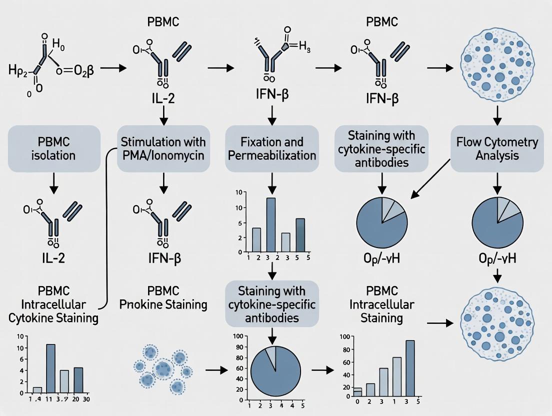
Mastering Intracellular Cytokine Staining: A Comprehensive PBMC Protocol Guide for Immune Response Analysis
This detailed protocol article provides researchers and drug development professionals with a complete, step-by-step guide for performing intracellular cytokine staining (ICS) on peripheral blood mononuclear cells (PBMCs).
Mastering Intracellular Cytokine Staining: A Comprehensive PBMC Protocol Guide for Immune Response Analysis
Abstract
This detailed protocol article provides researchers and drug development professionals with a complete, step-by-step guide for performing intracellular cytokine staining (ICS) on peripheral blood mononuclear cells (PBMCs). Covering foundational principles, an optimized methodological workflow, common troubleshooting strategies, and validation techniques, this guide serves as an essential resource for accurately profiling antigen-specific T-cell responses in immunological research, vaccine development, and immunotherapy assessment. The protocol emphasizes critical steps from cell preparation and stimulation to fixation, permeabilization, staining, and flow cytometric analysis, enabling reliable and reproducible detection of key cytokines like IFN-γ, TNF-α, and IL-2.
Understanding ICS on PBMCs: Core Principles and Research Applications
What is Intracellular Cytokine Staining (ICS) and Why Use PBMCs?
Within the broader thesis investigating optimization strategies for PBMC-based intracellular cytokine staining (ICS) protocols, this application note delineates the fundamental principles and practical applications of ICS. ICS is a flow cytometry-based technique that enables the detection and quantification of cytokine production at the single-cell level within defined immune cell populations. Peripheral Blood Mononuclear Cells (PBMCs) serve as the primary ex vivo model system due to their physiological relevance, heterogeneity, and practicality in clinical and translational research. This document details protocols, key reagents, and data analysis frameworks essential for robust ICS assays in drug development and immune monitoring.
ICS is a cornerstone technique for evaluating antigen-specific T-cell responses. Following ex vivo stimulation, proteins that inhibit cellular secretion (e.g., Brefeldin A) are added, leading to the accumulation of cytokines within the Golgi apparatus and endoplasmic reticulum. Cells are then fixed, permeabilized, and stained with fluorescently-labeled antibodies against specific cytokines and surface markers, allowing for the identification of functional subsets (e.g., IFN-γ-producing CD4+ T cells).
Rationale for Using PBMCs in ICS Assays
PBMCs, isolated via density gradient centrifugation, consist of lymphocytes (T cells, B cells, NK cells) and monocytes. Their use in ICS is justified by several factors:
- Physiological Relevance: They represent the circulating immune compartment directly accessible from blood.
- Functional Viability: PBMCs maintain metabolic and functional capacity for ex vivo stimulation.
- Translational Bridge: Essential for longitudinal monitoring in clinical trials and vaccine studies.
- Practicality: Standardized isolation and cryopreservation enable batch testing.
Table 1: Quantitative Comparison of Common ICS Cell Sources
| Cell Source | Typical Yield per 10 mL Blood | Key Advantages for ICS | Primary Limitations |
|---|---|---|---|
| PBMCs | 10-20 x 10^6 cells | Contains diverse immune subsets; ideal for polyfunctional analysis. | Requires stimulation; does not include granulocytes. |
| Whole Blood | N/A (assay in whole matrix) | Minimal processing; preserves physiological context. | High background; consumes more reagents; complex staining. |
| Sorted/Purified T Cells | 2-5 x 10^6 cells (CD3+) | Reduced non-specific signal; focused analysis. | Lengthy preparation may alter cell state; loses cellular interactions. |
Detailed ICS Protocol for PBMCs
Materials & Pre-Stimulation
- Isolated PBMCs: Fresh or viably cryopreserved.
- Stimulation Cocktail: Choose based on target:
- Positive Control: PMA (e.g., 50 ng/mL) + Ionomycin (e.g., 1 µg/mL).
- Antigen-Specific: Peptide pools (e.g., CEF pool) or specific antigens.
- Secretion Inhibitor: Brefeldin A (5 µg/mL) or Monensin.
- Culture Medium: RPMI-1640 with 10% FBS, L-Glutamine.
Protocol:
- Thaw and rest PBMCs overnight in complete medium at 37°C, 5% CO2.
- Plate 0.5-1 x 10^6 cells per well in a 96-well U-bottom plate.
- Add stimulation agent and secretion inhibitor. Include an unstimulated control (with inhibitor only).
- Incubate 4-6 hours (for strong activators) or 6-18 hours (for antigen-specific responses) at 37°C, 5% CO2.
Cell Staining for Flow Cytometry
- Surface Stain: Perform with antibodies against CD3, CD4, CD8, etc., in PBS+2% FBS for 20-30 min at 4°C.
- Fixation/Permeabilization: Use commercial fixation/permeabilization buffers (e.g., BD Cytofix/Cytoperm). Fix for 20 min at 4°C, then wash with 1X Permeabilization Buffer.
- Intracellular Stain: Add anti-cytokine antibodies (e.g., anti-IFN-γ, IL-2, TNF-α) in permeabilization buffer for 30 min at 4°C.
- Acquisition: Resuspend cells in PBS and acquire on a flow cytometer within 24 hours.
The Scientist's Toolkit: Key Research Reagent Solutions
Table 2: Essential Materials for PBMC ICS
| Item | Function & Importance | Example Product/Component |
|---|---|---|
| Density Gradient Medium | Isolates PBMCs from whole blood via centrifugation. | Ficoll-Paque PLUS |
| Cell Stimulation Cocktail | Activates T-cell receptor signaling to induce cytokine production. | PMA/Ionomycin; Peptide pools (CEF/CEFT) |
| Protein Transport Inhibitor | Blocks cytokine secretion, enabling intracellular accumulation. | Brefeldin A, Monensin |
| Fixation/Permeabilization Buffer | Fixes cells and permeabilizes membranes for intracellular antibody access. | BD Cytofix/Cytoperm Kit, Foxp3/Transcription Factor Staining Buffer Set |
| Fluorochrome-conjugated Antibodies | Specific detection of surface markers and intracellular cytokines. | Anti-human CD3, CD4, CD8, IFN-γ, IL-4, IL-17A |
| Viability Dye | Distinguishes live from dead cells to improve data quality. | Zombie Aqua, 7-AAD |
| Flow Cytometer | Instrument for acquiring multi-parameter single-cell data. | BD FACSymphony, Beckman CytoFLEX |
Data Analysis & Representation
Data is typically analyzed using flow cytometry software (e.g., FlowJo, FCS Express). Key steps include gating on lymphocytes, single cells, live cells, T-cell subsets (CD3+/CD4+ or CD3+/CD8+), and finally, cytokine-positive populations within these subsets.
This application note details protocols for the intracellular detection of key T-cell cytokines—IFN-γ, TNF-α, IL-2, IL-4, and IL-17—within the context of PBMC-based assays for immunophenotyping and drug discovery. These cytokines define major T-helper (Th) cell subsets (Th1, Th2, Th17) and effector functions, making their precise measurement critical for evaluating immune responses in vaccine development, autoimmune disease, and cancer immunotherapy research.
Table 1: Key Cytokines, Their Cellular Sources, Primary Functions, and Secretion Kinetics
| Cytokine | Primary Cellular Source(s) in T Cells | Major Functions in Immunity | Typical Secretion Onset Post-Activation |
|---|---|---|---|
| IFN-γ | Th1 cells, CD8+ Tc1 cells, NK cells | Macrophage activation, MHC class I/II upregulation, antiviral & antibacterial defense, Th1 differentiation. | 4-6 hours |
| TNF-α | Th1 cells, Macrophages, Mast cells | Pro-inflammatory, induces fever & apoptosis, activates neutrophils & endothelial cells, anti-tumor activity. | 1-2 hours |
| IL-2 | Activated CD4+ T cells (primarily) | T-cell proliferation & differentiation, regulatory T cell (Treg) function, immune tolerance. | 4-8 hours |
| IL-4 | Th2 cells, Mast cells, Basophils | B-cell class switching to IgE, Th2 differentiation, alternative macrophage activation, anti-helminthic. | 8-16 hours |
| IL-17 | Th17 cells, γδ T cells, Innate lymphoid cells | Neutrophil recruitment, epithelial/endothelial defense, pathogenesis in autoimmunity & inflammation. | 8-24 hours |
Table 2: Common Stimulation Cocktails for Intracellular Cytokine Staining (ICS) of Human PBMCs
| Target Cytokines | Recommended Stimulus | Co-Stimulatory Signal | Protein Transport Inhibitor | Incubation Duration |
|---|---|---|---|---|
| IFN-γ, TNF-α, IL-2 | PMA (10-50 ng/mL) + Ionomycin (0.5-1 µg/mL) | Optional (inherent) | Brefeldin A (5-10 µg/mL) or Monensin | 4-6 hours |
| IL-4, IL-17 | PMA + Ionomycin (as above) | Optional (inherent) | Brefeldin A (5-10 µg/mL) | 6-12 hours (optimize for IL-4) |
| Antigen-Specific (e.g., IFN-γ) | Peptide Pools (1-2 µg/mL) or Antigen | Anti-CD28/CD49d (1 µg/mL each) | Brefeldin A (10 µg/mL) | 12-16 hours |
Detailed Protocol: PBMC Intracellular Cytokine Staining by Flow Cytometry
Protocol 1: Stimulation and Staining for Th1/Th2/Th17 Cytokines
Day 1: PBMC Isolation and Plating
- Isolate PBMCs from fresh heparinized or EDTA blood using density gradient centrifugation (e.g., Ficoll-Paque).
- Count cells and adjust viability and concentration. Seed 0.5-1 x 10^6 PBMCs per well in a 96-well U-bottom plate in 200 µL of complete RPMI-1640 medium (with 10% FBS, L-glutamine, penicillin/streptomycin).
- Prepare stimulation cocktails (see Table 2). For a positive control, use PMA/Ionomycin. Include an unstimulated control (medium only) and a negative control with protein transport inhibitor only.
- Add stimuli to respective wells. Add protein transport inhibitor (e.g., Brefeldin A) to all wells except the unstimulated control (for surface staining only).
- Incubate plate at 37°C, 5% CO2 for the durations specified in Table 2.
Day 1: Cell Surface Staining
- Post-incubation, centrifuge plate at 300 x g for 5 minutes. Decant supernatant.
- Resuspend cells in 200 µL of cold PBS and centrifuge. Repeat wash.
- Resuspend cell pellet in 100 µL of FACS buffer (PBS + 2% FBS + 0.09% Azide) containing a pre-titrated cocktail of surface antibodies (e.g., anti-CD3, CD4, CD8, CD45RA).
- Vortex gently and incubate for 20-30 minutes at 4°C in the dark.
- Wash cells twice with 200 µL cold FACS buffer.
Day 1: Fixation and Permeabilization
- Thoroughly resuspend cell pellet in 100 µL of Fixation/Permeabilization solution (commercial kit, e.g., BD Cytofix/Cytoperm or equivalent). Incubate for 20 minutes at 4°C in the dark.
- Centrifuge at 500 x g for 5 minutes. Decant supernatant.
- Wash cells twice with 200 µL of 1X Permeabilization/Wash Buffer (from commercial kit). Centrifuge at 500 x g for 5 minutes.
Day 1: Intracellular Staining
- Resuspend cell pellet in 50-100 µL of Permeabilization/Wash Buffer containing pre-titrated intracellular antibodies (e.g., anti-IFN-γ, IL-2, TNF-α, IL-4, IL-17). Use matched isotype controls.
- Vortex gently and incubate for 30 minutes at 4°C in the dark.
- Wash cells twice with 200 µL Permeabilization/Wash Buffer, then once with FACS buffer.
- Resuspend cells in 200-300 µL of FACS buffer or fixation buffer (1-4% PFA). Acquire data on a flow cytometer within 24-48 hours, or store fixed samples at 4°C in the dark for up to a week.
Protocol 2: Critical Experimental Controls for ICS
- Unstimulated Control: PBMCs with medium only (no inhibitor). Sets baseline for surface markers and autofluorescence.
- Activation Control: PMA/Ionomycin stimulation. Determines maximum cytokine production capacity.
- Inhibitor Control: PBMCs with protein transport inhibitor only (no stimulus). Controls for inhibitor-induced artifacts.
- Fluorescence Minus One (FMO) Controls: Essential for accurate gating, especially for low-abundance cytokines like IL-4.
- Isotype Controls: Antibodies of the same IgG subclass but irrelevant specificity. Account for non-specific antibody binding.
The Scientist's Toolkit
Table 3: Essential Research Reagent Solutions for PBMC ICS
| Item | Function & Critical Notes |
|---|---|
| Ficoll-Paque PLUS | Density gradient medium for isolation of viable PBMCs from whole blood. |
| Cell Stimulation Cocktail | Contains PMA (protein kinase C activator) and Ionomycin (calcium ionophore) for potent, receptor-independent T-cell activation. |
| Protein Transport Inhibitors (Brefeldin A/Monensin) | Disrupts Golgi apparatus, preventing cytokine secretion and allowing intracellular accumulation. |
| Flow Cytometry Antibody Panels | Fluorochrome-conjugated monoclonal antibodies against surface markers (CD3, CD4, CD8) and intracellular cytokines. Require careful panel design for spectral overlap. |
| Fixation/Permeabilization Kit | Commercial buffers (e.g., BD Cytofix/Cytoperm, Foxp3/Transcription Factor Staining Buffer Set) that preserve cell structure and allow antibody access to intracellular epitopes. |
| Viability Dye (e.g., LIVE/DEAD Fixable Stain) | Distinguishes live from dead cells, critical for excluding false-positive signals from apoptotic cells. |
| 96-well U-bottom Microplates | Ideal format for low-volume staining and high-throughput sample processing. |
| High-Speed Flow Cytometer | Analyzer with ≥3 lasers to resolve multiple fluorochromes simultaneously. Data analysis software (e.g., FlowJo, FACSDiva) is essential. |
Signaling Pathways and Experimental Workflow
T-Cell Activation and ICS Detection Pathway
PBMC ICS Experimental Workflow
The Role of ICS in Immunology, Vaccine, and Immunotherapy Research
Intracellular Cytokine Staining (ICS) coupled with flow cytometry is a cornerstone technique for quantifying antigen-specific T-cell responses by measuring cytokine production at the single-cell level. Within the broader thesis on PBMC ICS protocol optimization, this technique is indispensable for evaluating cellular immunology in vaccine candidates (e.g., against HIV, malaria, SARS-CoV-2) and immunotherapies (e.g., checkpoint inhibitors, CAR-T cells). It enables the simultaneous assessment of cytokine polyfunctionality, memory phenotypes, and activation states, providing a multidimensional view of immune efficacy.
Table 1: Key Applications of ICS in Research and Development
| Application Field | Primary Measured Cytokines | Key Readout | Typical Cell Population |
|---|---|---|---|
| Vaccine Immunology | IFN-γ, TNF-α, IL-2, IL-4, IL-5, IL-13 | Magnitude and quality of antigen-specific T-cell response; Th1/Th2/Th17 skewing. | CD4+, CD8+ T cells |
| Cancer Immunotherapy | IFN-γ, TNF-α, Granzyme B, Perforin, IL-10 | Cytotoxic potential of tumor-infiltrating lymphocytes (TILs) or circulating T cells; Exhaustion markers (PD-1, TIM-3). | CD8+ T cells, CD4+ T cells |
| Autoimmune & Infectious Disease | IL-17A, IFN-γ, TNF-α, IL-6 | Pathogenic vs. regulatory cytokine profiles; Response to pathogen antigens. | Th17, Th1, Treg cells |
| Immunomodulatory Drug Screening | IFN-γ, IL-2, IL-4, IL-10 | Shift in cytokine profiles pre- and post-treatment; On-target immune modulation. | Pan-T cells, specific subsets |
Detailed PBMC ICS Protocol
This protocol details the critical steps for detecting antigen-specific T-cell responses from human Peripheral Blood Mononuclear Cells (PBMCs).
Part A: PBMC Stimulation
- Materials: Fresh or viably frozen PBMCs, sterile tissue culture plates/ tubes, complete RPMI-1640 media, antigenic peptides (e.g., viral peptide pools), positive control stimuli (PMA/Ionomycin or SEB), protein transport inhibitors (Brefeldin A/Monensin).
- Procedure:
- Thaw and rest PBMCs overnight in complete media at 37°C, 5% CO₂.
- Plate 0.5-1 x 10⁶ PBMCs per well in a 96-well U-bottom plate.
- Stimulate: Add specific peptide (e.g., 1 µg/mL) or positive control. Include an unstimulated control (media only). Final volume: 200 µL/well.
- Incubate for 2 hours at 37°C.
- Add Protein Transport Inhibitor: Add Brefeldin A (final conc. 10 µg/mL). Critical: For positive controls (PMA/Ionomycin), add Brefeldin A at the start.
- Incubate for an additional 4-16 hours (typically 6 hours) at 37°C, 5% CO₂.
Part B: Cell Surface Staining
- Materials: FACS buffer (PBS + 2% FBS), fluorescently conjugated antibodies against surface markers (e.g., anti-CD3, CD4, CD8, CD69), viability dye (e.g., fixable viability dye eFluor 506).
- Procedure:
- Transfer cells to FACS tubes. Centrifuge at 500 x g for 5 min. Decant supernatant.
- Resuspend pellet in 100 µL FACS buffer containing a viability dye. Incubate for 20-30 min at 4°C in the dark.
- Wash with 2 mL FACS buffer. Centrifuge. Decant.
- Resuspend in 100 µL FACS buffer containing titrated antibodies against surface markers. Incubate for 30 min at 4°C in the dark.
- Wash with 2 mL FACS buffer. Centrifuge. Decant. Proceed to fixation.
Part C: Fixation, Permeabilization, and Intracellular Staining
- Materials: Fixation/Permeabilization buffer (commercial kit, e.g., BD Cytofix/Cytoperm or Foxp3/Transcription Factor Staining Buffer Set), permeabilization wash buffer, intracellular cytokine antibodies (e.g., anti-IFN-γ, IL-2, TNF-α).
- Procedure:
- Fix & Permeabilize: Resuspend cell pellet in 250 µL of fixation/permeabilization buffer. Incubate for 20-60 min at 4°C in the dark.
- Wash: Add 1-2 mL of permeabilization wash buffer. Centrifuge at 500 x g for 5 min. Decant. Note: Higher g-force may be needed due to fixation.
- Intracellular Staining: Resuspend cell pellet in 100 µL of permeabilization wash buffer containing pre-titrated intracellular antibodies. Incubate for 30-60 min at 4°C in the dark.
- Final Wash: Wash with 2 mL permeabilization wash buffer. Centrifuge. Decant.
- Resuspend: Resuspend cells in 200-300 µL of FACS buffer or stabilizing fixative. Acquire on a flow cytometer within 24-48 hours.
The Scientist's Toolkit: Key Research Reagent Solutions
Table 2: Essential Materials for ICS Experiments
| Reagent Category | Specific Example | Function & Importance |
|---|---|---|
| Protein Transport Inhibitor | Brefeldin A, Monensin | Blocks Golgi-mediated export, causing cytokines to accumulate intracellularly for detection. |
| Viability Dye | Fixable Viability Dye eFluor 506, Zombie NIR | Distinguishes live from dead cells, crucial for excluding false-positive signals from dying cells. |
| Surface Stain Antibodies | Anti-human CD3, CD4, CD8, CD45RA, CCR7 | Identifies major lymphocyte lineages and defines memory/effector subsets (e.g., naïve, central memory). |
| Cytokine Antibodies | Anti-human IFN-γ, TNF-α, IL-2, IL-4, IL-17A | Directly detects the cytokines produced, enabling functional profiling. |
| Fixation/Permeabilization Kit | BD Cytofix/Cytoperm, eBioscience Foxp3 Buffer Set | Preserves cell structure while making the intracellular cytokine accessible to antibodies. |
| Activation Stimuli | PMA/Ionomycin, Staphylococcal Enterotoxin B (SEB) | Positive control to demonstrate maximal cellular potential and assay functionality. |
| Synthetic Antigens | Peptide pools (e.g., CEF, SARS-CoV-2 PepTivator) | Antigen-specific stimulation to probe pathogen- or vaccine-induced memory T cells. |
Diagrams
ICS Experimental Protocol Workflow
Mechanism of T Cell Activation and ICS Detection
Application Notes
Intracellular cytokine staining (ICS) of peripheral blood mononuclear cells (PBMCs) is a cornerstone technique in immunology and drug development, enabling the functional characterization of T-cell subsets. The protocol's success hinges on the precise use of specific reagents, from stimulation inhibitors to detection antibodies, and on sophisticated analytical instrumentation. This note details the critical components within the context of optimizing a PBMC ICS protocol for thesis research on T-helper cell responses.
The core principle involves stimulating PBMCs with a mitogen or antigen in the presence of protein transport inhibitors like Brefeldin A (BFA) and/or Monensin. These reagents disrupt Golgi apparatus function, causing cytokines to accumulate within the cell, thereby enhancing detection sensitivity during subsequent staining and flow cytometric analysis. The choice between BFA and Monensin, or their combination, is empirically determined based on the target cytokine and cell type.
Key Considerations:
- Stimulation: Phorbol 12-myristate 13-acetate (PMA) with Ionomycin is a potent polyclonal stimulant for T-cells but can downregulate the CD4 receptor. Antigen-specific stimulation (e.g., peptide pools) is used for antigen-reactive T-cell studies but yields lower frequency populations.
- Inhibition: BFA is effective for most cytokines (e.g., IFN-γ, TNF-α, IL-2). Monensin is often preferred for IL-4, IL-5, and IL-10. Combining both can provide broad-spectrum inhibition.
- Staining: Successful intracellular staining requires robust cell fixation and permeabilization. Commercial kits with optimized buffers are essential for maintaining cell morphology and antibody accessibility while minimizing background fluorescence.
- Analysis: Modern multi-laser flow cytometers with 5+ fluorescence detectors are standard. Spectral flow cytometers are increasingly valuable for high-parameter panels, reducing spillover and enabling deeper immunophenotyping alongside cytokine detection.
Table 1: Comparison of Common Protein Transport Inhibitors in ICS
| Reagent | Primary Mechanism | Typical Working Concentration | Key Target Cytokines | Notes & Considerations |
|---|---|---|---|---|
| Brefeldin A (BFA) | Inhibits ER-to-Golgi transport by inhibiting ARF1 activation. | 1-10 µg/mL (often 5 µg/mL) | IFN-γ, TNF-α, IL-2, IL-6 | Can be toxic with prolonged incubation (>12h). May reduce CD8 staining. |
| Monensin | Na+/H+ ionophore, disrupts Golgi and trans-Golgi network pH gradient. | 2-5 µM | IL-4, IL-5, IL-10, IFN-γ | Often used in combination with BFA for broader inhibition. |
| BFA + Monensin | Combined action of both mechanisms. | BFA (5 µg/mL) + Monensin (2 µM) | Broad spectrum (Th1, Th2, Th17) | May increase cellular toxicity and background. Empirical optimization required. |
Table 2: Typical PMA/Ionomycin Stimulation Conditions for T-Cell ICS
| Component | Function | Typical Concentration | Incubation Time |
|---|---|---|---|
| PMA | Protein Kinase C activator, provides Signal 1. | 20-50 ng/mL | 4-6 hours (with inhibitors) |
| Ionomycin | Calcium ionophore, provides Signal 2. | 0.5-1 µM | 4-6 hours (with inhibitors) |
| Protein Transport Inhibitor | Retains cytokines intracellularly. | See Table 1 | Co-incubated with stimulants |
Detailed Protocols
Protocol 1: Standard PBMC ICS for Th1/Th2 Cytokines
Objective: To detect intracellular IFN-γ and IL-4 in CD4+ T-cells after polyclonal stimulation.
Materials: See "The Scientist's Toolkit" below.
Method:
- PBMC Preparation: Thaw cryopreserved PBMCs in pre-warmed complete RPMI-1640 medium. Rest for 4-6 hours at 37°C, 5% CO₂.
- Stimulation Setup: Aliquot 0.5-1 x 10⁶ PBMCs per well into a 96-well U-bottom plate. Centrifuge (300 x g, 5 min), aspirate supernatant.
- Stimulation & Inhibition: Resuspend cells in 200 µL complete medium containing:
- PMA (25 ng/mL final)
- Ionomycin (1 µM final)
- Brefeldin A (5 µg/mL final)
- Optional: Add CD28/CD49d co-stimulatory antibodies (1 µg/mL each) to enhance response.
- Negative Control: Medium with BFA only (no stimulants).
- Incubate for 4-6 hours at 37°C, 5% CO₂.
- Surface Staining: Post-incubation, add 20 µL of 20 mM EDTA per well, mix, incubate 15 min at RT. Wash with PBS + 2% FBS (FACS buffer). Block Fc receptors with human Fc block for 10 min on ice. Without washing, add directly titrated surface antibody cocktail (e.g., anti-CD3, CD4, CD8, viability dye). Incubate 30 min in the dark on ice. Wash twice with FACS buffer.
- Fixation & Permeabilization: Resuspend cell pellet thoroughly in 100 µL of commercial fixation/permeabilization concentrate. Incubate 20-30 min at 4°C in the dark. Wash twice with 1x permeabilization/wash buffer.
- Intracellular Staining: Resuspend fixed/permeabilized cells in 50-100 µL of permeabilization/wash buffer containing titrated intracellular antibody cocktail (e.g., anti-IFN-γ, IL-4). Incubate 30-60 min at 4°C in the dark. Wash twice with permeabilization/wash buffer.
- Acquisition: Resuspend final cell pellet in FACS buffer or fixation buffer. Acquire on a flow cytometer within 24 hours. Collect a minimum of 50,000 events in the lymphocyte gate.
Protocol 2: Titration of Protein Transport Inhibitors
Objective: To empirically determine the optimal concentration of BFA and/or Monensin for a specific cytokine/cell system.
Method:
- Prepare PBMCs and stimulation medium as in Protocol 1.
- In a 96-well plate, set up a matrix of stimulation conditions. Columns: Varying concentrations of BFA (0, 1, 2.5, 5, 10 µg/mL). Rows: Varying concentrations of Monensin (0, 1, 2, 5 µM). Include unstimulated controls for each inhibitor combination.
- Add cells and stimuli (e.g., PMA/Ionomycin) to all test wells. Incubate for the standard 6 hours.
- Perform surface staining, fixation/permeabilization, and intracellular staining for your target cytokines (e.g., IFN-γ and IL-4) following Protocol 1 steps 4-7.
- Analysis: On the flow cytometer, gate on live CD3+CD4+ lymphocytes. Compare the mean fluorescence intensity (MFI) and percentage of cytokine-positive cells across the matrix. The optimal condition maximizes the signal-to-noise ratio (positive population MFI / negative control MFI) without excessive cell death (as measured by viability dye).
Visualizations
Title: Mechanism of Brefeldin A in Intracellular Cytokine Staining
Title: ICS Protocol Workflow for PBMCs
The Scientist's Toolkit
Table 3: Essential Reagents and Equipment for PBMC ICS
| Category | Item | Primary Function in ICS Protocol |
|---|---|---|
| Cell Source | Cryopreserved PBMCs | Primary human immune cells for ex-vivo functional assays. |
| Stimulation | PMA & Ionomycin | Potent pharmacological activators inducing cytokine production in T-cells. |
| Protein Transport Inhibitors | Brefeldin A, Monensin | Block cytokine secretion, causing intracellular accumulation for detection. |
| Co-stimulation | Anti-CD28/CD49d antibodies | Provides additional co-stimulatory signal, enhancing activation. |
| Viability Dye | e.g., Fixable Viability Stain | Distinguishes live from dead cells during flow analysis, critical for accuracy. |
| Surface Stain Antibodies | Anti-CD3, CD4, CD8 | Define major lymphocyte subsets for downstream gating. |
| Fixation/Permeabilization Kit | Commercial buffer system (e.g., Cytofix/Cytoperm) | Fixes cells and creates pores in membranes allowing intracellular antibody access. |
| Intracellular Antibodies | Anti-cytokine (IFN-γ, IL-2, TNF-α, IL-4, etc.) | Directly conjugate to fluorochromes for detection of accumulated cytokines. |
| Flow Cytometer | Analyzer with ≥2 lasers (488nm, 640nm) and 5+ detectors. | Instrument for single-cell analysis of light scatter and fluorescence. |
| Analysis Software | e.g., FlowJo, FCS Express | Software for visualizing, gating, and quantifying flow cytometry data. |
Within the broader thesis investigating optimization of Peripheral Blood Mononuclear Cell (PBMC) intracellular cytokine staining (ICS) protocols, the rigorous definition of stimulation conditions and corresponding controls is paramount. This document provides detailed application notes and protocols for establishing these critical experimental parameters, enabling precise measurement of antigen-specific T-cell responses in research and drug development contexts.
Core Stimulation Strategies for PBMC ICS
Effective ICS requires activation of specific immune pathways to induce cytokine production. The choice of stimulant dictates the nature of the response measured.
Common Stimulation Agents & Their Targets
Table 1: Quantitative Summary of Common PBMC Stimulants for ICS
| Stimulant Category | Specific Agent | Typical Working Concentration | Incubation Time (hr) | Primary Target Cell | Key Induced Cytokine(s) |
|---|---|---|---|---|---|
| Polyclonal Activators | PMA + Ionomycin | 50 ng/mL + 1 µg/mL | 4-6 | T cells (all) | IFN-γ, IL-2, TNF-α, IL-4 |
| Anti-CD3/CD28 beads | 1 bead:1 cell ratio | 6-18 | T cells | IFN-γ, IL-2 | |
| Antigen-Specific | Peptide Pools (e.g., CEF) | 1-2 µg/mL per peptide | 6-18 | Memory T cells | IFN-γ, IL-2 |
| Viral Lysates | 1-10 µg/mL | 12-18 | Antigen-specific T cells | IFN-γ, TNF-α | |
| Toll-like Receptor (TLR) Ligands | LPS (TLR4) | 100 ng/mL - 1 µg/mL | 6-18 | Monocytes, B cells | IL-1β, IL-6, TNF-α |
| R848 (TLR7/8) | 1-5 µg/mL | 6-18 | Monocytes, DCs | IFN-α, IL-12 | |
| Cytokine Stimulation | IL-12 + IL-18 | 10 ng/mL + 100 ng/mL | 6-18 | NK, T cells | IFN-γ |
Key Signaling Pathways Induced by Common Stimulants
Diagram 1: Signaling pathways for TCR and mitogen stimulation.
The Critical Role of Controls
Appropriate controls are non-negotiable for data interpretation, enabling discrimination between specific and non-specific staining, and accounting for background cytokine production.
Essential Control Conditions
Table 2: Mandatory Control Conditions for PBMC ICS Experiments
| Control Type | Purpose | Recommended Composition | Acceptable Background Cytokine+ (%) |
|---|---|---|---|
| Unstimulated | Measures baseline activation/background. | Cells + media only, with equivalent [DMSO] if used as solvent. | <0.1% (CD4/8 T cells); <0.5% (NK cells) |
| Solvent Control | Accounts for effects of stimulant solvent (e.g., DMSO). | Cells + media with highest [solvent] used in assay. | Should match unstimulated. |
| Positive Control | Assesss cell viability/functionality & assay performance. | PMA + Ionomycin (strong); SEB (superantigen). | Expect high % (e.g., 5-30% IFN-γ+ in CD4). |
| Stimulus Backbone | For complex stimuli (e.g., peptide pools in DMSO). | All non-active components of the stimulus. | Should match solvent control. |
| Compensation Control | For flow cytometry color compensation. | Single-stained beads or cells. | N/A |
| Fluorescence Minus One (FMO) | Determines positive staining gates. | All antibodies except the one of interest. | N/A |
Experimental Workflow for Stimulation & Control Setup
Diagram 2: ICS workflow with control condition integration.
Detailed Protocols
Protocol 4.1: Setup of a Standard Antigen-Specific ICS Assay with Controls
Objective: To detect antigen-specific CD4+ and CD8+ T-cell responses via IFN-γ and IL-2 production. Materials: See Scientist's Toolkit below. Procedure:
- PBMC Preparation: Isolate PBMCs via density gradient centrifugation. Rest overnight (12-18h) in complete RPMI (10% FBS, 1% Pen/Strep) at 37°C, 5% CO₂.
- Stimulation Plate Preparation: In a sterile 96-well U-bottom plate, pre-dilute all stimulants and controls in complete RPMI to 2X final concentration.
- Well A1-A3: Unstimulated Control (100 µL media + 0.1% DMSO).
- Well A4-A6: Positive Control (100 µL media containing 100 ng/mL PMA + 2 µg/mL Ionomycin, from stock solutions).
- Well B1-H6: Experimental Stimuli (e.g., 100 µL media containing 2 µg/mL peptide pool, 10 µg/mL protein antigen, or TLR ligand).
- Cell Addition: Count rested PBMCs. Add 100 µL of cell suspension (2 x 10⁶ cells/mL, so 2 x 10⁵ cells/well) to each well containing 100 µL of 2X stimulus. This achieves a final volume of 200 µL/well and the desired 1X stimulus concentration.
- Protein Transport Inhibition: Immediately add 2 µL of 1 mg/mL Brefeldin A stock (or Monensin per manufacturer's instructions) to each well for a final concentration of 10 µg/mL. Mix gently by tapping.
- Incubation: Incubate plate for 12-16 hours (for antigen recall) at 37°C, 5% CO₂.
- Post-Stimulation Processing: Proceed to surface staining, followed by fixation/permeabilization and intracellular staining per optimized ICS protocol.
Protocol 4.2: Preparation and Use of Fluorescence Minus One (FMO) Controls
Objective: To accurately set positivity gates for each fluorescent channel in the flow cytometry panel. Procedure:
- Design Panel: For a 7-color panel (CD3, CD4, CD8, CD69, IFN-γ, IL-2, Viability Dye), prepare 8 staining tubes for a single unstimulated or positive control sample.
- Tube Setup:
- Tube 1 (Full Panel): Contains all 7 antibodies.
- Tube 2 (FMO IFN-γ): Contains all antibodies except anti-IFN-γ. Replace with an isotype control or buffer.
- Tube 3 (FMO IL-2): Contains all antibodies except anti-IL-2.
- Tubes 4-8: Repeat for each other marker (CD69, etc.) if the spread of negative/positive populations is not well established.
- Staining: Aliquot an equal number of cells (from the same stimulated control sample) into each tube. Stain, fix, and permeabilize simultaneously under identical conditions.
- Gating: During analysis, use the FMO control to define the boundary between negative and positive populations for its omitted antibody. The "Full Panel" tube is used for final data collection, not for setting gates.
The Scientist's Toolkit: Research Reagent Solutions
Table 3: Essential Materials for PBMC ICS Stimulation & Control Experiments
| Item / Reagent Solution | Function in Experiment | Example Product/Catalog # (Reference) |
|---|---|---|
| Cell Preparation | ||
| Lymphocyte Separation Medium | Density gradient medium for PBMC isolation. | Ficoll-Paque PLUS (Cytiva) |
| Complete Cell Culture Medium | Supports cell viability during rest & stimulation. | RPMI-1640 + 10% FBS + 1% Pen/Strep |
| Stimulation Agents | ||
| Phorbol 12-Myristate 13-Acetate (PMA) | PKC activator, mitogen (used with Ionomycin). | Sigma-Aldrich, P1585 |
| Ionomycin Calcium Salt | Calcium ionophore (used with PMA). | Sigma-Aldrich, I3909 |
| Anti-CD3/CD28 Activator Beads | Polyclonal T-cell activator mimicking TCR engagement. | Gibco, Dynabeads Human T-Activator CD3/CD28 |
| Peptide Pools (e.g., CEF, CEFX) | Overlapping peptides from common viral antigens; positive control for memory T cells. | JPT, PM-CEFX |
| Inhibition & Staining | ||
| Brefeldin A Solution | Inhibits protein transport from Golgi, accumulates cytokines intracellularly. | BioLegend, 420601 |
| Monensin Solution | Alternative protein transport inhibitor (e.g., for chemokines). | BioLegend, 420701 |
| BD Cytofix/Cytoperm Kit | Widely used solution set for fixation and permeabilization. | BD Biosciences, 554714 |
| Foxp3/Transcription Factor Staining Buffer Set | Alternative for nuclear or difficult cytokines. | Thermo Fisher, 00-5523-00 |
| Flow Cytometry Controls | ||
| Compensation Bead Set | Negative and positive beads for multicolor compensation. | Thermo Fisher, UltracComp eBeads, 01-2222-42 |
| Isotype Control Antibodies | Matched to primary antibody host, subclass, and fluorochrome. | Various manufacturers |
| Viability Assessment | ||
| Fixable Viability Dye (e.g., Zombie NIR) | Distinguishes live/dead cells prior to fixation. | BioLegend, 423106 |
Step-by-Step Optimized PBMC ICS Protocol: From Cell Harvest to Data Acquisition
Within the broader research thesis investigating intracellular cytokine staining (ICS) protocols for Peripheral Blood Mononuclear Cells (PBMCs), the initial isolation step is critical. The quality, viability, and functional purity of the isolated PBMC population directly impact downstream ICS results, affecting the accuracy of immunophenotyping and cytokine detection. This application note details optimized, current best practices for density gradient centrifugation to ensure high-yield, high-viability PBMC isolation.
Key Quantitative Parameters for Optimal Isolation
The following tables summarize target metrics and the impact of key variables on PBMC isolation outcomes, based on current literature and manufacturer guidelines.
Table 1: Target Metrics for High-Quality PBMC Isolation
| Parameter | Optimal Target Range | Importance for ICS |
|---|---|---|
| Viability | ≥ 95% | Dead cells increase nonspecific staining and background fluorescence. |
| PBMC Yield | 0.5 - 2.0 x 10^6 cells / mL of whole blood | Ensures sufficient cells for multi-panel staining and experimental replicates. |
| Lymphocyte Recovery | ≥ 85% of total PBMCs | Lymphocytes (T, B, NK cells) are primary targets for cytokine analysis. |
| Granulocyte Contamination | < 5% | Myeloid cells can nonspecifically bind antibodies and alter assay background. |
| Platelet Contamination | < 10 platelets per lymphocyte | Excessive platelets can mask surface antigens and block antibody binding. |
Table 2: Effect of Centrifugation Parameters on Isolation Purity & Viability
| Variable | Recommended Setting | Effect of Deviation |
|---|---|---|
| Centrifugation Force (g) | 400 - 500 g | Too low: Poor separation. Too high: Pelleted granulocytes contaminate PBMC layer. |
| Brake Setting | OFF (or LOW) | Brake use disrupts the gradient layer, mixing cells and reducing purity. |
| Centrifugation Time | 20 - 30 minutes | Insufficient time compromises separation; excessive time may reduce viability. |
| Temperature | 18 - 22°C (Room Temp) | Cold temperatures can increase platelet aggregation and reduce monocyte viability. |
| Sample: Medium Ratio | 1:1 to 1:2 (Blood:Diluent) | Proper dilution reduces viscosity and improves separation efficiency. |
Detailed Protocol: PBMC Isolation via Ficoll-Paque
Materials & Reagents
- Anticoagulated Blood: Human peripheral blood collected in sodium heparin or EDTA vacutainers.
- Density Gradient Medium: Ficoll-Paque Plus (ρ = 1.077 ± 0.001 g/mL).
- Dilution/Wash Buffer: Phosphate-Buffered Saline (PBS), sterile, without Ca2+/Mg2+.
- Complete Culture Medium: RPMI-1640 supplemented with 10% Fetal Bovine Serum (FBS) or Human AB Serum.
- Equipment: Centrifuge with swing-out rotor, sterile centrifuge tubes, pipettes, biological safety cabinet.
Method
- Blood Dilution: Dilute anticoagulated whole blood 1:1 with room temperature PBS or wash buffer. Mix gently by inversion.
- Layering: In a sterile 50 mL conical tube, carefully layer 15 mL of Ficoll-Paque. Slowly and gently overlay 25-30 mL of the diluted blood onto the Ficoll-Paque, maintaining a clear interface. Avoid mixing the layers.
- Centrifugation: Centrifuge at 400-500 g for 20-30 minutes at room temperature (18-22°C) with the brake OFF. This allows for isopycnic separation without disrupting the gradient.
- PBMC Collection: After centrifugation, four distinct layers will be visible (top to bottom: plasma/platelets, PBMC ring, Ficoll-Paque, granulocytes/erythrocytes). Using a sterile pipette, carefully harvest the opaque PBMC interface layer. Transfer to a new 50 mL tube.
- Washing: Fill the tube containing PBMCs with wash buffer (at least 3x the volume of harvested cells). Centrifuge at 300-350 g for 10 minutes at room temperature with a low brake setting. Decant supernatant completely.
- Platelet Removal (Optional but Recommended for ICS): Resuspend the cell pellet in 10 mL of wash buffer or serum-free medium. Centrifuge at 200 g for 10 minutes at room temperature. This softer spin pellets lymphocytes/monocytes while leaving platelets in suspension. Repeat if necessary.
- Final Resuspension & Counting: Resuspend the final PBMC pellet in complete culture medium or staining buffer. Count cells using an automated cell counter or hemocytometer with trypan blue exclusion to determine viability and concentration.
The Scientist's Toolkit: Research Reagent Solutions
Table 3: Essential Materials for PBMC Isolation & Viability Assessment
| Item | Function & Relevance |
|---|---|
| Ficoll-Paque Plus | Polysucrose-sodium diatrizoate solution with optimized density (1.077 g/mL) for selective separation of mononuclear cells from other blood components. |
| Density Gradient Tubes (Leucosep) | Tubes with a porous barrier that simplifies layering, preventing mixing of blood and Ficoll, improving reproducibility. |
| Heparin or EDTA Tubes | Anticoagulant blood collection tubes. Heparin is preferred for functional assays like ICS, as EDTA can chelate calcium required for cell activation. |
| Trypan Blue Stain (0.4%) | Vital dye used to distinguish live (unstained) from dead (blue-stained) cells for viability assessment post-isolation. |
| Automated Cell Counter | Provides rapid, reproducible cell count and viability analysis, superior to manual hemocytometer counts for consistency. |
| DNAse I (Optional) | Reduces cell clumping caused by free DNA released from lysed cells during processing, improving cell recovery. |
| Human AB Serum | Serum supplement for culture media that minimizes background activation of PBMCs compared to some FBS lots. |
Visualizing the Workflow and Critical Relationships
PBMC Isolation Workflow for ICS
How Viability Affects ICS Results
Within the broader thesis on optimizing PBMC intracellular cytokine staining (ICS) protocols, a critical comparative analysis of T-cell stimulation methods is required. Antigen-specific stimulation, pharmacologic activation (PMA/Ionomycin), and superantigen engagement (SEB) represent three fundamentally distinct approaches, each with unique applications and experimental outcomes. This application note details these protocols, providing researchers with the methodologies necessary to select the appropriate stimulation strategy for specific immunology and drug development research questions.
Table 1: Core Characteristics of T-Cell Stimulation Protocols
| Feature | Antigen-Specific | PMA/Ionomycin | Staphylococcal Enterotoxin B (SEB) |
|---|---|---|---|
| Mechanism | TCR-pMHC interaction | Protein Kinase C (PKC) activation & calcium influx | Superantigen; bridges TCR Vβ region and MHC-II |
| Target Population | Rare, antigen-specific clones (~0.01-1% of T cells) | All T cells, especially CD4+ and CD8+ | Polyclonal, Vβ-specific subsets (up to 20% of T cells) |
| Typical Cytokine Output | Low to moderate (requires amplification) | Very high, polyfunctional | High, polyclonal |
| Requires Antigen-Presenting Cells (APCs) | Yes | No | Yes |
| Optimal Duration | Long (6-16 hours) | Short (4-6 hours) | Intermediate (6-12 hours) |
| Key Application | Vaccine research, infectious disease, cancer immunotherapy | Maximal cytokine induction for immunophenotyping | Broad polyclonal response; positive control |
Table 2: Typical Reagent Concentrations and Incubation Times
| Stimulus | Typical Working Concentration | Protein Transport Inhibitor Added At | Total Incubation Time | Temperature |
|---|---|---|---|---|
| Peptide Pools (e.g., CEF) | 1-2 µg/mL per peptide | 0-2 hours post-stimulation | 6-16 hours | 37°C, 5% CO2 |
| PMA/Ionomycin | PMA: 10-50 ng/mL; Ionomycin: 0.5-1 µg/mL | At stimulation start | 4-6 hours | 37°C, 5% CO2 |
| SEB | 0.1-1 µg/mL | 0-2 hours post-stimulation | 6-12 hours | 37°C, 5% CO2 |
Detailed Experimental Protocols
Protocol 1: Antigen-Specific Stimulation of PBMCs for ICS
This protocol is designed to detect low-frequency, antigen-specific T-cell responses, crucial for vaccine immunogenicity studies.
Materials:
- Fresh or properly thawed PBMCs.
- Antigen of interest: peptide pools (e.g., CEF, viral peptides), recombinant proteins.
- Co-stimulatory antibodies: anti-CD28 and anti-CD49d (1 µg/mL each).
- Protein transport inhibitor: Brefeldin A (BFA, 5-10 µg/mL) or Monensin.
- Complete RPMI-1640 culture medium.
- 96-well U-bottom or V-bottom plates.
Procedure:
- Cell Plating: Resuspend PBMCs in complete medium and plate 0.2-1 x 10^6 cells per well in a 96-well plate.
- Stimulation Setup:
- Test Wells: Add antigen peptide/protein at optimal concentration (see Table 2).
- Positive Control Wells: Add PMA/Ionomycin at concentrations from Table 2.
- Negative Control Wells: Add medium only or an irrelevant peptide.
- Add Co-stimulation: Add anti-CD28/anti-CD49d antibodies to all wells except the negative control.
- Incubate: Place plate in a humidified 37°C, 5% CO2 incubator for 2 hours.
- Inhibit Protein Transport: Add Brefeldin A (or Monensin) to all wells. Return plate to the incubator for an additional 4-14 hours (typical total incubation: 6-16 hours).
- Harvest: Proceed to surface and intracellular staining for flow cytometry. Cells are now ready for the staining procedures outlined in the overarching thesis.
Protocol 2: Polyclonal Stimulation with PMA and Ionomycin for ICS
This protocol provides a strong, universal stimulus for detecting cytokine production capacity across most T cells, useful for immunophenotyping and functional assays.
Materials:
- PBMCs.
- Phorbol 12-myristate 13-acetate (PMA) stock solution.
- Ionomycin calcium salt stock solution.
- Protein transport inhibitor (BFA/Monensin).
- Complete medium.
- 96-well plate.
Procedure:
- Cell Plating: Plate PBMCs as in Protocol 1.
- Stimulation Setup: Prepare a master mix containing PMA (final 10-50 ng/mL) and Ionomycin (final 0.5-1 µg/mL) in complete medium. Add to cell wells.
- Simultaneous Inhibition: Add protein transport inhibitor (BFA/Monensin) at the same time as stimulants.
- Incubate: Incubate plate at 37°C, 5% CO2 for 4-6 hours. Do not exceed 6 hours, as PMA can cause significant downregulation of surface receptors like CD4 and TCR.
- Harvest: Cells are ready for staining. Note that surface marker staining may be affected; prioritize intracellular targets or use special staining panels.
Protocol 3: Polyclonal Stimulation with Superantigen SEB for ICS
SEB provides a broad, polyclonal stimulation bridging TCR and MHC-II, suitable for robust positive controls and studies of T-cell repertoire.
Materials:
- PBMCs.
- Staphylococcal Enterotoxin B (SEB).
- Co-stimulatory antibodies (anti-CD28/49d).
- Protein transport inhibitor.
- Complete medium.
- 96-well plate.
Procedure:
- Cell Plating: Plate PBMCs.
- Stimulation: Add SEB at a final concentration of 0.1-1 µg/mL to test wells.
- Add Co-stimulation: Add co-stimulatory antibodies to all wells (including SEB wells) except the negative control.
- Incubate & Inhibit: Incubate for 2 hours, then add BFA/Monensin. Continue incubation for an additional 4-10 hours (total 6-12 hours).
- Harvest: Proceed to staining.
Signaling Pathways and Workflow Visualizations
T Cell Stimulation Signaling Pathways Comparison
PBMC Stimulation Workflow for ICS
The Scientist's Toolkit: Research Reagent Solutions
Table 3: Essential Materials for Cell Stimulation and ICS
| Reagent Category | Specific Example(s) | Function in Protocol | Key Consideration |
|---|---|---|---|
| Stimulants | Peptide pools (CEF, viral), PMA, Ionomycin, SEB | Activate T-cells via distinct mechanisms to induce cytokine production. | Titrate for optimal signal-to-noise; PMA downregulates CD4/CD3. |
| Protein Transport Inhibitors | Brefeldin A (BFA), Monensin | Inhibit Golgi transport, causing cytokines to accumulate intracellularly for detection. | Add at correct timepoint; toxicity increases with incubation time. |
| Co-stimulatory Antibodies | Anti-CD28, Anti-CD49d | Provide secondary signal required for robust antigen-specific activation. | Omit in negative control; not required for PMA/Ionomycin. |
| Cell Culture Medium | RPMI-1640 + FBS + Pen/Strep + L-Glutamine | Maintains cell viability and health during stimulation period. | Use serum from same species as APCs if present. |
| Blocking Reagent | Human Fc Receptor Blocking Solution | Reduces nonspecific antibody binding via Fc receptors. | Apply before surface staining for cleaner flow results. |
| Viability Dye | Fixable Viability Dye (e.g., Zombie NIR) | Distinguishes live from dead cells during flow cytometry analysis. | Use before fixation/permeabilization for best results. |
| Fixation/Permeabilization Buffer | Commercial ICS Kit (e.g., BD Cytofix/Cytoperm) | Fixes cells and permeabilizes membranes to allow intracellular antibody access. | Must be compatible with fluorochromes and target cytokines. |
| Intracellular Antibodies | Anti-IFN-γ, Anti-IL-2, Anti-TNF-α, Anti-IL-4, etc. | Detect and quantify cytokine production at the single-cell level. | Titrate and validate; check clone compatibility with fixation. |
Within the broader research thesis on optimizing Peripheral Blood Mononuclear Cell (PBMC) intracellular cytokine staining (ICS) protocols, the selection and application of secretion inhibitors is a critical determinant of success. Brefeldin A (BFA) and monensin are the principal pharmacological agents used to block cytokine egress, thereby enabling their intracellular accumulation for detection by flow cytometry. This Application Note provides detailed protocols and comparative data to guide researchers in their optimal use.
Mechanism of Action & Comparative Pharmacology
Brefeldin A: A fungal metabolite that disrupts the Golgi apparatus and endoplasmic reticulum (ER) structure by inhibiting ADP-ribosylation factor (ARF) guanine nucleotide exchange factors (GEFs). This prevents the formation of COP-I-coated vesicles, blocking anterograde transport from the ER to the Golgi and causing a reversible disintegration of the Golgi complex.
Monensin: A carboxylic ionophore that exchanges monovalent cations (Na+/H+, K+/H+) across membranes. In the Golgi apparatus, it disrupts ionic gradients, leading to osmotic swelling of Golgi cisternae and inhibition of secretory vesicle transport. It primarily blocks transport at the trans-Golgi network (TGN).
Diagram Title: Mechanisms of Brefeldin A and Monensin Action on Secretory Pathway
Quantitative Comparison & Optimization Data
Table 1: Comparative Profile of Brefeldin A and Monensin
| Parameter | Brefeldin A (BFA) | Monensin |
|---|---|---|
| Primary Target | ARF-GEFs (e.g., GBF1) | Na+/H+ & K+/H+ exchange |
| Main Site of Action | ER-Golgi Interface | trans-Golgi Network (TGN) |
| Typical Working Concentration | 1-10 µg/mL (3.6-36 µM) | 2-10 µM |
| Standard Incubation Time | 2-6 hours (last 4-6h of stimulation) | 4-6 hours (last 4-6h of stimulation) |
| Key Cytokines Affected | TNF-α, IL-2, IL-4, IL-6, IFN-γ (broad spectrum) | IFN-γ, IL-1β, IL-6, Chemokines (MIP-1β) |
| Cellular Toxicity | Moderate (time-sensitive) | Lower (but can affect pH-sensitive processes) |
| Reversibility | Reversible upon washout (4-12h) | Slowly reversible |
| Compatibility with Surface Staining | Excellent post-permeabilization | Can increase cellular autofluorescence |
Table 2: Optimization Guide for PBMC ICS Protocols
| Experimental Goal | Recommended Inhibitor | Concentration | Timing (Relative to Stimulus) | Notes |
|---|---|---|---|---|
| General Th1/Th2 Cytokines (IFN-γ, IL-4) | BFA or Monensin | BFA: 5 µg/mLMonensin: 2 µM | Added at time of stimulation or 1-2h after. Incubate 4-6h total. | BFA may give more robust signals for IL-2. |
| TNF-α Detection | Brefeldin A | 10 µg/mL | Added concurrently with stimulus. Incubate 4-5h. | Monensin is less effective for TNF-α. |
| Chemokine Detection (MIP-1β) | Monensin | 5-10 µM | Last 4-6h of stimulation. | Superior to BFA for many chemokines. |
| Prolonged Stimulation (>12h) | Brefeldin A | 5 µg/mL, refresh if >12h | Add for the final 4-6h only to reduce toxicity. | Avoid continuous monensin >12h. |
| Multiparameter Panel | BFA | 5 µg/mL | Standard 4-6h co-incubation. | Preferred for consistency and lower autofluorescence. |
Detailed Experimental Protocols
Protocol 1: Standard PBMC Stimulation with Brefeldin A for Th1 Cytokine Detection
- Objective: To detect intracellular IFN-γ and IL-2 in CD4+ T cells after polyclonal stimulation.
- Materials: Fresh or cryopreserved human PBMCs, RPMI-1640/10% FBS, cell culture plates, PMA/Ionomycin stimulus cocktail, Brefeldin A (1000x stock in DMSO or ethanol), fluorochrome-conjugated antibodies (surface: CD3, CD4, CD8; intracellular: IFN-γ, IL-2), fixation/permeabilization buffer kit.
- Procedure:
- Cell Preparation: Seed PBMCs in a 96-well U-bottom plate at 1-2 x 10^6 cells/mL in 200 µL complete medium.
- Stimulation & Inhibition: Add PMA (e.g., 50 ng/mL) and Ionomycin (e.g., 1 µg/mL). Immediately add Brefeldin A to a final concentration of 5 µg/mL. Include an unstimulated control with BFA (negative control) and a stimulated sample without BFA (secretion control).
- Incubation: Incubate plate at 37°C, 5% CO2 for 4-6 hours.
- Surface Staining: Transfer cells to a V-bottom plate. Wash with PBS/BSA. Stain with surface antibody cocktail for 20-30 minutes at 4°C in the dark. Wash.
- Fixation & Permeabilization: Fix cells using IC fixation buffer (e.g., 4% PFA) for 20 min at RT. Wash. Permeabilize cells with 1X permeabilization buffer (saponin-based) for 10 min.
- Intracellular Staining: Stain with intracellular antibody cocktail in permeabilization buffer for 30 min at 4°C in the dark. Wash in permeabilization buffer, then final wash in PBS/BSA.
- Acquisition: Resuspend cells in fixation buffer or staining buffer and acquire on a flow cytometer within 24 hours.
Protocol 2: Comparison of BFA vs. Monensin for Chemokine vs. Cytokine Detection
- Objective: To compare inhibitor efficacy for IFN-γ versus MIP-1β in CD8+ T cells.
- Materials: As in Protocol 1, plus monensin (1000x stock in ethanol), anti-MIP-1β antibody.
- Procedure:
- Set up PBMC stimulation as in Protocol 1, Step 1.
- Prepare three conditions: a) Stimulus + 5 µg/mL BFA, b) Stimulus + 2 µM Monensin, c) Stimulus only (secretion control). Add inhibitors at time of stimulation.
- Incubate for 5 hours at 37°C.
- Process all samples identically for surface staining (CD3, CD8), fixation, and permeabilization.
- Perform intracellular staining with cocktails containing either IFN-γ or MIP-1β.
- Analyze by flow cytometry. Compare the Mean Fluorescence Intensity (MFI) and percentage of positive cells in the BFA vs. Monensin conditions.
The Scientist's Toolkit: Essential Research Reagents
Table 3: Key Reagent Solutions for Secretion Inhibition Studies
| Reagent / Material | Function & Rationale | Example Product/Catalog |
|---|---|---|
| Brefeldin A (Solution or Powder) | Gold-standard protein transport inhibitor targeting ARF-GEFs. Essential for retaining most cytokines. | BioLegend #420601, Sigma #B7651 |
| Monensin (Solution or Powder) | Ionophore inhibitor optimal for trans-Golgi block. Often superior for chemokines (e.g., MIP-1β). | BioLegend #420701, Sigma #M5273 |
| Protein Transport Inhibitor Cocktail | Pre-mixed BFA and Monensin for broad-spectrum inhibition. Convenient but less flexible. | BD #554715 |
| Cell Activation Cocktail (PMA/Ionomycin) | Polyclonal stimulator for maximal cytokine induction in T cells. Used as a positive control stimulus. | BioLegend #423301 |
| Ionomycin Calcium Salt | Calcium ionophore used in conjunction with PMA. Critical component of the stimulation signal. | Sigma #I3909 |
| Fixation/Permeabilization Buffer Kit | Allows antibody access to intracellular epitopes after secretion inhibition. Saponin-based buffers are standard. | BD Cytofix/Cytoperm, Foxp3/Transcription Factor Staining Buffer Set |
| Fc Receptor Blocking Agent | Reduces non-specific antibody binding, critical for clean intracellular staining. | Human TruStain FcX, purified anti-CD16/32 |
| Viability Dye | Distinguish live from dead cells; crucial as inhibitors can affect cell viability. | Zombie UV, 7-AAD, Propidium Iodide |
This application note details the critical process of marker selection and panel design for cell surface staining, framed within a broader thesis research project focusing on intracellular cytokine staining (ICS) in peripheral blood mononuclear cells (PBMCs). Accurate surface phenotyping is a prerequisite for downstream functional assays, such as ICS, as it allows for the precise identification and isolation of specific immune cell subsets prior to cytokine analysis.
Key Considerations for Marker Selection and Panel Design
Panel Design Parameters
The success of a multicolor flow cytometry panel relies on balancing multiple experimental and technical parameters.
Table 1: Key Panel Design Parameters and Considerations
| Parameter | Consideration | Impact on Panel Design |
|---|---|---|
| Instrument Configuration | Number of lasers and detectors; filter sets. | Defines the available fluorescent channels (e.g., 3-laser/10-color vs. 4-laser/18-color). |
| Antigen Density | High, Medium, Low expression level on target cell. | Pair bright fluorochromes with low-density antigens and dim fluorochromes with high-density antigens. |
| Fluorochrome Brightness | Relative brightness index (e.g., PE, BV421 are bright; FITC, Alexa Fluor 700 are dim). | Must match antigen density. Avoid using two dim fluorochromes on co-expressed markers. |
| Spectral Overlap | Spillover Spread (SS) matrix values. | Use compensation controls and software tools (e.g., SpectraViewer) to minimize spillover into critical detectors. |
| Biological Context | Co-expression patterns; cellular subsets. | Ensure markers for rare populations are in well-resolved channels. Use exclusion markers (e.g., CD14, CD19) in bright channels. |
| Experimental Goal | Phenotyping, sorting, phospho-flow, ICS. | For ICS, surface staining is often done prior to fixation/permeabilization. Validate that fixation does not quench fluorochromes. |
Core PBMC Surface Marker Panel for ICS Research
Within the context of PBMC ICS protocols, a foundational surface staining panel is required to identify major lymphocyte populations before fixing, permeabilizing, and staining for intracellular cytokines.
Table 2: Example Core 8-Color PBMC Phenotyping Panel for CD4+ T Cell ICS
| Specificity | Clone Example | Fluorochrome | Purpose & Antigen Density |
|---|---|---|---|
| CD3 | UCHT1 | BV421 (Bright) | Pan-T cell marker (High). Essential gate. |
| CD4 | RPA-T4 | AF700 (Medium) | Helper T cell subset (High). |
| CD8 | RPA-T8 | APC-Cy7 (Bright) | Cytotoxic T cell subset (Medium). |
| CD45RA | HI100 | FITC (Dim) | Naïve/Memory subsetting (Medium). |
| CD197 (CCR7) | G043H7 | PE (Bright) | Central/Effector Memory (Low). |
| CD14 | M5E2 | PerCP-Cy5.5 (Medium) | Monocyte exclusion (High). Use bright channel. |
| CD19 | HIB19 | PerCP-Cy5.5 (Medium) | B cell exclusion (High). Co-stain with CD14. |
| Viability Dye | - | Fixable Viability Dye eFluor 506 | Dead cell exclusion. Must be fixable. |
Experimental Protocol: Cell Surface Staining for PBMC Samples Prior to ICS
Materials and Reagents
- Freshly isolated or cryopreserved human PBMCs.
- Flow cytometry staining buffer (e.g., PBS + 2% FBS + 1 mM EDTA).
- Fc receptor blocking solution (e.g., Human TruStain FcX).
- Titrated antibody cocktail in staining buffer.
- Fixable Viability Dye (e.g., Zombie UV, LIVE/DEAD Fixable stains).
- 1.5mL microcentrifuge tubes or 96-well U-bottom plates.
- Refrigerated centrifuge.
- Ice or 4°C refrigerator.
Step-by-Step Procedure
- Cell Preparation: Thaw cryopreserved PBMCs or use fresh isolates. Wash twice with warm complete media, rest for 1 hour at 37°C, then wash with cold staining buffer. Count and assess viability.
- Viability Staining: Resuspend cell pellet (~1-2x10^6 cells/test) in 100 µL of PBS. Add 1 µL of fixable viability dye (pre-titrated), mix, and incubate for 15-20 minutes at room temperature in the dark. Wash with 2 mL of staining buffer.
- Fc Receptor Blocking: Resuspend cell pellet in 100 µL of staining buffer containing Fc block (5 µL per test). Incubate for 10 minutes on ice.
- Surface Antibody Staining: Add pre-mixed, titrated surface antibody cocktail directly to the cells (no wash step). Typical final volume is 100 µL. Mix thoroughly by pipetting.
- Incubation: Incubate for 30 minutes in the dark on ice.
- Washing: Wash cells twice with 2 mL of cold staining buffer. Centrifuge at 400-500 x g for 5 minutes at 4°C.
- Fixation (for downstream ICS): Resuspend cells in 100-200 µL of IC fixation buffer (e.g., BD Cytofix). Incubate for 20 minutes at room temp in the dark. Proceed to permeabilization and intracellular staining per ICS protocol.
- Data Acquisition: If surface staining only, resuspend in 200-300 µL staining buffer and acquire on flow cytometer within 24 hours. For ICS, acquire after intracellular staining is complete.
The Scientist's Toolkit: Research Reagent Solutions
Table 3: Essential Reagents for Cell Surface Staining
| Reagent / Solution | Function & Key Feature |
|---|---|
| Fluorochrome-Conjugated Antibodies | Specific detection of surface antigens. Must be titrated for optimal S/N. |
| Fixable Viability Dyes | Distinguishes live from dead cells. Impermeable amine-reactive dyes that are retained after fixation. |
| Fc Receptor Blocking Reagent | Reduces non-specific antibody binding via Fcγ receptors, lowering background. |
| Flow Cytometry Staining Buffer | PBS-based buffer with protein (FBS/BSA) and EDTA to minimize cell clumping and non-specific binding. |
| Cell Fixation Buffer | Typically a formaldehyde-based solution that crosslinks proteins to preserve surface stain and inactivate pathogens. Required before permeabilization for ICS. |
Diagrams
Title: Cell Surface Staining Workflow for ICS
Title: Flow Cytometry Panel Design Strategy
Within the context of optimizing a Peripheral Blood Mononuclear Cell (PBMC) intracellular cytokine staining (ICS) protocol for immunophenotyping, fixation and permeabilization are critical, sequential steps. They enable antibodies to access and bind intracellular targets like cytokines, transcription factors, or other antigens. Fixation halts cellular processes and preserves cell morphology, while permeabilization renders the lipid membranes porous. The choice and execution of these steps profoundly impact signal-to-noise ratio, epitope integrity, and downstream flow cytometry data quality.
Key Considerations and Quantitative Data
The efficacy of fixation and permeabilization is influenced by several variables. Data from recent literature is summarized below.
Table 1: Comparison of Common Fixation Agents
| Fixative | Mechanism | Optimal Concentration | Incubation Time | Key Advantages | Key Drawbacks for ICS |
|---|---|---|---|---|---|
| Paraformaldehyde (PFA) | Crosslinks proteins | 1-4% | 10-30 min at RT | Excellent morphology preservation; consistent. | Can mask some epitopes; requires careful quenching. |
| Formaldehyde | Crosslinks proteins | 1-4% | 10-30 min at RT | Rapid penetration; widely available. | Less pure than PFA; potential batch variability. |
| Methanol | Precipitates proteins & dissolves lipids | 90-100% (ice-cold) | 10-20 min at -20°C | Excellent for nuclear targets (e.g., FoxP3); strong permeabilization. | Can disrupt light scatter; destroys some protein conformations. |
| Acetone | Precipitates proteins & dissolves lipids | 100% (ice-cold) | 5-10 min at -20°C | Fast; good for phosphorylated epitopes. | Harsh; can severely disrupt morphology and scatter. |
Table 2: Permeabilization Buffer Components & Effects
| Component | Typical Concentration | Function in Permeabilization | Notes for Cytokine Staining |
|---|---|---|---|
| Saponin | 0.1-0.5% (w/v) | Creates cholesterol pores in membranes. | Reversible; staining must be done in saponin-containing buffer. Preferred for labile epitopes. |
| Triton X-100 | 0.1-0.5% (v/v) | Non-ionic detergent dissolving lipid membranes. | Strong, permanent permeabilization. Can disrupt some protein structures. |
| Tween-20 | 0.1-0.2% (v/v) | Mild non-ionic detergent. | Often used in wash buffers post-permeabilization. |
| Methanol | >90% | Precipitates proteins and dissolves lipids. | Acts as both fixative and permeabilizer (see Table 1). |
Table 3: Impact of Protocol Variations on ICS Signal (Mean Fluorescence Intensity, MFI)
| Protocol Step | Variation | Typical Impact on Target MFI (vs. Standard) | Reference Cell Type |
|---|---|---|---|
| Fixation | Longer duration (60 min vs. 15 min) | IFN-γ: -15 to -25%; TNF-α: -10 to -20% | Activated Human PBMCs |
| Permeabilization | Saponin vs. Triton X-100 | Nuclear Factor (FoxP3): +40% with Saponin; Cytokine (IL-2): Comparable | Human Tregs & Teffs |
| Fixation Temp. | RT vs. 4°C | Most cytokines: <±10% change; Phospho-proteins: Significant loss at RT | Mouse Splenocytes |
| Permeabilization Time | Extended (30 min vs. 10 min) | Moderate increase (~+15%) for some intracellular antigens; risk of cell loss. | Jurkat Cell Line |
Detailed Protocols
Protocol 1: Standard PBMC ICS using PFA/Saponin
Application: Detection of cytokines (e.g., IFN-γ, IL-2, IL-4, IL-17) in stimulated human PBMCs. Reagents: See "The Scientist's Toolkit" below. Workflow:
- Stimulation: Culture PBMCs with stimulus (e.g., PMA/Ionomycin) and protein transport inhibitor (Brefeldin A/Monensin) for 4-6 hours.
- Surface Staining: Harvest cells, wash with FACS buffer. Stain with surface marker antibodies for 20-30 min at 4°C in the dark. Wash twice.
- Fixation: Resuspend cell pellet in 100-200µL of 4% PFA (in PBS). Incubate for 20 minutes at room temperature (RT), protected from light.
- Wash: Add 2mL of FACS buffer, centrifuge. Decant supernatant. Optional: Quench residual PFA with 100mM Glycine for 10 min.
- Permeabilization: Resuspend cell pellet in 100-200µL of permeabilization buffer (0.5% Saponin in FACS buffer). Incubate for 10 min at RT.
- Intracellular Staining: Add intracellular antibody cocktail prepared in permeabilization buffer. Incubate for 30-45 min at 4°C or RT in the dark.
- Final Wash: Wash cells twice with permeabilization buffer, then once with standard FACS buffer.
- Acquisition: Resuspend in FACS buffer and acquire on a flow cytometer within 24 hours (or fix in 1% PFA for later acquisition).
Protocol 2: Transcription Factor Staining (e.g., FoxP3) using Commercial Kit
Application: Staining of nuclear antigens requiring harsher permeabilization. Workflow:
- Surface Staining: Perform as per Protocol 1, steps 1-2.
- Fixation/Permeabilization: Use a commercial fixation/permeabilization concentrate (e.g., based on PFA with non-ionic detergents). Follow manufacturer's instructions (typically fix/permeabilize for 30-60 min at 4°C).
- Wash: Use the proprietary wash buffer provided (often contains permeabilizing agents).
- Intracellular Staining: Dilute nuclear target antibodies in the wash/permeabilization buffer. Incubate as recommended.
- Final Wash & Acquisition: Wash with proprietary buffer, then resuspend in FACS buffer for acquisition.
Diagrams
Title: PBMC ICS Protocol Workflow
Title: Mechanism of Cellular Access for Staining
The Scientist's Toolkit: Research Reagent Solutions
Table 4: Essential Materials for PBMC Intracellular Staining
| Item | Function | Example/Note |
|---|---|---|
| Protein Transport Inhibitors (Brefeldin A, Monensin) | Block Golgi-mediated export, causing cytokine accumulation intracellularly. | Critical for cytokine detection. Use during stimulation. |
| Paraformaldehyde (PFA), 4% Solution | Primary fixative. Crosslinks proteins, preserving structure and halting activity. | Pre-formulated ampules ensure consistency and safety. |
| Saponin-Based Permeabilization Buffer | Creates temporary pores in membranes for antibody access. | Must be present in all antibody and wash steps post-fixation. |
| Commercial Fix/Perm Kit (e.g., FoxP3/Transcription Factor Staining Buffer Set) | Integrated, optimized solutions for demanding nuclear targets. | Standardizes harsh permeabilization steps for reproducibility. |
| Fluorochrome-Conjugated Antibodies | Specific detection of surface and intracellular antigens. | Validate for use in ICS; some clones perform poorly post-permeabilization. |
| FACS Buffer (PBS + 2% FBS + 0.09% Azide) | Standard washing and staining buffer. Maintains cell viability and reduces non-specific binding. | |
| 96-Well U- or V-Bottom Plates | Facilitate efficient staining and washing with minimal cell loss. | Ideal for low cell number experiments. |
| Flow Cytometer with ≥3 Lasers | Enables multiplex detection of multiple cytokines and cell subsets. | Required for high-parameter immunoprofiling. |
1. Introduction Within the broader research thesis investigating standardized PBMC intracellular cytokine staining (ICS) protocols, this document details the critical application notes for three foundational steps: antibody titration, incubation, and wash procedures. Optimizing these steps is paramount for achieving specific, reproducible, and high-signal-to-noise data in multiparametric flow cytometry, directly impacting the validity of immunogenicity and drug mechanism-of-action studies.
2. Research Reagent Solutions Toolkit
| Item | Function in ICS Protocol |
|---|---|
| Fixation Buffer (e.g., 4% PFA) | Cross-links proteins, stabilizing cell structure and trapping intracellular cytokines. Halts all cellular processes. |
| Permeabilization Buffer | Disrupts the cell membrane, allowing fluorescently conjugated antibodies to access intracellular epitopes. |
| Intracellular Staining Antibodies | Fluorochrome-conjugated monoclonal antibodies targeting specific cytokines (e.g., IFN-γ, IL-2) or transcription factors. |
| Fc Receptor Blocking Reagent | Reduces nonspecific antibody binding via Fc receptors, lowering background fluorescence. |
| Cell Stimulation Cocktail | Activates cells (e.g., PMA/Ionomycin + Protein Transport Inhibitor) to induce cytokine production. |
| Flow Cytometry Staining Buffer | PBS-based buffer with serum or protein to block nonspecific binding during surface staining steps. |
| Viability Dye | Distinguishes live from dead cells, critical for excluding false-positive signals from compromised cells. |
3. Core Protocols
3.1. Protocol: Antibody Titration for Optimal Staining Index
- Objective: Determine the optimal concentration of each intracellular antibody that yields the highest signal-to-noise ratio (Staining Index).
- Method:
- Cell Preparation: Use stimulated, fixed, and permeabilized PBMCs from a known positive control (e.g., a strong cytokine producer).
- Antibody Dilutions: Prepare a series of 2-fold dilutions of the target intracellular antibody (e.g., 1:50, 1:100, 1:200, 1:400, 1:800) in permeabilization buffer.
- Staining: Aliquot cells into tubes. Add the titration series to respective tubes. Include an unstained and fluorescence-minus-one (FMO) control.
- Incubation: Incubate for 30 minutes in the dark at 4°C.
- Wash & Acquisition: Wash cells twice with 2 mL of permeabilization buffer, resuspend in staining buffer, and acquire on a flow cytometer.
- Data Analysis: Calculate the Staining Index (SI) for each dilution:
SI = (Median Positive - Median Negative) / (2 * Robust SD of Negative). The dilution yielding the highest SI is optimal.
3.2. Protocol: Standardized Incubation & Wash Procedure
- Objective: Perform consistent intracellular staining post-permeabilization.
- Method:
- Antibody Cocktail Prep: Prepare the master mix of titrated intracellular antibodies in permeabilization buffer. Include viability dye if not added prior to fixation.
- Antibody Addition: Thoroughly resuspend the fixed/permeabilized cell pellet. Add the appropriate volume of antibody cocktail. Vortex gently.
- Incubation: Incubate in the dark for 30 minutes at 4°C. Avoid room temperature incubations to minimize nonspecific binding.
- First Wash: Add 2-3 mL of permeabilization buffer. Centrifuge at 300-500 x g for 5 minutes. Decant supernatant completely.
- Second Wash: Repeat Step 4.
- Resuspension: Resuspend the final cell pellet in 200-300 µL of flow cytometry staining buffer or PBS for immediate acquisition. For delayed acquisition, resuspend in 1% PFA in PBS.
4. Data Summary Tables
Table 1: Representative Titration Data for Anti-Human IFN-γ Antibody
| Antibody Dilution | Median Fluorescence (Positive) | Median Fluorescence (Negative) | Staining Index |
|---|---|---|---|
| 1:50 | 45,200 | 850 | 58.1 |
| 1:100 | 42,100 | 520 | 78.5 |
| 1:200 | 38,500 | 480 | 65.2 |
| 1:400 | 25,000 | 450 | 34.1 |
| 1:800 | 12,300 | 430 | 13.8 |
Table 2: Impact of Wash Stringency on Staining Quality
| Wash Buffer Volume | Number of Washes | Non-Specific Binding (MFI of FMO) | % Signal Retention |
|---|---|---|---|
| 1 mL | 1 | 1,250 | 100% |
| 2 mL | 2 | 650 | 98% |
| 3 mL | 2 | 320 | 96% |
5. Visualized Workflows and Pathways
Title: Intracellular Cytokine Staining Core Workflow
Title: Key Signaling Pathway for Cytokine Production in ICS
This application note provides detailed protocols for flow cytometry setup, focusing on compensation, gating strategies, and acquisition, within the context of a broader thesis investigating intracellular cytokine staining (ICS) in human Peripheral Blood Mononuclear Cells (PBMCs). Accurate setup is critical for generating reliable, reproducible data for immunophenotyping and functional analyses in drug development.
Key Research Reagent Solutions
| Reagent/Tool | Function in PBMC ICS Protocol |
|---|---|
| Viability Dye (e.g., Zombie NIR) | Distinguishes live from dead cells; crucial as dead cells bind antibodies non-specifically. |
| Surface Stain Antibody Cocktail | Labels extracellular markers (e.g., CD3, CD4, CD8) for immunophenotyping prior to fixation. |
| BD Cytofix/Cytoperm Buffer | Fixes cells and permeabilizes membranes to allow intracellular antibody access. |
| Intracellular Antibody Cocktail | Detects target cytokines (e.g., IFN-γ, IL-2, TNF-α) post-permeabilization. |
| Cell Stimulation Cocktail (PMA/Ionomycin) | Activates T-cells to induce cytokine production during the stimulation step. |
| Protein Transport Inhibitor (Brefeldin A) | Blocks cytokine secretion, allowing intracellular accumulation. |
| Compensation Beads (UltraComp eBeads) | Single-stain controls for accurate fluorescence compensation. |
| Flow Cytometry Setup Beads (CS&T Beads) | Standardizes instrument performance for day-to-day reproducibility. |
Experimental Protocols
Preparation of Compensation Controls
Objective: To create single-color controls for calculating spectral overlap (compensation) matrices.
- For each fluorophore used in the panel (including viability dye), prepare one tube of compensation beads.
- Add 1 drop of anti-mouse/anti-rat Igκ Negative Control Compensation Beads to a labeled microtube.
- Add the corresponding antibody or viability dye at the same volume/concentration used in the experimental stain.
- Vortex and incubate for 15 minutes at room temperature (RT), protected from light.
- Add 1 mL of PBS, centrifuge at 500 x g for 5 minutes, and decant supernatant.
- Resuspend in 0.5 mL of PBS for acquisition. Note: For a viability dye control, stain PBMCs or beads as per manufacturer's instructions.
PBMC Intracellular Cytokine Staining Protocol
Objective: To stain PBMCs for surface markers and intracellular cytokines.
- Stimulation: Resuspend isolated PBMCs in complete RPMI with stimulation cocktail (e.g., PMA/Ionomycin + Brefeldin A) at 1x10^6 cells/mL. Incubate for 4-6 hours at 37°C, 5% CO₂.
- Surface Staining: a. Transfer cells to a V-bottom plate, wash with PBS. b. Resuspend cells in viability dye diluted in PBS. Incubate for 15 min at RT, protected from light. c. Wash with PBS + 2% FBS (FACS Buffer). d. Resuspend in surface antibody cocktail in FACS Buffer. Incubate for 30 min at 4°C, protected from light. e. Wash with FACS Buffer.
- Fixation/Permeabilization: a. Resuspend cells in 100 µL of BD Cytofix/Cytoperm solution. Incubate for 20 min at 4°C. b. Wash with 1X BD Perm/Wash Buffer.
- Intracellular Staining: a. Resuspend cell pellet in intracellular antibody cocktail prepared in Perm/Wash Buffer. b. Incubate for 30 min at 4°C, protected from light. c. Wash with Perm/Wash Buffer, then resuspend in FACS Buffer for acquisition.
Flow Cytometer Setup and Acquisition
Objective: To standardize instrument settings and acquire compensated data.
- Daily Startup & QC: Run startup and quality control beads (e.g., CS&T Beads) as per manufacturer's protocol to ensure laser delays and photomultiplier tube (PMT) voltages are standardized.
- Voltage Optimization: Using unstained PBMCs, adjust PMT voltages so that negative populations are on-scale in the first log decade.
- Compensation Setup: a. Acquire each single-stained compensation control tube. b. Use the flow cytometer's compensation software to calculate the compensation matrix. Apply the matrix to the experimental samples.
- Acquisition: a. Create a sample acquisition template including all fluorophores and scatter parameters. b. Set a stopping gate (e.g., total number of live lymphocytes) to ensure consistent event collection across samples. c. Acquire experimental samples using a consistent flow rate (e.g., low or medium).
- Post-Run: Export FCS files and relevant instrument settings for downstream analysis.
Data Presentation
Table 1: Typical Gating Strategy for CD4+ T-cell Cytokine Analysis
| Gating Step | Parameter 1 | Parameter 2 | Purpose | Typical Yield* |
|---|---|---|---|---|
| Live Cells | FSC-A | Viability Dye | Exclude dead cells | 85-95% of total events |
| Singlets | FSC-A | FSC-H | Exclude doublets/aggregates | 95-99% of live cells |
| Lymphocytes | FSC-A | SSC-A | Enrich for lymphoid population | 60-80% of singlets |
| CD3+ T-cells | Lymphocytes | CD3 | Identify T lymphocytes | 70-85% of lymphocytes |
| CD4+ T-cells | CD3+ | CD4 | Identify helper T-cell subset | ~50% of CD3+ cells |
| Cytokine+ | CD4+ | Cytokine (e.g., IFN-γ) | Identify antigen-responsive cells | 1-20% of CD4+ cells |
Yields are approximate and depend on donor health and stimulation efficiency. *Percentage varies significantly based on stimulus and donor.
Visualized Workflows and Strategies
Title: Sequential Gating Strategy for ICS Analysis
Title: PBMC ICS Experimental Workflow
Title: Compensation Setup and Verification Process
PBMC ICS Troubleshooting: Solving Common Issues and Enhancing Signal Quality
Application Notes
This document, framed within a broader thesis on PBMC intracellular cytokine staining protocol research, addresses the critical challenges of low specific signal or high background noise. These issues compromise data integrity in immunophenotyping and functional assays crucial for translational research and drug development.
I. Quantitative Causes and Effects
Table 1: Common Causes and Quantitative Impact on Staining Quality
| Category | Specific Cause | Primary Effect | Typical Impact on MFI/Detection |
|---|---|---|---|
| Biological | Low antigen expression | Low Signal | >50% reduction vs. positive control |
| Cell autofluorescence (e.g., monocytes) | High Background | Can increase background MFI by 2-5x | |
| Sample & Fixation | Over-fixation ( >20 min, 4% PFA) | Low Signal | Antigen loss up to 70% for some epitopes |
| Under-fixation | High Background | Non-specific binding increase up to 40% | |
| Permeabilization | Incomplete permeabilization | Low Signal | Intracellular target detection failure |
| Harsh permeabilization (Triton X-100 >0.5%) | High Background & Signal Loss | Altered antibody specificity; increased noise | |
| Antibody & Staining | Antibody concentration too low | Low Signal | Linear reduction in MFI with dilution |
| Antibody concentration too high | High Background | Non-specific binding; plateau or drop in MFI | |
| Insufficient washing | High Background | Can double background MFI | |
| Instrument & Detection | PMT voltage too low | Low Signal | Poor resolution of dim populations |
| PMT voltage too high | High Background | Spectral spillover and noise amplification | |
| Inadequate compensation | High Background | False positive rates >15% in adjacent channels |
II. Detailed Experimental Optimization Protocols
Protocol 1: Titration of Surface and Intracellular Antibodies Objective: To determine the optimal antibody concentration for maximal signal-to-noise ratio (SNR).
- Prepare stained PBMCs (from human whole blood, Ficoll-purified) at 1x10^7 cells/mL.
- Aliquot 100 µL of cell suspension (1x10^6 cells) per flow tube.
- For each antibody (surface or intracellular), prepare a 2-fold serial dilution series in staining buffer (PBS + 2% FBS). Test a range (e.g., 0.125 µg/test to 2.0 µg/test).
- Perform surface and intracellular staining (as per Protocol 2), using the same lot of cells and fixation/permeabilization buffers.
- Acquire data on a flow cytometer with constant PMT voltages.
- Calculate SNR: (MFI of stained sample) / (MFI of FMO control). The optimal concentration is the point before the SNR plateaus or declines.
Protocol 2: Optimized Standard PBMC Intracellular Cytokine Staining Materials: See "Research Reagent Solutions" below. Procedure:
- Stimulation: Resuspend fresh or cryopreserved PBMCs in complete RPMI at 1-2x10^6 cells/mL. Stimulate with PMA (50 ng/mL) + Ionomycin (1 µg/mL) in the presence of a protein transport inhibitor (e.g., Brefeldin A, 10 µg/mL) for 4-6 hours at 37°C, 5% CO2. Include an unstimulated control.
- Surface Staining: a. Transfer cells to V-bottom plates, wash with cold PBS. b. Resuspend in Live/Dead viability dye (diluted in PBS) for 15 min at RT, protected from light. Wash with staining buffer. c. Block Fc receptors with human Fc block (5 min, RT). d. Add titrated surface antibody cocktail in 100 µL staining buffer. Incubate 30 min at 4°C, protected from light. Wash twice.
- Fixation/Permeabilization: a. Fix cells with 100 µL of 4% PFA for exactly 20 min at RT. Critical: Do not exceed. b. Wash twice with staining buffer. c. Permeabilize cells with 100 µL of ice-cold, genuine intracellular staining permeabilization wash buffer (e.g., Foxp3/Transcription Factor Buffer) for 30 min at 4°C.
- Intracellular Staining: a. Centrifuge, decant supernatant. b. Add titrated intracellular antibody cocktail in 50 µL of permeabilization buffer. Incubate 30-45 min at 4°C, protected from light. c. Wash twice with permeabilization buffer, then once with staining buffer. d. Resuspend in PBS + 1% PFA for acquisition. Acquire on flow cytometer within 24 hours.
III. Visualization of Key Concepts
Title: Primary Causes of Low Signal and High Background
Title: Optimized ICS Protocol Workflow
IV. The Scientist's Toolkit
Table 2: Research Reagent Solutions for PBMC ICS
| Reagent / Material | Function / Purpose | Key Consideration |
|---|---|---|
| Cell Stimulation Cocktail | Activates T-cells and induces cytokine production (e.g., PMA/Ionomycin). | Use at optimal concentration/time to prevent apoptosis and modulation of surface markers. |
| Protein Transport Inhibitor | Blocks Golgi-mediated export, accumulating cytokines intracellularly (e.g., Brefeldin A, Monensin). | Brefeldin A is standard for most cytokines; Monensin is preferred for some (e.g., IL-4). |
| Viability Dye | Distinguishes live from dead cells (e.g., Fixable Viability Dye eFluor 506). | Critical pre-fixation step; dead cells bind antibodies non-specifically. |
| Fc Receptor Blocking Reagent | Reduces non-specific antibody binding via Fcγ receptors. | Use species-specific IgG or commercial blocking reagents (e.g., Human TruStain FcX). |
| Cross-link Fixative | Preserves cellular structure and antigenicity (e.g., 4% Paraformaldehyde). | Over-fixation masks epitopes; standardized time (20 min) is crucial. |
| Commercial Permeabilization Buffer | Creates pores in membrane for intracellular antibody access (e.g., Foxp3/Transcription Factor Staining Buffer Set). | Provides consistent, gentle saponin-based permeabilization. Avoid harsh detergents. |
| Titrated Antibody Panels | Specific detection of surface markers and intracellular cytokines. | Requires pre-optimization for ICS; clones can perform differently post-permeabilization. |
| Compensation Beads | Single-stained controls for accurate spectral spillover compensation. | Use antibody-capture beads matching the host species of your conjugated antibodies. |
Within the framework of research optimizing a Peripheral Blood Mononuclear Cell (PBMC) intracellular cytokine staining (ICS) protocol, a critical and recurrent challenge is the significant loss of cell viability following in vitro stimulation. This application note addresses the primary factors contributing to poor post-stimulation viability and provides evidence-based, detailed protocols to enhance culture conditions, thereby improving assay sensitivity and data reliability for researchers and drug development professionals.
Key Factors Impacting Viability Post-Stimulation
Recent investigations and literature highlight several controllable variables whose optimization can markedly improve cell health during the demanding stimulation phase required for ICS.
Table 1: Primary Factors and Their Impact on PBMC Viability Post-Stimulation
| Factor | Sub-Optimal Condition | Typical Viability Impact | Optimized Approach |
|---|---|---|---|
| Culture Medium | RPMI 1640 alone, high glucose, no supplements | 40-60% viability | RPMI 1640 + 5-10% human AB serum, 1% GlutaMAX, 1% HEPES |
| Serum Source | Fetal Bovine Serum (FBS) | 50-70% viability (possible xenogeneic response) | Human AB Serum or Autologous Plasma (≥5%) |
| Cell Density | Too low (<1x10⁶/mL) or too high (>3x10⁶/mL) | Increased apoptosis or nutrient depletion | Optimal Density: 1-2 x 10⁶ cells/mL |
| Stimulation Duration | Prolonged (>16 hours) with strong stimuli (e.g., PMA/Iono) | Severe toxicity, viability <30% | Limit strong stimuli to 4-6 hours. For antigen-specific, use 12-16h max. |
| Brefeldin A/Monensin | Concentration too high (>10 µg/mL BFA), added at start | Accelerated cell death, disrupted metabolism | Add after 2h of stimulation; Use 1:1000 GolgiPlug/Stop. |
| Physical Conditions | Incorrect CO₂ (not 5%), humidity fluctuations, plate shaking | Increased stress, medium evaporation | Use humidified, 5% CO₂ incubator; avoid shaking unless required. |
| PBMC Quality | Poor isolation, cryopreservation artifacts, high granulocyte contamination | Inherently low viability | Use Ficoll-Paque PLUS; >95% viability post-thaw; rest cells 4-8h pre-stim. |
Detailed Optimized Protocols
Protocol 3.1: Pre-Stimulation PBMC Resting and Culture Setup
Objective: To ensure PBMCs recover from isolation/cryopreservation stress before stimulation.
- Isolate/Thaw PBMCs: Isolate using Ficoll-Paque density gradient or rapidly thaw cryopreserved vials in a 37°C water bath.
- Wash: Gently transfer cells to 10mL pre-warmed complete medium. Centrifuge at 300 x g for 10 minutes.
- Rest Culture: Resuspend cell pellet in Complete Resting Medium (RPMI 1640, 10% human AB serum, 1% L-Glutamine, 1% HEPES, 1% Penicillin-Streptomycin) at a density of 1-2 x 10⁶ cells/mL.
- Incubate: Culture in a T25 flask or non-tissue culture treated plate for 4-8 hours in a humidified 37°C, 5% CO₂ incubator.
- Count & Adjust: After rest, perform a viability count (e.g., Trypan Blue). Adjust to the desired density for stimulation.
Protocol 3.2: Optimized Stimulation for Intracellular Cytokine Staining
Objective: To stimulate cells while maximizing viability for subsequent staining. Materials: Pre-rested PBMCs, Complete Stimulation Medium (as above), stimulus (e.g., Peptide pools, PMA/Ionomycin), Protein Transport Inhibitor (e.g., GolgiPlug containing Brefeldin A), 96-well U-bottom plate.
- Plate Cells: Aliquot 1-2 x 10⁵ cells (100µL) per well into a 96-well U-bottom plate. Include unstimulated and positive control (PMA 50 ng/mL + Ionomycin 1 µg/mL) wells.
- Add Stimulus: Add 100µL of 2X stimulus prepared in Complete Stimulation Medium to appropriate wells. For unstimulated control, add medium only.
- For antigen-specific responses: Use peptide pools (e.g., CEF) at 1-2 µg/mL per peptide final concentration.
- For polyclonal stimulation: Use PMA/Ionomycin (final conc. as above).
- Delayed Transport Inhibition: Incubate plate for 2 hours at 37°C, 5% CO₂.
- Add Brefeldin A/Monensin: After 2h, add Protein Transport Inhibitor (e.g., 0.7µL GolgiPlug per well) directly to each well. Mix gently by pipetting.
- Continue Stimulation:
- For PMA/Ionomycin stimulation, incubate for an additional 4 hours (6 hours total).
- For antigenic stimulation, incubate for an additional 10-14 hours (12-16 hours total).
- Proceed to Staining: After stimulation, cells are ready for surface and intracellular staining per ICS protocol. Do not exceed the recommended stimulation times.
The Scientist's Toolkit: Research Reagent Solutions
Table 2: Essential Reagents for Optimizing PBMC Viability
| Item | Function & Rationale | Example Product/Catalog |
|---|---|---|
| Human AB Serum | Provides species-specific growth factors and adhesion proteins. Reduces background activation vs. FBS. | Sigma H3667; Gemini Bio 100-512 |
| CTL Anti-Aggregate Wash | Buffered solution containing DNase to reduce cell clumping post-stimulation, improving staining accuracy. | CTL-AntiAggregate Wash (CTL-AA-005) |
| ImmunoCult Human CD3/CD28 T Cell Activator | Defined, gentle polyclonal stimulator for extended cultures; improves viability vs. antibody-bound beads. | Stemcell Tech 10971 |
| GolgiPlug / GolgiStop | Optimized, pre-titrated concentrations of Brefeldin A or Monensin. Critical for delayed addition protocol. | BD Biosciences 555029 / 554724 |
| Cell Viability Dyes (Fixable) | Distinguishes live from dead cells prior to fixation, crucial for accurate analysis of fragile post-stim cells. | Invitrogen L34966 (Zombie Aqua) |
| Ficoll-Paque PLUS | Low endotoxin, high density gradient medium for consistent, high-viability PBMC isolation. | Cytiva 17144002 |
| CryoStor CS10 | Serum-free, GMP-manufactured cryopreservation medium. Minimizes ice crystal formation, improves post-thaw viability. | Stemcell Tech 07930 |
Visualizing Key Concepts
Workflow for Optimized PBMC Stimulation
Factors Impacting Cell Viability
Application Notes
In the systematic investigation of PBMC intracellular cytokine staining (ICS) protocols, a critical quality control challenge is the validation of adequate T-cell function upon polyclonal stimulation. The phorbol 12-myristate 13-acetate (PMA) and ionomycin stimulation model is a cornerstone assay, bypassing T-cell receptor (TCR) signaling to directly activate protein kinase C (PKC) and calcium flux, respectively. An inadequate response—characterized by low frequencies of cytokine-positive T cells (e.g., IFN-γ, TNF-α)—compromises assay sensitivity and can lead to false-negative results in downstream antigen-specific assays. This note details the troubleshooting framework and validation protocols essential for confirming T-cell functional competence.
Primary causes of inadequate PMA/ionomycin response are categorized in Table 1.
Table 1: Common Causes of Inadequate PMA/Ionomycin Response in PBMC ICS
| Category | Specific Cause | Typical Impact on Cytokine+ % | Evidence/Solution |
|---|---|---|---|
| Cell Health & Viability | Excessive PBMC cryopreservation/thawing stress | 50-80% reduction vs. fresh | Low viability (<85%); trypan blue or flow viability dye. |
| Inhibitory Reagents | Carryover of kinase inhibitors from prior culture | >90% suppression | Audit cell pre-treatment history; include wash steps. |
| Brefeldin A/Monensin added concurrently with stimulants | 60-90% suppression | Confirm secretory blockade added after 1-2 hour stimulation lag. | |
| Stimulation Conditions | Suboptimal PMA or Ionomycin concentration | Variable, non-linear suppression | Titration required (see Protocol I). |
| Inadequate stimulation duration (<4 hours) | Linear reduction with time | Standardize to 4-6 hours with protein transport inhibitor. | |
| Assay Execution | Over-fixation/permeabilization damaging epitopes | Up to 70% loss of signal | Optimize fixation/permeabilization time/temp; validate antibodies. |
| Biological Factors | Immunosuppression (e.g., patient samples, corticosteroids) | Variable, donor-dependent | Include healthy donor control in every run. |
| T-cell exhaustion or anergy in chronic disease states | Donor-dependent reduction | Assess multiple cytokines (IFN-γ, TNF-α, IL-2). |
Quantitative benchmarks for a valid response are essential. Data from 50 healthy donor PBMC assays (6-hour PMA/ionomycin stimulation) establish expected ranges (Table 2).
Table 2: Expected Response Ranges in Healthy Donor PBMCs
| T-Cell Subset | Stimulus | Target Cytokine | Expected % Positive (Mean ± SD) | Minimum Acceptable % (Mean - 2SD) |
|---|---|---|---|---|
| CD4+ T cells | PMA (10 ng/mL) + Ionomycin (1 µM) | IFN-γ | 45.2% ± 12.1% | 21.0% |
| CD8+ T cells | PMA (10 ng/mL) + Ionomycin (1 µM) | IFN-γ | 62.8% ± 15.6% | 31.6% |
| CD4+ T cells | PMA (10 ng/mL) + Ionomycin (1 µM) | TNF-α | 55.7% ± 14.3% | 27.1% |
| CD8+ T cells | PMA (10 ng/mL) + Ionomycin (1 µM) | TNF-α | 58.3% ± 13.9% | 30.5% |
Experimental Protocols
Protocol I: Titration of PMA and Ionomycin for Optimal Stimulation
- Prepare PBMCs: Thaw cryopreserved PBMCs from a healthy donor, rest overnight in complete RPMI (10% FBS) at 37°C, 5% CO₂.
- Plate Cells: Seed 1 x 10⁶ viable PBMCs per well in a 96-well U-bottom plate. Centrifuge (300 x g, 5 min) and aspirate supernatant.
- Prepare Stimulant Matrix: Create a 4x4 matrix of PMA (final conc: 0.1, 1, 10, 50 ng/mL) and Ionomycin (final conc: 0.1, 0.5, 1, 5 µM) in complete RPMI. Include an unstimulated control.
- Stimulate: Add 50 µL of 4X stimulant mix to 150 µL of cells in medium (total 200 µL/well). Incubate for 1 hour at 37°C.
- Add Secretion Inhibitor: Add 20 µL of Brefeldin A (5 mg/mL stock, 1:100 dilution) for a final concentration of 10 µg/mL. Incubate for an additional 5 hours.
- Process for ICS: Proceed with standard surface staining, fixation/permeabilization (using BD Cytofix/Cytoperm or equivalent), and intracellular staining for IFN-γ and TNF-α.
- Analysis: Acquire on flow cytometer. Gate on live, singlet, CD3+CD4+ or CD3+CD8+ cells. Plot cytokine frequency vs. stimulant concentration to identify plateau.
Protocol II: Validation of T-Cell Function in Patient Samples
- Parallel Processing: Always run a healthy donor control PBMC sample in parallel with the test (e.g., patient) PBMC sample.
- Stimulation: Stimulate both samples with optimized PMA/ionomycin concentration (e.g., 10 ng/mL, 1 µM) and an unstimulated control, following Protocol I steps 2-6.
- Staining Panel: Include surface markers (CD3, CD4, CD8, viability dye) and intracellular cytokines (IFN-γ, TNF-α, IL-2).
- Acceptance Criterion: The healthy donor control must meet the "Minimum Acceptable %" for CD4+ and CD8+ IFN-γ response (Table 2). If it fails, the entire experiment is invalid.
- Interpretation: If control passes but patient sample shows <50% of the control's response frequency for all cytokines, report as "global T-cell hyporesponsiveness." If control fails, investigate causes from Table 1.
Mandatory Visualization
Title: Troubleshooting Inadequate PMA/Ionomycin Response
Title: PMA & Ionomycin T-Cell Activation Pathway
The Scientist's Toolkit
Table 3: Key Research Reagent Solutions for PMA/Ionomycin ICS Assays
| Reagent/Material | Function & Role in Validation | Example/Catalog Consideration |
|---|---|---|
| PMA (Phorbol 12-myristate 13-acetate) | Synthetic activator of Protein Kinase C (PKC), bypassing TCR. Critical for polyclonal stimulation. | Sigma-Aldrich P8139; aliquot in DMSO, store at -20°C. |
| Ionomycin, Calcium Salt | Calcium ionophore that elevates cytosolic Ca²⁺, synergizing with PMA for full T-cell activation. | Thermo Fisher I24222; aliquot in DMSO or ethanol. |
| Brefeldin A (or Monensin) | Protein transport inhibitor that blocks cytokine secretion, allowing intracellular accumulation. | BioLegend 420601 (Brefeldin A); must be added post-stimulation initiation. |
| Cell Viability Dye | Distinguishes live from dead cells for accurate flow cytometry analysis of functional cells. | Fixable Viability Dye eFluor 506 (Thermo) or Zombie NIR (BioLegend). |
| Fluorochrome-conjugated Antibodies | Surface (CD3, CD4, CD8) and intracellular (IFN-γ, TNF-α, IL-2) markers for phenotyping and functional readout. | Validate clones for compatibility with fixation/permeabilization (e.g., BD Biosciences, BioLegend). |
| Fixation/Permeabilization Buffer Kit | Essential for cell membrane fixation and permeabilization to allow intracellular antibody access. | BD Cytofix/Cytoperm or Foxp3/Transcription Factor Staining Buffer Set (eBioscience). |
| Cryopreserved Healthy Donor PBMCs | Essential control for validating assay performance and establishing response benchmarks. | Commercial vendors (e.g., STEMCELL Technologies, AllCells) or in-house banked samples. |
Context: This application note is part of a broader thesis research optimizing intracellular cytokine staining (ICS) protocols for peripheral blood mononuclear cells (PBMCs) to evaluate T-cell function in immunomodulatory drug development.
Successful intracellular cytokine staining requires a delicate balance between fixing cells to stabilize intracellular components and permeabilizing membranes to allow antibody entry, all while preserving the structural integrity of target epitopes. Common pitfalls include over-fixation, which can mask epitopes, and aggressive permeabilization, which can lead to loss of intracellular proteins and compromised cell morphology. This document outlines critical parameters, provides optimized protocols, and presents data to guide researchers.
Quantitative Comparison of Common Fixation/Permeabilization Agents
Table 1: Performance Metrics of Common Fixation Agents in PBMC ICS
| Fixative Agent | Typical Concentration | Incubation Time (RT) | Epitope Preservation Score (1-5)* | Cell Morphology Impact | Key Cytokines Affected |
|---|---|---|---|---|---|
| Paraformaldehyde (PFA) | 1-4% | 10-30 min | 4 | Low | Consistent across IFN-γ, TNF-α, IL-2 |
| Formaldehyde | 2% | 20 min | 3 | Moderate | Potential reduction in IL-4 signal |
| Methanol-free PFA | 4% | 15 min | 5 | Very Low | Optimal for IL-10, IL-17A |
| Acetone | 100% (cold) | 5 min | 2 | High | Disruptive; not recommended for cytokines |
*1=Poor, 5=Excellent; based on comparative flow cytometry MFI.
Table 2: Permeabilization Buffer Composition and Efficacy
| Buffer Type | Detergent/Saponin | Typical Incubation | Intracellular Access (MFI Ratio) | Impact on Surface Marker Staining | Best For |
|---|---|---|---|---|---|
| Saponin-based | 0.1-0.5% saponin | 10-15 min | 1.0 (reference) | Reversible; good preservation | Live-cell permeabilization, delicate epitopes |
| Triton X-100 | 0.1-0.5% | 5-10 min | 1.8 | Permanent; can degrade surface epitopes | Robust staining for nuclear targets |
| Tween-20 | 0.1-0.2% | 10 min | 0.7 | Minimal impact | Mild permeabilization, combined protocols |
| Commercial Perm Buffer (e.g., FoxP3) | Proprietary | 30-60 min | 2.1 | Variable; often requires post-fix | Transcription factors (FoxP3, NF-κB) |
| Methanol | 90-100% (cold) | 20 min (on ice) | 2.5 | Severe degradation; requires careful titration | Viral antigens, difficult intracellular targets |
Ratio of target cytokine MFI relative to standard saponin buffer control.
Detailed Optimized Protocols
Protocol 3.1: Standard Two-Step Fixation/Permeabilization for PBMC Cytokines
Objective: To detect intracellular cytokines (e.g., IFN-γ, IL-2, TNF-α) in stimulated PBMCs with optimal signal-to-noise ratio.
Materials:
- Stimulated and protein transport inhibitor-treated PBMCs.
- PBS (1X), sterile.
- Flow cytometry staining buffer (PBS + 2% FBS).
- Fixation Solution: 4% methanol-free, ultrapure PFA in PBS, pH 7.4.
- Permeabilization/Wash Buffer: 0.5% saponin, 1% BSA, 0.05% sodium azide in PBS.
- Fluorescently conjugated anti-cytokine and surface marker antibodies.
- Refrigerated centrifuge.
Procedure:
- Surface Stain (Optional): Resuspend cell pellet in staining buffer with desired surface antibodies. Incubate for 20 minutes at 4°C in the dark. Wash with 2 mL staining buffer. Centrifuge at 500 x g for 5 min. Decant supernatant.
- Fixation: Resuspend cell pellet thoroughly in 1 mL of 4% PFA. Incubate for 15 minutes at room temperature (20-25°C). Do not exceed 20 minutes.
- Wash: Add 2 mL of staining buffer. Centrifuge at 500 x g for 5 min. Decant supernatant. Repeat once.
- Permeabilization: Resuspend cells in 1 mL of permeabilization/wash buffer. Incubate for 10 minutes at room temperature.
- Intracellular Stain: Centrifuge at 500 x g for 5 min. Decant. Resuspend pellet in 100 µL permeabilization/wash buffer containing pre-titrated intracellular antibodies. Incubate for 30 minutes at 4°C in the dark.
- Final Wash: Add 2 mL of permeabilization/wash buffer. Centrifuge at 500 x g for 5 min. Decant. Wash once with 2 mL standard staining buffer.
- Resuspension: Resuspend cells in 200-300 µL staining buffer for acquisition on a flow cytometer. Analyze within 24 hours for best results.
Protocol 3.2: One-Step Fix/Perm for Transcription Factors (e.g., FoxP3)
Objective: To stain for nuclear transcription factors which require stronger permeabilization. Procedure: Follow manufacturer's instructions for commercial fix/perm buffers (e.g., eBioscience FoxP3/Transcription Factor Staining Buffer Set). Typically, this involves a single 30-60 minute incubation in a fix/perm solution, followed by washing and staining in a perm buffer. Critical Note: Surface staining must be performed after fixation and permeabilization with these kits.
Visualization of Workflows and Pathways
Title: PBMC Intracellular Staining Workflow Decision Tree
Title: Fixation Impact on Epitope-Antibody Binding
The Scientist's Toolkit: Research Reagent Solutions
Table 3: Essential Reagents for Fixation and Permeabilization
| Reagent | Example Product/Catalog # | Primary Function | Critical Consideration for ICS |
|---|---|---|---|
| Protein Transport Inhibitor | Brefeldin A (BFA), Monensin | Blocks Golgi transport, causing cytokine accumulation within the cell. | Titrate concentration and incubation time (typically 4-6h) to balance signal and cell viability. |
| Methanol-Free PFA | Thermo Fisher 28906, Polysciences 18814 | Cross-links proteins, stabilizing cellular structure without denaturing many epitopes. | Use fresh or freshly thawed aliquots. pH must be 7.4. Methanol-free grade reduces background. |
| Saponin | Sigma-Aldrich 47036 | Cholesterol-dependent detergent creating pores in membranes. Permeabilization is reversible. | Must be present in all wash and antibody incubation buffers after fixation to maintain access. |
| Commercial Fix/Perm Buffer | eBioscience FoxP3 Kit, BD Cytofix/Cytoperm | Integrated, optimized solutions for specific targets (e.g., transcription factors). | Often requires surface staining after fixation, unlike standard cytokine protocols. |
| Permeabilization Enhancer | Triton X-100, Tween-20 | Non-ionic detergents for stronger, permanent membrane permeabilization. | Can destroy surface epitopes and light scatter properties. Use at low concentrations (<0.2%). |
| Blocking Agent | Normal Serum, BSA, Fc Block (anti-CD16/32) | Reduces non-specific antibody binding via Fc receptor interaction and charge. | Include in permeabilization buffer. Species should match secondary antibody host if used. |
| Nucleic Acid Stain | DAPI, 7-AAD, PI | Allows for live/dead discrimination and cell cycle analysis post-permeabilization. | Many are permeant and require titration post-permeabilization. Vital for excluding dead cells. |
Fluorophore Bleed-Through and Compensation Challenges
Within the broader research on optimizing PBMC intracellular cytokine staining (ICS) protocols, managing fluorophore spectral overlap—commonly termed bleed-through—is a critical technical hurdle. This phenomenon, if not properly corrected via compensation, leads to misinterpretation of data, false-positive signals, and compromised conclusions regarding immune cell functionality. This application note details the challenges and provides structured protocols for effective compensation in multicolor ICS panels.
Quantifying Spectral Overlap
The degree of bleed-through is fluorophore- and instrument-dependent. The following table summarizes typical spillover spreading values (SSM, a modern metric superior to traditional spillover percentage) for common fluorophores used in ICS, measured on a standard 3-laser flow cytometer.
Table 1: Typical Spillover Spreading Matrix (SSM x 100) for Common ICS Fluorophores
| Fluorophore | Laser/Detector | PE-Cy7 (780/60) | APC (660/20) | AF488 (530/30) | BV421 (450/50) |
|---|---|---|---|---|---|
| FITC | 488/530/30 | 0.5 | 0.1 | 100 | 0.1 |
| PE | 561/586/15 | 12.5 | 1.2 | 5.5 | 0.3 |
| PE-Cy7 | 561/780/60 | 100 | 0.8 | 0.5 | 0.2 |
| APC | 640/660/20 | 2.5 | 100 | 0.2 | 1.5 |
| BV421 | 405/450/50 | 0.3 | 0.5 | 0.1 | 100 |
Note: Values are illustrative. Actual SSM must be determined empirically for each instrument configuration.
Protocol 1: Single-Stained Control Preparation for Compensation
Objective: Generate high-quality, bright single-positive controls to calculate compensation coefficients.
Materials:
- PBMCs or compensation beads (anti-mouse/anti-rat/human Ig κ)
- Individual antibody conjugates for each fluorophore in the panel
- Cell staining buffer (PBS + 2% FBS)
- Flow cytometer
Procedure:
- Sample Allocation: For each fluorophore in your ICS panel (e.g., CD4-BV421, IFN-γ-FITC, IL-2-APC), prepare one dedicated tube. Include one unstained control.
- Staining: Use compensation beads OR fixed PBMCs.
- Bead Method: Add 1 drop of beads to each tube. Add 0.5-2 µL of the corresponding antibody conjugate to its tube. Incubate 15-20 min at RT, protected from light.
- Cell Method: Use PBMCs fixed/permeabilized as per your ICS protocol. Aliquot cells and stain each aliquot with a single antibody conjugate.
- Wash & Resuspend: Add 1 mL of cell staining buffer, centrifuge, decant supernatant. Resuspend in 300-500 µL of buffer.
- Acquisition: Acquire samples on the flow cytometer using the same settings as for experimental samples. Ensure the positive population for each control is bright and has a high signal-to-noise ratio.
Protocol 2: Post-Acquisition Compensation Using Software
Objective: Apply calculated compensation matrix to experimental ICS data.
Procedure:
- Load Single-Stained Controls: Import the FCS files for all single-stained controls and the unstained control into flow cytometry analysis software (e.g., FlowJo, FCS Express).
- Gate for Analysis: For bead controls, gate on the single bead population. For cell controls, gate on the relevant positive lymphocyte population.
- Calculate Matrix: Use the software's compensation tool. Select the appropriate unstained control and assign each single-stained file to its corresponding fluorescent channel. The software will calculate a spillover matrix.
- Review & Apply: Examine the compensated controls. The median fluorescence intensity (MFI) of the positive population in the off-target channels should match the MFI of the unstained control. Apply the calculated matrix to all experimental ICS files.
- Validation: Verify compensation on experimental samples using biological internal controls (e.g., cytokine-negative cell populations).
Diagram Title: Compensation Workflow for ICS
The Scientist's Toolkit: Research Reagent Solutions
Table 2: Essential Materials for Managing Bleed-Through in ICS
| Item | Function in Compensation |
|---|---|
| UltraComp eBeads / Compensation Beads | Provide a consistent, bright negative and positive signal for each fluorophore, independent of biological variability, for robust matrix calculation. |
| Viability Dye (Fixable, e.g., Zombie NIR) | Distinguishes live from dead cells. Must be included in compensation as it often has significant spillover. |
| Fluorophore-Conjugated Antibodies (Identical Clones) | The same antibody clones used in the full panel must be used for single stains to ensure identical binding characteristics. |
| Intracellular Fixation & Permeabilization Buffer Kit | Essential for processing PBMCs for ICS. The fixation step can alter fluorophore brightness and spillover, necessitating post-fix compensation. |
| Pre-titrated Antibody Panels | Using optimally titrated antibody reduces spreading error due to overly bright stains, improving compensation accuracy. |
| ArC Amine Reactive Beads | Beads for capturing and validating spillover from amine-reactive dyes (viability dyes) on specific instruments. |
Advanced Protocol: Spillover Spreading Matrix (SSM) Calculation
Objective: Quantify spillover spreading error, which persists even after compensation.
Methodology:
- Acquire fully compensated single-stained controls from Protocol 1.
- For each control, measure the median fluorescence intensity (MFI) in all detection channels.
- For a given stain in channel i, calculate the SSM value for its spread into channel j using the formula: SSM(i,j) = robust standard deviation (rSD) of the population in channel j / rSD of the population in channel i.
- Construct an n x n matrix (where n = number of parameters). Modern software (e.g., FlowJo v10.8+) automates this calculation.
- Use the SSM to guide panel design—avoid pairing high SSM fluorophores where the affected channel is critical for downstream analysis.
Diagram Title: Data Correction via Compensation
Effective management of fluorophore bleed-through through meticulous control preparation and software compensation is non-negotiable for accurate PBMC intracellular cytokine analysis. Incorporating spillover spreading metrics into panel design and validation further refines data quality, directly supporting the integrity of research conclusions in immunology and drug development.
Within the broader thesis on PBMC intracellular cytokine staining (ICS) protocol research, the optimization of high-parameter fluorescent panels is paramount. This application note details the systematic management of spectral overlap and spillover—critical factors that define the resolution and accuracy of polychromatic flow cytometry. Effective spillover compensation is non-negotiable for precise quantification of rare cytokine-producing T-cell subsets from stimulated PBMCs.
Understanding Spillover and Spread: Quantitative Assessment
Spectral spillover is measured via the spillover spreading matrix (SSM). The key metric is the spillover spread coefficient (SSC), calculated as the median absolute deviation of the uncompensated signal in a negative population. Below is a summary of typical spillover values for common fluorochromes in a blue (488 nm) laser configuration.
Table 1: Spillover Spread Coefficients for Common Fluorochromes (488 nm Laser)
| Primary Fluorochrome | Primary Detector (nm) | Major Spillover Detector (nm) | Typical SSC (%) | Impact on Panel Design |
|---|---|---|---|---|
| FITC | 530/30 | 585/42 | 15-25 | High. Separate from PE. |
| PE | 585/42 | 670/LP | 30-45 | Very High. Key for PerCP-Cy5.5. |
| PE-Cy7 | 780/60 | 710/50 | 5-15 | Moderate. Manage with BV421. |
| PerCP-Cy5.5 | 695/40 | 780/60 | 10-20 | High. Critical for PE-Cy7. |
Core Protocols for Spillover Management
Protocol: Single Stain Control Preparation for Compensation
Purpose: To generate high-quality data for calculating compensation matrices. Materials: See "Research Reagent Solutions" table. Procedure:
- Sample Preparation: Aliquot at least 5x10^5 viability dye-negative PBMCs or UltraComp eBeads into separate tubes for each fluorochrome in the panel.
- Staining: Add the optimal predetermined concentration of each antibody-fluorochrome conjugate to its respective tube. Include one unstained control.
- Incubation: Protect from light, incubate at 4°C for 30 minutes.
- Wash: Add 2 mL of PBS + 0.5% BSA, centrifuge at 500xg for 5 min. Decant supernatant.
- Fixation (for cells): Resuspend in 200 µL of 1% formaldehyde-based fixative. Acquire on flow cytometer within 24 hours.
- Acquisition: Collect a minimum of 10,000 positive events for bright markers and 20,000 for dim markers, using the same voltage settings as the experimental panel.
Protocol: Post-Acquisition Compensation Using Spillover Spreading Matrix (SSM)
Purpose: To apply precision compensation that accounts for spreading error. Procedure:
- Load single-stain control FCS files into flow cytometry analysis software (e.g., FlowJo v10.9, Cytobank).
- Generate a classical compensation matrix using the software's automated tool.
- Apply this matrix to the single-stain controls. For each control, create a 2D plot of the primary detector vs. every other detector.
- Quantify the spread of the negative population in each secondary detector using the SSC (median absolute deviation).
- For pairs where SSC exceeds an acceptable threshold (e.g., >15% for dim signals, >5% for bright signals), consider panel redesign (fluorochrome substitution, conjugate choice) or apply bi-exponential transformation during analysis to better resolve dim positive populations.
Diagram: Spectral Overlap & Compensation Workflow
Diagram Title: Flow Cytometry Spillover Management Workflow
Diagram: Key Interactions in a 6-Color Cytokine Panel
Diagram Title: 6-Color ICS Panel Spillover Map
The Scientist's Toolkit: Research Reagent Solutions
Table 2: Essential Materials for ICS Spillover Optimization
| Item & Example Product | Function in Spillover Management |
|---|---|
| UltraComp eBeads (Thermo Fisher) | Synthetic antibody-capture beads for generating consistent, cell-free single-stain controls, eliminating biological variability. |
| Viability Dye (e.g., Zombie NIR, BioLegend) | Distinguishes live/dead cells. Chosen for far-red emission to minimize spillover into cytokine detection channels. |
| PE/Cyanine7 Antibody Conjugates | Bright tandem fluorochrome for detecting low-abundance cytokines (e.g., IL-2). Requires careful compensation due to donor (PE) emission. |
| Brilliant Violet 421 (BV421) Conjugates | Polymer-based dye with narrow emission, ideal for high-abundance markers (e.g., CD4) to minimize spill into PE-Cy7 detector. |
| Cell Activation Cocktail (with Brefeldin A, BioLegend) | Stimulates cytokine production in PBMCs for ICS. Brefeldin A blocks secretion, allowing intracellular accumulation. |
| Flow Cytometry Setup & Tracking Beads (CS&T, BD) | Standardizes instrument performance daily, ensuring laser alignment and fluorescence intensities are stable for reproducible compensation. |
| Fc Receptor Blocking Solution (Human TruStain FcX, BioLegend) | Reduces non-specific antibody binding, lowering background fluorescence and improving signal-to-noise ratio. |
| Permeabilization Buffer (Foxp3/Transcription Factor Staining Kit, Thermo Fisher) | Robust, consistent buffer for intracellular staining. Variability here can affect fluorochrome brightness and spillover. |
Validating Your ICS Assay: Controls, Standards, and Comparative Methodologies
Application Notes
Intracellular Cytokine Staining (ICS) by flow cytometry is a cornerstone technique in immunology and immunotherapy development for probing the functional state of immune cells, particularly within Peripheral Blood Mononuclear Cells (PBMCs). The accurate interpretation of ICS data is critically dependent on the implementation of appropriate controls. These controls are essential for distinguishing true cytokine-positive cell populations from background noise, spectral overlap artifacts, and non-specific staining.
Within the context of a broader thesis optimizing PBMC ICS protocols, this document details the three fundamental controls: Unstimulated, Stimulation (Positive), and Fluorescence Minus One (FMO). Their combined use establishes the thresholds for positivity, validates assay performance, and enables precise immunophenotyping.
- Unstimulated Control: This sample contains cells processed identically to test conditions but without the addition of any activating stimulus (e.g., no PMA/Ionomycin, peptides, or antigens). It measures the baseline level of cytokine production or marker expression due to incidental cell activation during handling. It sets the background threshold for defining positive events in stimulated samples.
- Stimulation (Positive) Control: This sample is treated with a polyclonal activator (e.g., PMA/Ionomycin, SEB) known to robustly induce cytokine production in specific cell subsets (e.g., CD4+ T cells). It verifies that the cell preparation is viable, the stimulation/blocking reagents are functional, and the intracellular staining protocol is working. It confirms assay sensitivity.
- FMO Control: For each fluorescent channel of interest, an FMO control is a sample stained with all antibodies except one. It defines the spread of signal into a detector due to spectral overlap from all other fluorochromes in the panel. This is crucial for setting accurate gating boundaries for dim markers or in highly multicolor panels, preventing false-positive calls.
Quantitative Impact of Controls on Data Analysis The following table summarizes typical data outcomes and their interpretation when these controls are employed in a standard PMA/Ionomycin-stimulated PBMC assay for IFN-γ in CD4+ T cells.
Table 1: Characteristic Values and Roles of Essential ICS Controls
| Control Type | Typical % IFN-γ+ CD4+ T Cells (Range) | Primary Function in Analysis | Consequence of Omission |
|---|---|---|---|
| Unstimulated | 0.01% - 0.2% | Sets the negative population/background. Defines lower limit of detection. | Overestimation of antigen-specific responses due to background inclusion. |
| Stimulation (P/I) | 15% - 40% | Validates cell functionality & staining protocol. Defines assay sensitivity and positive population. | Inability to distinguish assay failure from true negative response. |
| FMO (for IFN-γ) | Varies by panel spillover | Precisely defines gate boundary for IFN-γ positivity by accounting for spillover. | False-positive identification of dim cytokine populations. |
Detailed Experimental Protocols
Protocol 1: Preparation of Essential Controls for PBMC ICS
This protocol outlines the parallel setup of Unstimulated, Stimulation, and FMO controls alongside antigen-specific test samples.
Materials:
- Cryopreserved or fresh PBMCs.
- Complete RPMI Culture Medium.
- Stimulation Cocktail: e.g., Leukocyte Activation Cocktail (containing PMA & Ionomycin) with Brefeldin A.
- Antigen of interest (e.g., peptide pools).
- Sterile 96-well U-bottom or V-bottom plates.
- CO₂ incubator (37°C, 5% CO₂).
Procedure:
- Cell Plating: Resuspend PBMCs and seed at 0.5-1x10⁶ cells per well in 200 µL of complete medium. Run all controls in duplicate or triplicate.
- Treatment:
- Unstimulated Control Wells: Add an equivalent volume of medium or DMSO vehicle only.
- Stimulation Control Wells: Add Leukocyte Activation Cocktail per manufacturer's instructions (typically 1 µL per 1 mL of cells).
- Test Wells: Add antigen (e.g., 1 µg/mL final concentration of peptide).
- Incubation: Incubate plate for 4-16 hours (typically 6 hours for most cytokines) at 37°C, 5% CO₂.
- Inhibition of Secretion: Brefeldin A (or Monensin) is typically included in the stimulation cocktail. For unstimulated controls, add Brefeldin A alone if required by the protocol.
Protocol 2: Staining for Surface, Intracellular Markers, and FMO Controls
Materials:
- Flow cytometry staining buffer (PBS + 0.5-1% BSA).
- Viability dye (e.g., Live/Dead Fixable Near-IR).
- Surface antibody cocktail (e.g., anti-CD3, CD4, CD8).
- Fixation/Permeabilization buffer kit (e.g., BD Cytofix/Cytoperm, Foxp3/Transcription Factor Staining Buffer Set).
- Intracellular antibody cocktail (e.g., anti-IFN-γ, IL-2, TNF-α).
- FMO Master Mixes: For each intracellular marker, prepare a master mix containing all antibodies except the one of interest.
Procedure:
- Post-incubation: Transfer cells to a V-bottom plate, wash once with cold stain buffer.
- Viability Staining: Resuspend cell pellets in viability dye diluted in PBS. Incubate 20 min on ice, protected from light. Wash.
- Surface Staining: Resuspend cells in surface antibody cocktail. Incubate 30 min on ice, protected from light. Wash.
- Fixation/Permeabilization: Resuspend cells thoroughly in 100 µL of fixation/permeabilization buffer. Incubate 20-45 min at 4°C in the dark. Wash twice with 1X permeabilization buffer.
- Intracellular Staining:
- Test, Unstimulated, & Stimulation Controls: Resuspend in the full intracellular antibody cocktail.
- FMO Controls: For marker "X", resuspend a dedicated cell aliquot (from the same stimulated condition) in the "FMO-X" master mix.
- Incubate 30-60 min at 4°C in the dark.
- Wash & Acquire: Wash cells twice in permeabilization buffer, then once in stain buffer. Resuspend in fixation buffer and acquire on a flow cytometer within 48 hours.
Visualization Diagrams
Title: ICS Experimental Workflow with Essential Controls
Title: Control Impact on Gating Accuracy
The Scientist's Toolkit: Research Reagent Solutions
Table 2: Essential Materials for PBMC ICS Controls
| Item | Function in ICS Controls | Example Product/Catalog |
|---|---|---|
| Protein Transport Inhibitor | Blocks cytokine secretion, allowing intracellular accumulation. Critical for all conditions. | Brefeldin A Solution (BioLegend, 420601); Monensin (eBioScience, 00-4505-51) |
| Polyclonal Stimulation Cocktail | Provides a robust positive control for T-cell function. | Cell Activation Cocktail (w/ Brefeldin A) (BioLegend, 423303) |
| Peptide Antigens/Pools | Antigen-specific stimulus for test conditions (e.g., viral peptides, CEF pool). | PepTivator CMV pp65 (Miltenyi, 130-093-435) |
| Fixation/Permeabilization Buffer Kit | Enables intracellular antibody access. Choice affects epitope recognition. | Foxp3/Transcription Factor Staining Buffer Set (eBioscience, 00-5523-00) |
| Titrated Antibody Panels | Pre-optimized antibody-fluorochrome conjugates for surface and intracellular targets. | TruStain FcX; Antibody Panels from manufacturers. |
| Viability Dye | Distinguishes live cells from dead cells to improve accuracy. | LIVE/DEAD Fixable Near-IR (Invitrogen, L34976) |
| FMO Control Antibody Mixtures | Custom cocktails prepared by the researcher by omitting one antibody from the full panel for each channel. | N/A (Prepared in-lab) |
This document provides detailed application notes and protocols for the validation of a phospho-specific flow cytometry assay for intracellular cytokine staining (ICS) in peripheral blood mononuclear cells (PBMCs). These validation studies are a core component of a broader thesis investigating the optimization of PBMC ICS protocols for robust immune monitoring in clinical trials and drug development. Validation of precision (repeatability and reproducibility), and sensitivity is critical to ensure data reliability for assessing immunomodulatory therapies.
Table 1: Summary of Assay Validation Metrics for PBMC ICS (IFN-γ CD8+ T Cells)
| Validation Parameter | Experimental Design | Result | Acceptance Criterion Met? |
|---|---|---|---|
| Repeatability (Intra-assay Precision) | 10 replicates of a stimulated sample in one run by one analyst. | CV = 8.2% | Yes (CV < 15%) |
| Intermediate Precision (Inter-assay Precision) | 3 replicates across 3 separate days by 2 analysts. | CV = 12.7% | Yes (CV < 20%) |
| Reproducibility | Same protocol across two laboratory sites. | Site-to-site CV = 15.1% | Yes (CV < 25%) |
| Assay Sensitivity (Lower Limit of Detection - LLOD) | Serial dilution of positive control cells into negative matrix. | 0.04% Frequency | Suitable for low-frequency responses |
| Assay Sensitivity (Minimum Detectable Concentration) | Titration of recombinant cytokine in spike-recovery. | 2.1 pg/mL | Yes |
Detailed Experimental Protocols
Protocol 1: Assessment of Intra- and Inter-Assay Precision
Objective: To determine the repeatability (intra-assay) and intermediate precision (inter-assay) of the frequency of antigen-specific, cytokine-positive T cells.
Materials (Research Reagent Solutions Toolkit):
- PBMCs: Cryopreserved human PBMCs from a healthy donor.
- Stimulant: Peptide pool (e.g., CEF pool) or PMA/Ionomycin cocktail.
- Protein Transport Inhibitor: Brefeldin A or Monensin.
- Surface Stain Antibodies: Anti-CD3, CD4, CD8.
- Intracellular Stain Antibodies: Anti-IFN-γ, TNF-α, IL-2.
- Fixation/Permeabilization Buffer: Commercially available kit (e.g., Foxp3/Transcription Factor Staining Buffer Set).
- Flow Cytometry Buffer: PBS with 2% FBS and 1mM EDTA.
- Flow Cytometer: Calibrated 8+ color instrument.
Methodology:
- Cell Stimulation: Thaw and rest PBMCs for 4-6 hours. Stimulate 1x10^6 cells per replicate in 96-well U-bottom plate with optimal concentration of stimulant and protein transport inhibitor. Include unstimulated controls.
- Staining: After 6-hour stimulation, stain cells with viability dye and surface antibodies. Fix and permeabilize cells according to kit instructions. Stain intracellularly with cytokine antibodies.
- Data Acquisition: Acquire data on flow cytometer, aiming for ≥ 100,000 live lymphocyte events per sample.
- Precision Study Design:
- Intra-Assay: One analyst processes 10 replicate samples of the same stimulated donor PBMCs in a single run.
- Inter-Assay: Two analysts each process 3 replicate samples of the same stimulated donor PBMCs across three separate days (total 18 samples).
- Analysis: Gate on live, singlet, CD3+CD8+ lymphocytes. Determine the frequency of cytokine-positive cells. Calculate the mean, standard deviation (SD), and coefficient of variation (CV%) for each condition.
Protocol 2: Assessment of Assay Sensitivity
Objective: To establish the Lower Limit of Detection (LLOD) for low-frequency antigen-specific T cells.
Materials: As in Protocol 1, plus a characterized positive control sample (e.g., a T-cell clone or a high-responding donor sample).
Methodology (Limiting Dilution Approach):
- Cell Mixture Preparation: Create a series of spiked samples by serially diluting the known positive control cells into a negative matrix of unstimulated, autologous PBMCs. Create mixtures spanning from an expected high frequency (e.g., 1%) down to a very low frequency (e.g., 0.01%).
- Assay Processing: Process each spiked sample (n=5 per dilution level) and unspiked negative controls (n=10) through the full ICS protocol as in Protocol 1.
- Data Analysis: For each dilution level, calculate the mean frequency and SD of cytokine-positive cells.
- LLOD Calculation: The LLOD is defined as the lowest concentration where the mean response is statistically greater than the negative control (p < 0.01 by t-test), with a CV < 30%. It can be calculated as: LLOD = Mean(Negative Control) + 3*SD(Negative Control).
Visualizations
Title: PBMC ICS Experimental Workflow
Title: Key Components of ICS Assay Validation
Table 2: Research Reagent Solutions Toolkit for PBMC ICS Validation
| Item | Function & Importance in Validation |
|---|---|
| Cryopreserved PBMC Panels | Provide standardized, renewable biological material for longitudinal precision studies. Critical for assessing inter-assay variability. |
| Lyophilized Peptide Pools (e.g., CEF, CMV) | Defined stimulants ensure consistent antigenic challenge across experiments and laboratories, key for reproducibility. |
| Validated Antibody Panels | Antibody cocktails pre-titrated for intracellular staining reduce optimization time and are essential for generating comparable data. |
| BD Cytofix/Cytoperm or equivalent | Reliable, consistent fixation/permeabilization is the most critical step for robust ICS. Kits minimize batch-to-batch variability. |
| Compensation Beads (Anti-Mouse/Rat Ig κ) | Essential for accurate multicolor panel setup and maintaining consistent fluorescence spillover correction across runs. |
| Process Control Cells (e.g., Stimulation Controls) | Fixed, stabilized cells or bead-based controls that verify the staining process worked, independent of biological variability. |
| Flow Cytometry Set-up & Tracking Beads | Daily quality control of instrument performance (laser delays, CVs, PMT voltages) is fundamental to reproducible quantitative data. |
Within the context of a comprehensive thesis on PBMC intracellular cytokine staining (ICS) protocol optimization, selecting the appropriate assay for cytokine detection is a critical foundational step. This application note provides a comparative analysis of three cornerstone technologies: ICS, Enzyme-Linked Immunospot (ELISpot), and Cytometric Bead Array (CBA). Each method offers distinct advantages, limitations, and applications in immunomonitoring and drug development. The following data, protocols, and visualizations are designed to guide researchers in aligning their specific experimental questions with the optimal technical platform.
Table 1: Core Characteristics of ICS, ELISpot, and CBA
| Feature | Intracellular Cytokine Staining (ICS) | Enzyme-Linked Immunospot (ELISpot) | Cytometric Bead Array (CBA) |
|---|---|---|---|
| Primary Readout | Cytokine production at single-cell level; cell phenotype. | Frequency of cytokine-secreting cells. | Quantification of soluble cytokines in supernatant (pg/mL). |
| Key Strength | Multiparametric; links function to specific cell subsets. | Highly sensitive for detecting low-frequency responders. | Broad dynamic range; multiplexing capability (up to 30+ analytes). |
| Throughput | Medium (flow cytometry dependent). | High (plate-based). | Very High (plate or flow-based). |
| Preserves Viability? | No (requires fixation/permeabilization). | Yes (cells remain viable). | N/A (measures supernatant). |
| Requires Cell Stimulation? | Yes (with protein transport inhibitors). | Yes. | Optional (can measure spontaneous or induced secretion). |
| Typical Sample | PBMCs, whole blood. | PBMCs. | Cell culture supernatant, serum, plasma. |
Table 2: Quantitative Performance Metrics
| Metric | ICS | ELISpot | CBA (Flow-Based) |
|---|---|---|---|
| Multiplexing Capacity | High (6-12+ cytokines with spectral cytometry) | Moderate (2-4 with dual-color kits) | Very High (15-30+ analytes) |
| Sensitivity | Moderate (50-100 pg/mL) | High (1-10 pg/mL) | High (1-20 pg/mL) |
| Sample Volume Required | Low (0.5-1 million cells/condition) | Low (0.1-0.3 million cells/well) | Low (25-50 µL supernatant) |
| Assay Time | ~24 hours (incubation + staining) | ~48 hours (incubation + development) | ~3-4 hours (incubation + acquisition) |
| Primary Instrumentation | Flow Cytometer | ELISpot Reader/Imager | Flow Cytometer or Dedicated Analyzer |
Detailed Experimental Protocols
Protocol 1: PBMC Intracellular Cytokine Staining (ICS) This protocol is central to the thesis research on optimizing PBMC-based ICS.
Day 1: Cell Stimulation & Inhibition
- PBMC Preparation: Isolate PBMCs from fresh blood via density gradient centrifugation (Ficoll-Paque). Count and adjust to 2-4 x 10⁶ cells/mL in complete RPMI-1640.
- Stimulation: Plate 0.5-1 million cells per well in a 96-well U-bottom plate.
- Positive Control: Add PMA (e.g., 50 ng/mL) + Ionomycin (e.g., 1 µg/mL).
- Antigen-Specific Stimulation: Add peptide pools (e.g., CEF pool) or antigens at optimized concentration.
- Negative Control: Add medium alone or an irrelevant peptide.
- Protein Transport Inhibition: Add Brefeldin A (1 µL/mL) or Monensin to each well. Incubate for 4-6 hours (for peptides) or 12-16 hours (for weaker antigens) at 37°C, 5% CO₂.
Day 1: Surface Staining & Fixation
- Harvest & Wash: Transfer cells to a V-bottom plate. Wash once with PBS.
- Viability Staining: Resuspend cell pellet in a viability dye (e.g., LIVE/DEAD Fixable Near-IR) diluted in PBS. Incubate for 20 min at 4°C in the dark. Wash with FACS Buffer (PBS + 2% FBS).
- Surface Staining: Resuspend cells in FACS Buffer containing fluorochrome-conjugated antibodies against surface markers (e.g., CD3, CD4, CD8, CD14, CD19). Incubate for 30 min at 4°C in the dark. Wash.
- Fixation: Fix cells using 4% paraformaldehyde (PFA) for 20 min at room temperature (RT) in the dark. Wash.
Day 1/2: Permeabilization & Intracellular Staining
- Permeabilization: Permeabilize cells using a saponin-based buffer (e.g., 0.1% saponin in FACS Buffer) for 10 min at RT.
- Intracellular Staining: Centrifuge, resuspend pellet in permeabilization buffer containing anticytokine antibodies (e.g., anti-IFN-γ, anti-IL-2, anti-TNF-α). Incubate for 30-45 min at 4°C in the dark.
- Wash & Resuspend: Wash twice with permeabilization buffer, then once with FACS Buffer. Resuspend in 1% PFA in PBS for analysis or store at 4°C in the dark for ≤ 48 hours.
- Acquisition: Acquire samples on a flow cytometer. Collect ≥ 100,000 live lymphocyte events per sample.
Protocol 2: ELISpot for IFN-γ Secreting Cells
- Plate Preparation: Coat a 96-well PVDF membrane plate with anti-IFN-γ capture antibody (5 µg/mL in PBS) overnight at 4°C.
- Blocking: Wash plate 3x with sterile PBS. Block with complete culture medium for 2 hours at 37°C.
- Cell Stimulation & Incubation: Add PBMCs (0.1-0.3 x 10⁶/well) in triplicate with stimuli (antigen, mitogen, medium control). Incubate for 24-48 hours at 37°C, 5% CO₂.
- Cell Removal & Detection: Discard cells, wash plate 5x with PBS/0.05% Tween-20. Add biotinylated detection antibody (2 µg/mL) and incubate 2 hours at RT.
- Streptavidin-Enzyme Conjugate: Wash, add Streptavidin-Alkaline Phosphatase (AP) for 1 hour at RT.
- Spot Development: Wash, add BCIP/NBT chromogenic substrate. Develop until distinct spots emerge (5-30 min). Stop reaction by rinsing with distilled water.
- Analysis: Air-dry plate and count spots using an automated ELISpot reader. Results expressed as spot-forming cells (SFC) per million input cells.
Protocol 3: Cytometric Bead Array (CBA) for Th1/Th2 Cytokines
- Standards & Sample Prep: Prepare serial dilutions of the cytokine standard mix. Dilute cell culture supernatants or serum/plasma as needed.
- Assay Mixture: In a V-bottom plate, mix 50 µL of sample or standard with 50 µL of the mixed capture bead suspension (beads coated with analyte-specific antibodies). Add 50 µL of the PE-conjugated detection antibody mixture.
- Incubation: Incubate for 2-3 hours at RT in the dark on a plate shaker.
- Wash & Centrifuge: Add 1 mL of Wash Buffer, centrifuge, aspirate supernatant.
- Resuspension: Resuspend bead pellet in 300 µL of Wash Buffer for acquisition.
- Acquisition & Analysis: Acquire on a flow cytometer. For each sample, collect ≥ 300 events per bead population. Generate a standard curve for each analyte using software (e.g., FCAP Array) to determine sample concentration (pg/mL).
Visualizations
Title: Decision Tree for Cytokine Assay Selection
Title: Comparative Workflow of ICS, ELISpot, and CBA
The Scientist's Toolkit: Research Reagent Solutions
Table 3: Essential Materials for Featured Protocols
| Item | Primary Function | Example/Note |
|---|---|---|
| Ficoll-Paque PLUS | Density gradient medium for isolating PBMCs from whole blood. | Maintain at room temperature for optimal separation. |
| Cell Stimulation Cocktail | Activates T-cells and induces cytokine production. | PMA/Ionomycin for strong positive control. |
| Protein Transport Inhibitors | Blocks cytokine secretion, allowing intracellular accumulation. | Brefeldin A or Monensin; concentration and timing are critical. |
| Fluorochrome-conjugated Antibodies | Detect surface markers and intracellular cytokines via flow cytometry. | Titrate for optimal signal-to-noise; consider tandem dye stability. |
| Fixation/Permeabilization Kit | Preserves cell structure and allows antibody access to intracellular targets. | Commercial kits (e.g., Foxp3/Transcription Factor kits) ensure reproducibility. |
| ELISpot PVDF Plate | Provides membrane surface for antibody coating and spot formation. | Pre-wet plates with 70% ethanol if using non-sterile versions. |
| ELISpot Antibody Pair | Matched capture and detection antibodies for specific cytokine. | Ensure high affinity and specificity to minimize background. |
| BCIP/NBT Substrate | Chromogen for AP enzyme, forms insoluble purple spots. | Protect from light; development time must be consistent. |
| CBA Flex Sets | Multiplexed bead arrays for soluble analyte detection. | Allows custom panel building; verify instrument compatibility. |
| PE-Conjugated Detection Antibody | Reporter for bead-captured cytokines in CBA. | Streptavidin-PE is common for biotinylated detection systems. |
| Flow Cytometer with Plate Sampler | High-throughput acquisition for CBA and ICS. | Configured lasers/filters must match fluorochrome panel. |
Within the broader thesis on PBMC intracellular cytokine staining (ICS) protocol research, a central challenge identified is the lack of standardization across laboratories. This inconsistency leads to data variability, hindering the comparison of results in multicenter clinical trials and translational research. These Application Notes provide a framework for standardizing the critical pre-analytical, analytical, and post-analytical phases of ICS, with a focus on PBMC-based assays in drug development.
Core Variables Impacting ICS Reproducibility
Quantitative data from inter-laboratory studies highlight key sources of variance.
Table 1: Major Sources of Inter-Laboratory Variability in ICS Assays
| Variable Category | Specific Parameter | Typical Range of Variation (Across Labs) | Impact on Cytokine+ Frequency (Reported CV%) |
|---|---|---|---|
| Pre-Analytical | Blood Hold Time (RT) before processing | 1 - 24 hours | CV: 15-40% for labile cytokines (e.g., IL-2) |
| PBMC Cryopreservation Method | DMSO concentration: 5-20%; Cooling rate: Variable | CV: 10-30% in post-thaw viability & recovery | |
| PBMC Thawing Protocol | Media, wash steps, rest duration | CV: 12-25% in functional responses | |
| Analytical | Stimulation Duration | 4 - 18 hours | CV: >50% for kinetic-sensitive cytokines |
| Protein Transport Inhibitor (Brefeldin A) Concentration | 1 - 10 μg/mL | CV: 20-35% in signal intensity | |
| Permeabilization Reagent & Time | Various commercial kits, 10-60 mins | CV: 25-45% in staining index | |
| Post-Analytical | Gating Strategy (Lymphocyte gate) | Manual vs. algorithm, hierarchy | CV: 10-20% in final population % |
| Compensation Matrix | Single vs. full panel, method | Critical for high-parameter panels |
Detailed Standardized Protocols
Protocol A: Standardized PBMC Processing & Cryopreservation for ICS
Objective: To generate consistent, high-viability PBMC batches for longitudinal ICS studies.
- Blood Collection & Hold: Collect whole blood in sodium heparin tubes. Process within 2 hours of draw (maximum 8 hours, held at RT).
- PBMC Isolation: Use density gradient centrifugation (e.g., Ficoll-Paque PLUS) with a fixed slow-brake setting. Wash cells twice in PBS + 0.5% BSA + 2mM EDTA.
- Cell Counting & Viability: Count using an automated cell counter with acridine orange/propidium iodide (AO/PI) staining. Accept only pre-cryopreservation viability >99%.
- Cryopreservation: Resuspend PBMCs at 10 x 10⁶ cells/mL in pre-chilled 90% FBS (or Human AB Serum) + 10% DMSO. Use a controlled-rate freezer (cool at -1°C/min to -80°C) before transfer to liquid nitrogen vapor phase. Do not use an uncontrolled -80°C freezer for long-term storage.
- Thawing: Rapidly thaw vial in 37°C water bath. Immediately transfer dropwise to 10mL pre-warmed complete RPMI (cRPMI: RPMI-1640, 10% FBS, 1% Pen/Strep). Centrifuge (300 x g, 5 min). Resuspend in cRPMI and rest in a tissue culture flask for 1 hour at 37°C, 5% CO₂ before counting and stimulation.
Protocol B: Standardized Stimulation, Staining & Acquisition
Objective: To minimize analytical variance in the detection of antigen-specific T-cell responses.
- Stimulation: Plate 1 x 10⁶ PBMCs/well in a 96-well U-bottom plate. Use positive controls: SEB (1 µg/mL) or CD3/CD28 antibodies. Use negative control (unstimulated, with protein transport inhibitor). Include a viability stain control (e.g., unstained, single-color).
- Incubation & Transport Inhibition: Stimulate for 6 hours at 37°C, 5% CO₂. Add protein transport inhibitor (e.g., Brefeldin A, final conc. 5 µg/mL) at the 1-hour mark.
- Surface Stain: After stimulation, wash with PBS. Stain with surface antibody cocktail (including CD4, CD8, CD3, CD69) in PBS + 0.5% BSA for 20 minutes at 4°C in the dark. Wash twice.
- Fixation & Permeabilization: Fix cells using 2% formaldehyde (PFA) for 20 minutes at RT. Wash once. Permeabilize using a single, validated commercial perm buffer (e.g., Foxp3/Transcription Factor Staining Buffer Set) for 30 minutes at 4°C. Do not vary the brand or time.
- Intracellular Stain: Add intracellular antibody cocktail (e.g., IFN-γ, IL-2, TNF-α) in the same perm buffer for 30 minutes at 4°C. Wash twice in perm buffer, then once in PBS.
- Acquisition: Resuspend in PBS + 1% PFA. Acquire on a flow cytometer within 24 hours. Perform instrument performance tracking daily using standardized beads. Use the same validated compensation matrix for the entire study panel.
Essential Diagrams
Title: Standardized PBMC ICS End-to-End Workflow
Title: Sequential Gating Hierarchy for ICS Analysis
The Scientist's Toolkit: Key Research Reagent Solutions
Table 2: Essential Materials for Standardized PBMC ICS
| Item | Function & Standardization Rationale |
|---|---|
| Sodium Heparin Blood Collection Tubes | Prevents coagulation while preserving cell viability and function better than EDTA for functional assays. |
| Ficoll-Paque PLUS (or PREMIUM) | Validated density gradient medium for consistent PBMC isolation. Lot-to-lot consistency is critical. |
| Controlled-Rate Freezer | Ensures a reproducible, optimal cooling rate (-1°C/min) for maximal post-thaw PBMC viability and function. |
| cGMP-grade Cell Stimulation Cocktail | Defined, lyophilized pools of peptides (e.g., CEFX) or mitogens (SEB) for consistent positive control responses. |
| Protein Transport Inhibitor (Brefeldin A) | Use a lyophilized, single-use vial format to avoid variability from freeze-thaw cycles of stock solutions. |
| Validated Permeabilization Buffer Kit | A single, lot-controlled commercial kit (e.g., Foxp3 kit) ensures consistent intracellular antibody access. |
| UltraComp eBeads / Compensation Beads | Essential for generating consistent, accurate compensation matrices for multicolor panels. |
| Daily QC Fluorospheres (e.g., CS&T Beads) | For daily instrument performance tracking (laser power, PMT voltages), ensuring longitudinal data stability. |
| Lyophilized Antibody Master Panels | Pre-mixed, lyophilized antibody cocktails eliminate pipetting error and improve inter-operator reproducibility. |
Within the context of a thesis investigating PBMC intracellular cytokine staining (ICS) protocol optimization, rigorous data analysis and reporting are paramount. This document details application notes and protocols for constructing reproducible gating hierarchies and applying appropriate statistical methods to flow cytometry data derived from ICS assays. The focus is on ensuring accurate quantification of antigen-specific T-cell responses for applications in vaccine development and immunomonitoring.
Gating Hierarchy Strategy for ICS
A logical, stepwise gating strategy is essential to isolate the population of interest (e.g., cytokine-positive T cells) while excluding debris, dead cells, and non-target populations.
Sequential Gating Protocol
Objective: To identify antigen-specific CD4+ T cells producing IFN-γ and TNF-α.
Detailed Methodology:
- Sample Acquisition: Acquire a minimum of 100,000 events per sample on a flow cytometer, ensuring instrument performance is validated using calibration beads.
- Initial Data Cleaning:
- Create a FSC-A vs. SSC-A plot. Draw a polygonal gate (P1) around the lymphocyte population, excluding debris and large aggregates.
- Single-Cell Selection:
- Plot FSC-H vs. FSC-A from the P1 population. Gate on the diagonal (P2) to select single cells, excluding doublets.
- Live/Dead Discrimination:
- From P2, plot the viability dye channel (e.g., Fixable Viability Dye) vs. SSC-A. Gate on the viability dye-negative population (P3) to exclude dead cells.
- Lymphocyte Refinement:
- From P3, create a second FSC-A vs. SSC-A plot with adjusted scaling to better resolve lymphocytes. Draw a tighter gate (P4).
- CD3+ T-Cell Selection:
- From P4, plot CD3 (typically Pacific Blue) vs. SSC-A. Gate on the CD3-positive population (P5).
- CD4+ Subset Isolation:
- From P5, plot CD4 (e.g., FITC) vs. CD8 (e.g., PerCP-Cy5.5). Gate on the CD4+ CD8- population (P6).
- Cytokine Analysis:
- From the final CD4+ T-cell gate (P6), create a plot of IFN-γ (e.g., PE) vs. TNF-α (e.g., APC).
- Set positivity thresholds using fluorescence minus one (FMO) controls or unstimulated controls. Gate on dual-positive and single-positive populations.
- Back-Gating: Verify the morphology and scatter properties of cytokine-positive events to ensure they align with expected cellular characteristics.
Diagram 1: Sequential Gating Strategy for ICS
Statistical Considerations for ICS Data
ICS data presents specific challenges: low-frequency events, non-normal distributions, and multiple experimental groups.
Key Statistical Tests and Applications
Table 1: Statistical Methods for ICS Data Analysis
| Statistical Question | Recommended Test | Application Example | Assumptions/Notes |
|---|---|---|---|
| Compare 2 groups (unpaired) | Mann-Whitney U test | Frequency of IFN-γ+ CD4+ cells in vaccine vs. placebo group. | Non-parametric. Use for skewed, low-frequency data. |
| Compare >2 groups (unpaired) | Kruskal-Wallis test with Dunn’s post-hoc | Comparing response across multiple dose cohorts. | Non-parametric omnibus test. |
| Compare paired samples | Wilcoxon signed-rank test | Pre- vs. post-vaccination response in same donor. | Non-parametric for paired data. |
| Correlate two variables | Spearman’s rank correlation | Relationship between cell frequency and antibody titer. | Non-parametric; monotonic relationship. |
| Analyze contingency tables | Fisher’s exact test | Proportion of responders vs. non-responders in two groups. | For categorical outcomes from low n. |
Protocol for Determining Response Positivity
Objective: Define an antigen-specific cytokine response as statistically greater than the background (negative control).
Detailed Methodology:
- Calculate Background: For each donor, determine the mean frequency (%) and standard deviation (SD) of the cytokine-positive population in negative control (unstimulated) wells.
- Define Threshold: Establish a positivity threshold. Common approaches include:
- Experimental: Mean(background) + 2 * SD(background).
- Statistical: The 99th percentile of the background distribution.
- Absolute: A pre-defined minimum frequency (e.g., 0.05%).
- Apply Threshold: A stimulated sample is considered positive if its cytokine frequency exceeds the donor-specific threshold AND the net frequency (stimulated - background) is ≥ a minimal relevant value (e.g., 0.02%).
- Responder Analysis: A donor is classified as a "responder" if one or more of their antigen-stimulated conditions meet the positivity criteria.
Diagram 2: Positivity Determination Workflow
The Scientist's Toolkit: Research Reagent Solutions
Table 2: Essential Materials for PBMC ICS & Data Analysis
| Item | Function | Example/Notes |
|---|---|---|
| Viability Dye | Distinguishes live from dead cells; critical for accuracy. | Fixable Viability Stain (FVS) dyes (e.g., Zombie NIR). Allows fixation post-stain. |
| Protein Transport Inhibitor | Accumulates cytokines intracellularly for detection. | Brefeldin A or Monensin. Added during stimulation. |
| Fluorochrome-Conjugated Antibodies | Specific detection of surface markers and intracellular cytokines. | CD3, CD4, CD8, IFN-γ, TNF-α, IL-2. Titrate for optimal S/N. |
| Cell Stimulation Cocktail | Positive control for T-cell function. | PMA/Ionomycin. Induces robust cytokine production. |
| Flow Cytometry Setup Beads | Daily instrument performance tracking and compensation. | Rainbow calibration beads; single-color antibody capture beads. |
| FMO Controls | Precisely set gates for dim markers and cytokine positivity. | Contains all antibodies except the one being gated. |
| Statistical Software | Perform advanced analysis and generate figures. | Prism, JMP, R (with flowCore, ggplot2 packages). |
| Flow Cytometry Analysis Software | Manual gating, batch analysis, and data visualization. | FlowJo, FCS Express, Cytobank. |
Conclusion
This comprehensive guide synthesizes the critical phases of performing robust intracellular cytokine staining on PBMCs, from grasping its foundational role in cell-mediated immunity to executing a meticulously optimized protocol. Effective ICS hinges on precise experimental design, careful attention to stimulation and permeabilization steps, rigorous troubleshooting, and thorough validation with appropriate controls. As a powerful tool for dissecting antigen-specific T-cell responses, a well-executed ICS assay provides indispensable insights for advancing immunology research, vaccine efficacy studies, and the development of novel immunotherapies. Future directions will likely involve increased multiplexing capacity with high-parameter spectral cytometry, improved standardization for clinical trials, and integration with single-cell transcriptomic analyses for a deeper functional understanding of immune cell subsets.