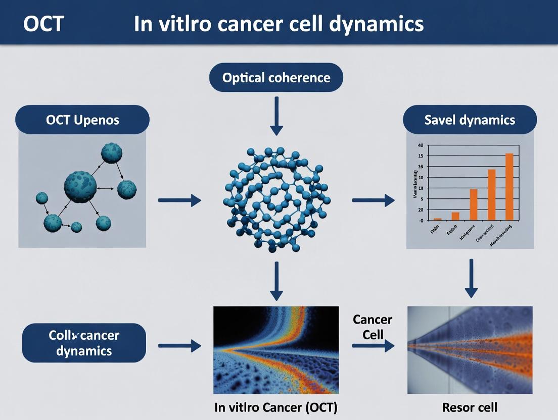
Mapping the Tumor Microenvironment: A Comprehensive Guide to OCT for 3D Cancer Cell Dynamics in Vitro
This article provides researchers, scientists, and drug development professionals with a comprehensive analysis of Optical Coherence Tomography (OCT) for non-invasive, label-free imaging of in vitro cancer cell dynamics.
Mapping the Tumor Microenvironment: A Comprehensive Guide to OCT for 3D Cancer Cell Dynamics in Vitro
Abstract
This article provides researchers, scientists, and drug development professionals with a comprehensive analysis of Optical Coherence Tomography (OCT) for non-invasive, label-free imaging of in vitro cancer cell dynamics. We explore OCT's foundational principles in oncology, detail methodologies for 3D spheroid and co-culture model imaging, address key challenges in signal interpretation and system optimization, and validate OCT against established modalities like confocal microscopy. The scope covers applications from basic tumor spheroid growth quantification to advanced drug response monitoring, empowering the integration of this powerful imaging tool into modern cancer research pipelines.
Unveiling the Mechanism: How OCT Visualizes Cancer Cell Behavior in 3D
This whitepaper details the core principles of Optical Coherence Tomography (OCT) as a label-free, interferometric imaging modality. Framed within a thesis on monitoring in vitro cancer cell dynamics, we elucidate the technical foundations that enable non-invasive, real-time, quantitative assessment of three-dimensional tissue morphology and cellular motion. The intrinsic contrast mechanism, reliant on endogenous light scattering, is paramount for long-term studies of cancer cell migration, invasion, and drug response without phototoxicity or label perturbation.
The Interferometric Principle: Capturing Coherence-Gated Scattering
OCT is an optical analog of ultrasound, using low-coherence interferometry to measure the time delay and intensity of backscattered light. The axial resolution (1-15 µm) is decoupled from the lateral resolution and is determined by the coherence length of the light source, while penetration depth (1-3 mm in scattering tissues) is primarily governed by the source's center wavelength.
Key Interferometer Configurations:
- Michelson Interferometer: The most common layout for Time-Domain OCT (TD-OCT).
- Fourier-Domain Interferometer: Encompasses Spectral-Domain OCT (SD-OCT) and Swept-Source OCT (SS-OCT), offering superior sensitivity and speed.
Mathematical Foundation
The detected interferometric signal for a single scattering site is proportional to:
I_D ∝ √(I_R I_S) ⋅ γ(Δl) ⋅ cos(2kΔl)
where I_R and I_S are reference and sample arm intensities, γ is the degree of coherence, Δl is the pathlength difference, and k is the wavenumber. In Fourier-Domain OCT, the depth-resolved A-scan is obtained via Fourier transform of the detected spectrum: S(z) = FT{I_D(k)}.
Table 1: Quantitative Comparison of OCT System Architectures
| Parameter | Time-Domain (TD-OCT) | Spectral-Domain (SD-OCT) | Swept-Source (SS-OCT) |
|---|---|---|---|
| Axial Resolution (in tissue) | 5-15 µm | 1-7 µm | 1-7 µm |
| A-scan Rate | 1-50 kHz | 20-500 kHz | 50,000 - 5,000,000 Hz |
| Sensitivity Advantage | Reference | ~20-30 dB higher | ~20-30 dB higher |
| Key Components | Broadband source, moving reference mirror | Broadband source, spectrometer | Wavelength-swept laser, photodetector |
| Primary Limitation | Speed/Sensitivity | Spectral detection limits max speed | Complex laser source, edge roll-off |
Application toIn VitroCancer Cell Dynamics: Core Protocols
Protocol: Quantitative Phase Imaging for Cell Motility & Membrane Fluctuations
Objective: To measure nanoscale cellular dynamics and dry mass changes in 2D/3D cancer cultures.
- Sample Preparation: Plate cells in glass-bottom dishes. For 3D, embed cells in Matrigel or collagen I matrix (e.g., 5 mg/mL concentration).
- System Calibration: Use a mirror sample to acquire reference spectrum. Remove fixed-pattern noise via background subtraction.
- Data Acquisition: Acquire time-series volumetric (4D) scans. For high-speed membrane fluctuation, use M-mode at a single lateral location.
- Signal Processing: Extract the complex-valued OCT signal. Phase stability is critical; use common-path or phase-stabilized setups.
- Analysis: Calculate quantitative phase data
Φ(x,y,z,t) = arg[F{S(k)}]. Derive cell displacement, motility metrics (e.g., mean squared displacement), and dry mass density via the relationship:Dry Mass = (Φ ⋅ λ) / (2πα), where α is the specific refractive index increment (~0.18 µm³/pg).
Protocol: Doppler-OCT for Microfluidic Flow Profiling in Tumor Mimics
Objective: To measure flow dynamics within microfluidic chips containing cancer cell spheroids.
- Chip Fabrication: Use PDMS-based devices with channels (e.g., 100 µm width) housing a central spheroid trapping region.
- System Setup: Align OCT beam perpendicular to flow direction. Ensure high phase stability.
- Acquisition: Perform repeated B-scans along the flow direction. The phase difference between consecutive A-scans
ΔΦis proportional to axial velocity:v_axial = (λ₀ ⋅ ΔΦ) / (4πn ⋅ Δt), wherenis refractive index,Δtis A-scan interval. - Processing: Apply phase unwrapping and bulk motion correction. Generate 2D/3D velocity vector maps.
Visualization of Core Concepts
Diagram 1: Basic OCT Michelson Interferometer Workflow
Diagram 2: Sources of Label-Free Contrast in OCT for Cell Assays
The Scientist's Toolkit: Key Research Reagent Solutions
Table 2: Essential Materials for In Vitro OCT Cancer Dynamics Research
| Item | Function & Relevance to OCT | Example/Notes |
|---|---|---|
| Glass-Bottom Culture Dishes | Provides optimal optical clarity and minimal aberrations for high-resolution imaging. | Delta TPG dishes, MatTek dishes. |
| Extracellular Matrix (ECM) Hydrogels | For 3D culture models. Scattering properties must be characterized. | Corning Matrigel, Rat Tail Collagen I (low concentration for clarity). |
| Phase-Stabilized Microfluidic Chips | Enables controlled flow and shear stress studies. Chip material must be OCT-transparent. | PDMS-based chips, Ibidi µ-Slides. |
| Refractive Index Matching Media | Reduces surface reflections and aberrations at interfaces. | Cell culture media with added agents (e.g., dextran) to match chip/ECM RI. |
| Calibration Standards | For system resolution, point spread function validation, and phase calibration. | Titanium dioxide/silica microsphere phantoms, mirrored surfaces. |
| Metabolism-Inert Spheroids | Positive controls for motility/death assays. | Agarose or polystyrene bead-based spheroids. |
Thesis Context: This whitepaper details the core technical advantages of Optical Coherence Tomography (OCT) for longitudinal in vitro studies of cancer cell dynamics, a critical methodology within a broader thesis investigating tumor spheroid evolution and drug response mechanisms.
Core Technical Principles & Quantitative Advantages
OCT is a low-coherence interferometry-based technique that provides depth-resolved, cross-sectional, and three-dimensional images from within scattering samples. Its key performance metrics for live-cell imaging are summarized below.
Table 1: Quantitative Performance Comparison of Live-Cell Imaging Modalities
| Imaging Modality | Axial Resolution | Lateral Resolution | Penetration Depth | Maximum Frame Rate | Key Limitation for Long-Term Imaging |
|---|---|---|---|---|---|
| Optical Coherence Tomography (OCT) | 1 - 15 µm | 1 - 15 µm | 1 - 3 mm | 100+ fps (spectral-domain) | Lower resolution vs. confocal. |
| Confocal Microscopy | 0.5 - 1.5 µm | 0.2 - 0.5 µm | 100 - 200 µm | 1 - 10 fps | Phototoxicity, photobleaching. |
| Two-Photon Microscopy | 0.8 - 1.5 µm | 0.3 - 0.8 µm | 500 - 1000 µm | 1 - 5 fps | High peak power, complex setup. |
| Brightfield/Phase Contrast | N/A | ~0.2 µm | Full sample | 10+ fps | No optical sectioning, 2D only. |
| Spinning Disk Confocal | 0.6 - 1.0 µm | 0.2 - 0.4 µm | 100 - 200 µm | 100 - 1000 fps | Reduced but present phototoxicity. |
Table 2: OCT System Parameters for Optimal Long-Term Cancer Cell Imaging
| Parameter | Typical Range for Cell Imaging | Impact on Long-Term Experiments |
|---|---|---|
| Central Wavelength | 1300 nm (biological window) | Reduced scattering, deeper penetration, minimal photodamage. |
| Average Power on Sample | 1 - 5 mW | Low energy prevents thermal stress, enabling multi-day imaging. |
| A-scan Rate | 20 - 500 kHz | Enables rapid volumetric capture, minimizing motion artifacts. |
| Dynamic Range | > 100 dB | Sensitive detection of weak reflections from cell membranes. |
| Cell Culture Compatibility | Integrated stage-top incubator | Maintains 37°C, 5% CO₂, and humidity for viability. |
Detailed Experimental Protocol: Longitudinal 3D Tumor Spheroid Growth Assay
This protocol is designed for quantifying the dynamic growth and morphological evolution of cancer spheroids using a spectral-domain OCT system.
Aim: To non-destructively monitor the 3D growth kinetics and structural changes of HCT-116 colorectal carcinoma spheroids over 14 days in response to a chemotherapeutic agent.
Materials & Setup:
- OCT System: Spectral-domain OCT with a 1300 nm central wavelength broadband source, axial resolution < 5 µm, lateral resolution < 7 µm.
- Scanning Lens: Telecentric, long working-distance objective compatible with culture dishes.
- Stage-Top Incubator: Enclosed chamber maintaining 37°C and 5% CO₂ on the motorized translation stage.
- Sample: HCT-116 spheroids formed in 96-well ultra-low attachment plates.
- Software: Custom LabVIEW or Python scripts for automated time-lapse acquisition and image processing.
Procedure:
- Spheroid Formation: Seed HCT-116 cells at 5,000 cells/well in a 96-well U-bottom plate. Centrifuge at 300 x g for 3 minutes to aggregate cells. Incubate for 72 hours to form compact spheroids.
- Baseline Imaging (Day 0): Transfer plate to the OCT stage-top incubator and allow thermal equilibration for 30 minutes. For each spheroid, acquire a 3D volumetric dataset (e.g., 1000 x 1000 x 512 pixels over 1.5 x 1.5 x 2 mm).
- Compound Administration: Add vehicle (control) or 5-Fluorouracil (5-FU, 10 µM final concentration) to respective wells.
- Long-Term Time-Lapse: Program the OCT system for automated, periodic imaging. Acquire 3D volumes of each spheroid every 12 hours for 14 days.
- Data Processing:
- Reconstruction: Generate 3D depth profiles (A-scans) into cross-sectional (B-scans) and volumetric data.
- Segmentation: Apply a semi-automatic level-set or machine learning algorithm to segment the spheroid boundary in each B-scan.
- Quantification: Calculate spheroid volume (V = Σ area_slice * slice thickness). Extract shape descriptors (e.g., sphericity index, surface roughness).
Key Metrics: Growth curve (Volume vs. Time), Doubling Time, Morphological Changes (core condensation, cavitation).
Visualizing the OCT Workflow & Data Pipeline
Diagram 1: Long-Term OCT Spheroid Assay Pipeline
Diagram 2: Spectral-Domain OCT System Schematic
The Scientist's Toolkit: Key Research Reagent Solutions
Table 3: Essential Materials for OCT-Based Live-Cell Assays
| Item | Function & Relevance to OCT | Example Product/Note |
|---|---|---|
| Ultra-Low Attachment (ULA) Microplates | Promotes 3D spheroid formation via forced aggregation or hanging drop. Critical for creating optically accessible samples. | Corning Spheroid Microplates, Nunclon Sphera plates. |
| Phenol Red-Free Medium | Eliminates background fluorescence and absorption interference at imaging wavelengths, improving OCT signal clarity. | Gibco FluoroBrite DMEM. |
| Matrigel/ECM Hydrogels | Provides a physiologically relevant 3D extracellular matrix for invasion assays. OCT visualizes cell migration within the gel. | Corning Matrigel Growth Factor Reduced. |
| Cell Viability Dye (Non-Fluorescent) | Post-experiment validation of non-destructive claim. Stains dead cells only after permeabilization at endpoint. | Trypan Blue (0.4%). |
| Stage-Top Gas & Temp Controller | Maintains physiological conditions (37°C, 5% CO₂, humidity) during long scans, ensuring cell health and experiment validity. | Tokai Hit or OKOLab stage-top incubators. |
| Fiducial Markers | Provides reference points for perfect registration of 3D volumes over time, enabling accurate tracking of single cells or regions. | Alignable plate holder or embedded polystyrene microbeads. |
| Image Analysis Software | Enables segmentation, volume rendering, and quantitative analysis of 3D OCT data stacks over time. | Open Source: ImageJ/Fiji with plugins. Commercial: Imaris, Amira. |
OCT's unique combination of label-free, non-destructive optical sectioning, deep penetration, and rapid 3D acquisition establishes it as an indispensable tool for longitudinal in vitro oncology research, providing unmatched kinetic and volumetric data essential for understanding cancer dynamics and therapeutic efficacy.
This technical guide details the quantitative analysis of optical scattering signatures to differentiate between cancer cells and stromal components using Optical Coherence Tomography (OCT). Framed within a thesis on in vitro cancer cell dynamics research, it provides methodologies for acquiring, processing, and interpreting scattering data, which is critical for non-invasive, label-free monitoring of tumor microenvironment interactions.
In OCT, backscattered light encodes information about subcellular morphology and tissue microstructure. Cancer cells, with their enlarged nuclei, increased nuclear-to-cytoplasmic ratio, and dense nucleoli, exhibit distinct scattering properties compared to stromal cells (e.g., fibroblasts) and extracellular matrix (ECM) components. Deciphering this contrast enables the tracking of tumor progression, stromal activation, and drug response dynamics in 3D in vitro models.
Theoretical Foundations of Scattering Contrast
The scattering coefficient (μs) and anisotropy factor (g) are primary parameters. The reduced scattering coefficient (μs' = μ_s(1-g)) is often used to describe the effective scattering in a diffusion regime. Key sources of contrast include:
- Cancer Cells: Higher refractive index inhomogeneity due to organelle crowding leads to elevated μ_s.
- Stromal Fibroblasts: Typically more elongated with less dense organelle packing, resulting in intermediate μ_s.
- Collagen-Rich ECM: Exhibits strong forward scattering (high g) with μ_s dependent on fibril density and alignment.
Table 1: Typical Scattering Parameters at 1300 nm Wavelength
| Cell/Component Type | Reduced Scattering Coefficient (μ_s') [mm⁻¹] | Anisotropy Factor (g) | Effective Scatterer Diameter (Inferred) [μm] | Refractive Index Contrast (Δn) |
|---|---|---|---|---|
| Pancreatic Cancer Cell (Panc-1) | 8.5 - 10.2 | 0.91 - 0.95 | 0.8 - 1.5 (Nuclear) | 0.04 - 0.06 |
| Breast Cancer Cell (MCF-7) | 7.8 - 9.5 | 0.90 - 0.94 | 0.7 - 1.2 | 0.03 - 0.05 |
| Pancreatic Stellate Cell (PSC) | 6.0 - 7.5 | 0.88 - 0.92 | 0.5 - 1.0 | 0.02 - 0.04 |
| Fibroblast (NHDF) | 5.5 - 7.0 | 0.87 - 0.91 | 0.4 - 0.8 | 0.02 - 0.03 |
| Type I Collagen Matrix (5 mg/mL) | 3.0 - 6.0 | 0.96 - 0.98 | 0.05 - 0.3 (Fibril) | 0.01 - 0.02 |
Table 2: Scattering-Based Metrics for Co-Culture Monitoring
| Metric | Calculation Method | Utility in Cancer vs. Stroma Discrimination |
|---|---|---|
| Scattering Intensity Variance | Variance of OCT signal amplitude within a region of interest (ROI). | Highlights regions of heterogeneous cell packing (tumor foci). |
| Texture Analysis (Entropy) | Gray-level co-occurrence matrix (GLCM) analysis of OCT B-scans. | Distinguishes chaotic cancer cell clusters from aligned stromal matrices. |
| Depth-Attentuation Rate (β) | Fitting A-scan decay: I(z) ∝ exp(-2βz). | Higher β indicates denser, more scattering cell clusters. |
Experimental Protocols
Protocol: OCT Imaging of 3D Co-Culture Spheroids
Objective: To acquire volumetric scattering data from in vitro tumor spheroids containing cancer and stromal cells.
- Model Preparation: Generate co-culture spheroids using a 1:1 ratio of fluorescently labeled cancer cells (e.g., MDA-MB-231 mCherry) and stromal fibroblasts (e.g., HS-5 GFP) in ultra-low attachment round-bottom plates. Culture for 72 hours until compact spheroids form (~500 µm diameter).
- OCT System Setup: Use a spectral-domain OCT system with a central wavelength of 1300 nm, axial resolution of ~5 µm in tissue, and lateral resolution of ~10 µm.
- Image Acquisition: Immerse spheroid in growth medium in a glass-bottom dish. Acquire a 3D volume scan (1x1x1 mm³) with 1024 x 512 x 512 (x,y,z) pixels. Maintain sample temperature at 37°C.
- Calibration: Acquire a reference scan of a uniform scattering phantom (μ_s' = 5 mm⁻¹) for system performance verification.
Protocol: Extraction of Scattering Coefficients
Objective: To quantify μ_s' from acquired OCT data.
- Pre-processing: Apply a logarithmic transform to linearize intensity data. Remove speckle noise using a 3D block-matching and filtering (BM3D) algorithm.
- Depth-Resolved Analysis: For each A-scan, fit the depth-dependent intensity decay I(z) to a single-backscattering model:
I(z) = K * μ_s' * exp(-2 * μ_s' * z). Use a least-squares fitting algorithm over a defined depth range (e.g., 100-400 µm). - Segmentation: Use the fluorescent labels (post-correlation) or texture-based segmentation to mask cancer cell regions and stromal cell regions within the spheroid volume.
- Quantification: Calculate the mean and standard deviation of μ_s' for all pixels within each segmented mask.
Visualization of Workflow and Signaling Context
OCT Scattering Analysis Workflow
Signaling Impact on Scattering Signatures
The Scientist's Toolkit: Essential Research Reagents & Materials
Table 3: Key Research Reagent Solutions for OCT-Based Scattering Studies
| Item Name / Category | Example Product / Specification | Primary Function in Experiment |
|---|---|---|
| Cancer Cell Line | MDA-MB-231 (Human Breast Adenocarcinoma), Panc-1 (Pancreatic) | Forms the neoplastic component of the model; genetically modifiable for labeling. |
| Stromal Cell Line | Human Pancreatic Stellate Cells (PSCs), Normal Human Dermal Fibroblasts (NHDF) | Represents the tumor microenvironment; can be activated to a cancer-associated fibroblast (CAF) phenotype. |
| Fluorescent Cell Label | CellTracker CM-Dil (Red) / CMFDA (Green), Lentiviral GFP/mCherry | Allows post-hoc correlation of OCT scattering regions with cell type for validation. |
| 3D Culture Matrix | Cultrex Basement Membrane Extract, Rat Tail Collagen I, Matrigel | Provides a biologically relevant 3D scaffold that scatters light, mimicking in vivo stroma. |
| OCT Calibration Phantom | Polystyrene Microsphere Suspension in Agarose (μ_s' = 3-10 mm⁻¹) | Essential for system calibration and verification of quantitative scattering measurements. |
| Live-Cell Imaging Medium | FluoroBrite DMEM, phenol red-free medium with HEPES | Reduces optical absorption and autofluorescence during live OCT and correlated fluorescence imaging. |
Within the thesis of advancing in vitro models for cancer research, the transition from traditional two-dimensional (2D) cell monolayers to three-dimensional (3D) tumor spheroids and organoids represents a paradigm shift. These 3D models recapitulate critical in vivo features such as cell-cell/cell-extracellular matrix interactions, nutrient and oxygen gradients, and heterogeneous proliferative zones. To validate and exploit these models, non-invasive, longitudinal, and quantitative imaging is essential. Optical Coherence Tomography (OCT), a label-free, interferometry-based technique providing micrometer-scale, cross-sectional, and 3D imaging, has emerged as a pivotal tool for this thesis. This technical guide details its critical role.
OCT Fundamentals & Advantages for 3D Models
OCT operates on low-coherence interferometry, analogous to ultrasound but using light (~1-1.3 µm wavelength common in biomedical OCT). It provides depth-resolved (axial resolution: 1-10 µm) and en-face structural images at rates suitable for live imaging. Key advantages for spheroid/organoid research include:
- Label-free, Non-destructive Monitoring: Enables long-term culture observation without phototoxicity or dye effects.
- Quantitative Analysis: Directly measures 3D morphology, volume, growth kinetics, and layer dynamics.
- Contrast Mechanisms: While primarily structural, variations in scattering intensity can indicate regions of differing cell density or necrosis. Advanced OCT (e.g., OCM, PS-OCT) can provide cellular-resolution or birefringence data.
Table 1: Quantitative Comparison of Imaging Modalities for 3D Models
| Modality | Resolution (Lateral/Axial) | Penetration Depth | Key Advantage | Key Limitation for 3D Models |
|---|---|---|---|---|
| Confocal Microscopy | ~0.2 µm / ~0.5 µm | ~100-200 µm (fixed) | High-res, molecular specificity | Photobleaching, shallow penetration, requires labeling |
| Two-Photon Microscopy | ~0.3 µm / ~0.5-1 µm | ~500 µm | Deeper penetration, reduced phototoxicity | Expensive, slow for large volumes, often requires labeling |
| Light-Sheet Microscopy | ~0.3-1 µm / ~0.5-1 µm | ~1-2 mm (cleared) | Very fast, low phototoxicity | Requires sample clearing/SPIM mounting for optimal use |
| Optical Coherence Tomography (OCT) | ~1-10 µm / 1-10 µm | ~1-2 mm | Fast, deep, label-free, live culture compatible | Lower resolution, primarily structural contrast |
| Micro-CT / MRI | 5-50 µm / 5-50 µm | Unlimited (sample size) | Whole-organoid contrast, 3D density mapping | Low resolution, expensive, often not live-cell compatible |
Experimental Protocols for Key OCT-based Assays
Protocol 3.1: Longitudinal Growth Kinetics Analysis
Objective: To quantify the volumetric growth dynamics of tumor spheroids in response to a therapeutic agent. Materials: U-bottom 96-well plates (for scaffold-free spheroids), Matrigel (for embedded organoids), cell culture medium, OCT system (e.g., spectral-domain OCT). Procedure:
- Model Generation: Seed 500-5000 cells/well in U-plate or embed in Matrigel dome. Allow spheroid formation (3-5 days).
- Baseline Scan (Day 0): Transfer plate to OCT stage with environmental control (37°C, 5% CO₂). Acquire 3D volumetric scan over the spheroid (e.g., 2x2x1 mm volume, 512x512x256 pixels).
- Treatment: Add compound or vehicle control directly to wells.
- Longitudinal Imaging: Repeat 3D OCT scans at 24, 48, 72, and 96 hours post-treatment. Maintain identical scan parameters and positions using a motorized stage.
- Data Analysis:
- Segmentation: Apply median filter, then auto-threshold (e.g., Otsu's method) or edge-detection algorithm to each B-scan to define spheroid boundaries.
- Volume Calculation: Reconstruct 3D binary mask. Calculate volume: V = (voxel count) * (x-dimension * y-dimension * z-dimension).
- Growth Curve: Plot normalized volume (V/V_day0) vs. time. Fit to exponential or logistic model to extract growth rate.
Protocol 3.2: Monitoring Core Necrosis Dynamics
Objective: To non-invasively track the formation and expansion of a necrotic core in large spheroids (>500 µm diameter). Procedure:
- Follow steps 1-4 from Protocol 3.1 to grow large, untreated spheroids.
- OCT Image Acquisition: Use higher axial resolution settings if possible. Ensure sufficient signal-to-noise ratio at the spheroid core.
- Contrast Analysis: The necrotic core exhibits lower scattering (appears darker) due to cell lysis and debris.
- Data Analysis:
- Manually or semi-automatically segment the hypointense core region in each central B-scan.
- Calculate necrotic core area or volume ratio relative to total spheroid area/volume over time.
- Correlate core appearance with spheroid diameter (e.g., necrotic core typically forms when diameter > ~500 µm, depending on cell line).
Signaling Pathways in 3D Tumor Models & OCT Correlates
3D architecture activates signaling pathways distinct from 2D culture. OCT can detect the morphological consequences of these pathways.
Diagram Title: Hypoxia-Driven Pathways & OCT Phenotypes in 3D Tumors
The Scientist's Toolkit: Essential Research Reagents & Materials
Table 2: Key Reagent Solutions for OCT-based 3D Model Research
| Item | Function & Relevance to OCT Imaging |
|---|---|
| U-bottom / Ultra-Low Attachment Plates | Promotes scaffold-free spheroid aggregation via forced floating. Essential for consistent, spherical model formation easily tracked by OCT. |
| Basement Membrane Extract (BME, e.g., Matrigel) | Provides in vivo-like ECM for embedded organoid growth. OCT visualizes organoid structures within the translucent BME dome. |
| Phenol Red-Free Medium | Eliminates background absorption and autofluorescence that can potentially interfere with OCT light penetration and signal. |
| Temperature & Gas Control Stage Top Chamber | Maintains 37°C and 5% CO₂ during prolonged OCT imaging sessions, ensuring physiologically relevant longitudinal data. |
| Fiducial Markers (e.g., polymer microbeads) | Mixed into BME or placed in adjacent wells to provide stable reference points for precise relocation and registration of samples over time. |
| Image Processing Software (e.g., Fiji, Amira) | For 3D segmentation, volume rendering, and quantitative analysis of OCT image stacks. Custom macros are often developed. |
Advanced Workflow: Integrating OCT with Other Modalities
A multi-modal approach leverages OCT's structural strengths with complementary techniques.
Diagram Title: Multi-modal Integration Workflow with OCT
OCT is not merely an imaging tool but a central enabling technology for the thesis that physiologically relevant 3D in vitro models are indispensable for modern cancer research. Its capacity for label-free, quantitative, and longitudinal structural analysis provides unmatched insights into the dynamic processes of tumor growth, treatment response, and microenvironmental evolution. As OCT technology advances towards higher resolution and functional extensions, its role as a critical pillar in the study of tumor spheroids and organoids is firmly established, bridging the gap between simple 2D assays and complex in vivo studies.
The study of in vitro cancer cell dynamics has traditionally relied on endpoint assays or low-throughput microscopy. Optical Coherence Tomography (OCT), historically a structural imaging modality, has undergone a transformative evolution with the advent of functional extensions, notably OCT Angiography (OCT-A) and Doppler OCT. These trends are revolutionizing oncology research by providing non-invasive, label-free, and quantitative volumetric imaging of live cell cultures, tumor organoids, and microtissues over extended time periods. Within the thesis context of probing cancer cell behavior—including proliferation, migration, angiogenesis, and drug response—high-resolution functional OCT offers a powerful, dynamic, and physiologically relevant data stream that bridges cellular-scale events with tissue-scale outcomes.
Core Functional OCT Technologies: Principles and Oncological Relevance
High-Resolution (HR-) OCT: Advancements in light sources and detection schemes have pushed axial resolutions to <1 µm, approaching cellular dimensions. This enables the visualization of individual cell clusters, spheroid boundaries, and intracellular architectural changes in 3D.
OCT Angiography (OCT-A): This functional extension detects static tissue from dynamic signals (e.g., moving red blood cells) by analyzing intensity or phase decorrelation between successive B-scans. In in vitro models, it maps the perfused vascular network within engineered tumors or angiogenic sprouts in co-culture systems without exogenous contrast agents.
Doppler OCT: Quantifies the axial velocity of moving scatterers by measuring the phase shift between sequential A-scans. In oncology research, it is used to measure flow velocity and volumetric flow rate within microfluidic channels or nascent vasculature, providing metrics on hemodynamic changes induced by therapeutic agents.
Table 1: Key Performance Metrics and Findings from Recent Functional OCT Studies in Oncology Models
| Study Focus | OCT Modality | Resolution (Axial x Lateral) | Key Quantitative Finding | Model Used |
|---|---|---|---|---|
| Anti-angiogenic Drug Screening | OCT-A | 1.3 µm x 3.0 µm | 40% reduction in vascular density after 72h treatment with VEGF inhibitor (p<0.01). | Glioblastoma spheroid-endothelial cell co-culture. |
| Tumor Spheroid Growth Kinetics | HR-OCT | 0.9 µm x 2.5 µm | Measured spheroid volume doubling time of 28.5 ± 3.2 hours. | Colorectal cancer spheroids. |
| Microfluidic Flow Analysis | Doppler OCT | 2.0 µm x 5.0 µm | Measured intraluminal flow velocity range of 0.5 - 4.0 mm/s in bio-printed channels. | Vascularized breast cancer microtissue in a chip. |
| Cell Migration & Invasion | HR-OCT + OCT-A | 1.5 µm x 3.5 µm | Quantified invasion front velocity of 15.2 ± 2.1 µm/day into a collagen matrix. | Pancreatic ductal adenocarcinoma organoid. |
Detailed Experimental Protocols
Protocol 1: Longitudinal Assessment of Anti-Angiogenic Therapy Using OCT-A Aim: To quantify the effect of a VEGF-inhibiting drug on vascular network formation in a 3D co-culture model.
- Model Preparation: Seed GFP-labeled human umbilical vein endothelial cells (HUVECs) with fluorescent cancer-associated fibroblasts (CAFs) in a fibrin gel. Form a central tumor spheroid (e.g., U87-MG glioblastoma).
- Culture & Treatment: Maintain in endothelial growth medium. At day 3, add therapeutic agent (e.g., Bevacizumab analog) to treatment group; control receives vehicle.
- OCT-A Imaging: Using a 1300 nm spectral-domain OCT system.
- Scan Protocol: 3D scan over 2x2 mm area, 1000 A-scans/B-scan, 500 B-scan positions.
- OCT-A Processing: Apply speckle decorrelation algorithm (e.g., Split-Spectrum Amplitude-Decorrelation Angiography) to consecutive B-scans at each position.
- Acquisition Schedule: Image at days 0 (pre-treatment), 1, 2, 3, and 4 post-treatment.
- Quantitative Analysis: Generate binary vascular masks from OCT-A data. Calculate metrics: Vascular Density (% volume occupied by vessels), Vessel Branch Points, and Mean Vessel Diameter.
Protocol 2: Measuring Intra-Spheroid Drug Diffusion via Doppler OCT Aim: To characterize changes in intratumoral fluid dynamics upon treatment with a chemotherapeutic agent.
- Model Preparation: Generate dense, >500 µm diameter spheroids from a patient-derived ovarian cancer cell line via the hanging-drop method.
- Microfluidic Setup: Immobilize a single spheroid in a low-shear perfusion chamber under continuous medium flow (50 µL/min).
- Doppler OCT Imaging: Using a swept-source OCT system with a high-NA objective.
- Scan Protocol: M-Mode scanning (repeated A-scans at one spatial location) for 5 seconds at multiple points within the spheroid and surrounding medium.
- Doppler Processing: Calculate phase difference ∆Φ between successive A-scans:
v = (λ₀ * ∆Φ) / (4πn * τ), wherevis velocity,λ₀is central wavelength,nis refractive index,τis A-scan time interval.
- Intervention & Analysis: Acquire baseline Doppler data. Introduce chemotherapeutic (e.g., Doxorubicin) into perfusion stream. Monitor Doppler shifts over 60 minutes. Correlate localized velocity changes with spheroid viability assays.
Visualization of Signaling Pathways and Workflows
Diagram 1: OCT-A anti-angiogenic drug assay workflow (83 chars)
Diagram 2: VEGF pathway & OCT readouts in angiogenesis (78 chars)
The Scientist's Toolkit: Key Research Reagent Solutions
Table 2: Essential Materials for Functional OCT Experiments in Cancer Cell Dynamics
| Item / Reagent | Function / Purpose in OCT Experiments |
|---|---|
| Fibrinogen / Thrombin Kit | Forms a natural, translucent 3D hydrogel for embedding cells, optimal for OCT penetration and supporting angiogenesis. |
| Matrigel (GFR, Phenol Red-Free) | Basement membrane matrix for organoid culture and invasion assays. Phenol-red-free version eliminates absorption interference. |
| Microfluidic Organ-on-Chip Devices | Provides controlled perfusion and physiological shear stress, enabling Doppler flow measurements in vascularized models. |
| Patient-Derived Cancer Organoid Media Kits | Enables long-term, physiologically relevant culture of tumor explants for longitudinal OCT monitoring. |
| Fluorescently-Labeled (GFP/RFP) Endothelial Cells | Allows correlation of OCT-A findings with standard fluorescence confocal microscopy for validation. |
| Collagen I, High Concentration (≥8 mg/mL) | Creates dense, scattering matrices for invasion assays, providing contrast for migrating cell fronts in HR-OCT. |
| Pharmacological Inhibitors (e.g., VEGF inhibitors, MMP inhibitors) | Tool compounds for perturbing biological processes (angiogenesis, invasion) and measuring functional OCT readouts. |
| Refractive Index Matching Solutions (e.g., PBS, Culture Media) | Standard immersion media for objectives; their known refractive index is critical for accurate size and Doppler velocity calibration. |
Protocols in Action: Implementing OCT for Drug Screening and Microenvironment Studies
Optical Coherence Tomography (OCT) has emerged as a pivotal, non-invasive tool for longitudinal, in vitro investigation of 3D cancer cell dynamics, including tumor spheroid growth, invasion, and response to therapeutic agents. The fidelity of these quantitative observations is fundamentally governed by the sample preparation methodology. Within the context of a broader thesis on OCT for in vitro cancer research, this guide details best practices for optimizing the two core physical scaffolds of any 3D culture OCT experiment: the extracellular matrix (ECM) mimic and the imaging chamber.
Suboptimal matrices can distort morphometric data, while poorly designed chambers introduce artifacts, limit nutrient exchange, or preclude high-resolution imaging. This technical whitepaper synthesizes current research to provide a standardized framework for preparing OCT-compatible samples that yield biologically relevant and quantitatively robust data.
Optimizing Extracellular Matrix (ECM) Mimics for OCT Imaging
The choice of hydrogel matrix is paramount, as it provides the 3D context for cell behavior and must be transparent to near-infrared OCT wavelengths (~800-1300 nm). Key parameters include concentration, polymerization method, and incorporation of biological cues.
Quantitative Comparison of Common OCT-Compatible Hydrogels
Table 1: Properties and OCT Performance of Common Hydrogel Matrices for 3D Cancer Models
| Matrix Type | Common Concentrations for OCT | Typical Gelation Method | Key Advantages for OCT | OCT Attenuation Coefficient (Approx. mm⁻¹)* | Ideal Cancer Model Use Case |
|---|---|---|---|---|---|
| Basement Membrane Extract (BME/Matrigel) | 4-8 mg/mL (diluted in cold media) | Thermal (37°C) | Contains native ECM proteins; supports complex morphogenesis. | 2.5 - 4.0 | Epithelial cancer organoids, ductal formation. |
| Collagen I | 1.5-3.0 mg/mL | Neutralization & Thermal (37°C) | Tunable stiffness; high clarity at low concentrations. | 1.8 - 3.5 (at 2 mg/mL) | Cell invasion, stromal co-culture, migration studies. |
| Fibrin | 3-5 mg/mL | Enzymatic (Thrombin + Ca²⁺) | Excellent for angiogenesis assays; controllable degradation. | 2.0 - 3.2 | Vascularized tumor models, patient-derived cell assays. |
| Synthetic (PEG-based) | Varies by formulation | Photo-polymerization (UV/vis light) | Defined chemistry; highly reproducible mechanical properties. | 1.5 - 2.5 (highly formulation-dependent) | Mechanobiology studies, controlled presentation of ligands. |
| Hyaluronic Acid (MeHA) | 1-2% (w/v) | Photo-polymerization | Relevant for many desmoplastic tumors; tunable viscosity. | 2.2 - 3.8 | Metastasis models, cancer stem cell niche studies. |
*Attenuation coefficients are approximate and depend on specific OCT system center wavelength (e.g., 1300 nm typically has lower scattering than 800 nm in biological tissue). Data compiled from recent literature.
Experimental Protocol: Preparing a Low-Scattering Collagen I Matrix for Spheroid Invasion
Objective: To embed pre-formed tumor spheroids in a translucent, mechanically defined collagen I gel for longitudinal OCT imaging of invasive protrusions.
Materials: Rat tail Collagen I, high concentration (e.g., ~8-10 mg/mL); Sterile 0.1M NaOH; 10X PBS; Cell culture medium; Pre-formed spheroids (e.g., in ultra-low attachment plates); Chilled 1.5 mL microcentrifuge tubes; OCT-compatible chamber (see Section 3).
Methodology:
- Calculation & Neutralization: On ice, calculate the required total volume of neutralized collagen solution. For a final concentration of 2 mg/mL in 200 µL, mix in a cold tube:
- Collagen I (8 mg/mL stock): 50 µL
- 10X PBS: 20 µL (to achieve 1X final)
- 0.1M NaOH: 5-7 µL (volume must be determined empirically for each batch to achieve pH ~7.4)
- Cell Culture Medium (with serum): 123-125 µL to reach 200 µL total.
- Keep the mixture on ice to prevent premature gelling.
- Spheroid Incorporation: Gently pellet 10-20 pre-formed spheroids (100-300 µm diameter). Aspirate supernatant. Resuspend the spheroid pellet in 50 µL of cold neutralized collagen mix by gentle pipetting.
- Chamber Loading: Transfer the spheroid-collagen suspension to the center of an OCT imaging chamber.
- Gelation: Place the chamber in a 37°C, 5% CO₂ incubator for 30-45 minutes for complete polymerization.
- Overlay with Medium: After gelation, gently add 200-500 µL of warm culture medium on top of the gel to prevent dehydration during imaging.
Selecting and Utilizing OCT Imaging Chambers
The imaging chamber must maintain sterility, permit gas exchange, be compatible with the microscope stage, and have optical properties that minimize signal loss and reflections.
Diagram: Workflow for Preparing a 3D Spheroid-in-Matrix Sample for OCT
Workflow for 3D Spheroid Sample Prep
Chamber Comparison and Selection Guide
Table 2: Comparison of Common Chamber Types for Longitudinal OCT Imaging of 3D Cultures
| Chamber Type | Material (Bottom) | Bottom Thickness | Key Pros | Key Cons | Best Suited For |
|---|---|---|---|---|---|
| Commercial Glass-Bottom Dish | #1.5 Cover Glass (170 µm) | ~0.17 mm | Excellent optical clarity; standard for high-NA objectives. | Cost; limited customization; potential for meniscus artifacts at edges. | High-resolution, long-term live imaging of single spheroids/organoids. |
| Coverslip-Based Assembly | #1.5 Cover Glass (170 µm) | ~0.17 mm | Inexpensive; highly customizable well size/shape using silicone gaskets. | Assembly requires skill; higher risk of leakage/contamination. | Multi-matrix comparisons, custom co-culture setups. |
| PDMS Microfluidic Chip | Glass or PDMS membrane | Variable (50 µm - mm) | Precise control over gradients (oxygen, drug); compartmentalization. | Specialized fabrication; potential for high OCT scattering from chip walls. | Angiogenesis, metastasis, and tumor microenvironment studies with controlled flow. |
| Multi-Well Plate (Optically Clear) | Polymer (e.g., PS, COP) | ~0.5 - 1.0 mm | High-throughput; standard format for compatibility with plate handlers. | Increased scattering from thick bottom; lower effective numerical aperture. | Medium-throughput drug screening with OCT endpoint/kinetic reads. |
The Scientist's Toolkit: Essential Research Reagent Solutions
Table 3: Key Reagents and Materials for Optimized OCT Sample Preparation
| Item | Function / Rationale | Example Product / Note |
|---|---|---|
| Phenol Red-Free Medium | Eliminates absorption of light by phenol red dye, reducing OCT signal attenuation and background. | Gibco DMEM/F-12, without phenol red. |
| High-Concentration Collagen I | Allows for precise, low-concentration gelling (1-3 mg/mL) while minimizing dilution of critical media components. | Corning Rat Tail Collagen I, 8-10 mg/mL. |
| Ice-Cold, Low-Binding Pipette Tips | Prevents premature gelling of collagen/Matrigel within the tip during aliquoting and mixing. | Tips treated to reduce protein adhesion. |
| Silicone Isolation Gaskets | Creates defined wells on coverslips for containing hydrogel samples, enabling custom chamber assembly. | Grace Bio-Labs SecureSeal gaskets. |
| Cell-Reconstituted Basement Membrane | Provides a biologically active, consistent, and xeno-free alternative to Matrigel for clinically relevant models. | Cultrex PathClear BME or equivalent. |
| Temperature-Controlled Chamber Lid | Maintains 37°C and 5% CO₂ environment on the microscope stage for extended longitudinal imaging. | Tokai Hit or PeCon stage-top incubators. |
Integrated Workflow: From Sample Prep to Data Acquisition
A robust protocol integrates matrix and chamber optimization. Below is a logic diagram outlining the decision pathway for configuring an OCT experiment based on biological question and practical constraints.
Diagram: Decision Logic for OCT Sample Configuration
Logic for OCT Sample Configuration
Optimizing the matrix and chamber is not merely a preparatory step but an experimental design cornerstone in OCT-based cancer cell dynamics research. By selecting hydrogels for both biological relevance and optical clarity, and pairing them with chambers that balance imaging performance with experimental needs, researchers can generate 3D in vitro data of exceptional quality and reliability. This standardization is critical for advancing the use of OCT as a quantitative, longitudinal tool in oncology drug development and fundamental cancer biology.
This protocol is framed within a broader thesis research program investigating the application of Optical Coherence Tomography (OCT) for in vitro cancer cell dynamics research. OCT provides a label-free, non-invasive, and quantitative method for longitudinally monitoring 3D tumor spheroid morphology, volume, and internal structure with micrometer resolution. This capability is critical for advancing kinetic studies of tumor growth and treatment response, bridging the gap between 2D cell culture and in vivo models. This guide details a standardized protocol for generating, maintaining, and quantitatively analyzing the growth kinetics of tumor spheroids, with integration points for OCT imaging specified throughout.
Materials & Research Reagent Solutions
The Scientist's Toolkit: Essential Materials for Tumor Spheroid Growth Kinetics
| Item | Function & Rationale |
|---|---|
| Ultra-Low Attachment (ULA) Plate (e.g., Corning Spheroid Microplate) | Coating prevents cell adhesion, forcing cells to aggregate into a 3D spheroid. Critical for consistent, single-spheroid-per-well formation. |
| Basement Membrane Extract (BME/Matrigel) | Extracellular matrix scaffold. Used for embedded or overlay cultures to promote complex morphology and relevant cell-ECM interactions. |
| High-Content Imaging System with confocal or OCT module | Enables automated, longitudinal imaging of spheroid size and viability without manual transfer. Maintains sterility. |
| Optical Coherence Tomography (OCT) System (e.g., spectral-domain) | Core thesis tool. Provides cross-sectional, label-free images for calculating 3D volume and monitoring internal necrotic core development. |
| Viability/Cytotoxicity Dual-Assay Kit (e.g., Calcein AM / Propidium Iodide) | Fluorescent stains for live/dead cell quantification. Validates OCT findings on viability and necrosis. |
| Glucose & Lactate Assay Kits | Metabolic profiling reagents. Used to correlate spheroid growth kinetics with glycolytic flux, a hallmark of tumor metabolism. |
| Liquid Handling Robot | Ensures precision and reproducibility in cell seeding, medium exchange, and reagent addition for high-throughput studies. |
| Advanced Analysis Software (e.g., ImageJ with 3D suite, MATLAB) | For processing OCT image stacks, segmenting spheroid boundaries, and calculating volumetric growth kinetics. |
Core Experimental Protocol
Part A: Spheroid Generation & Culture
Method: Liquid Overlay Technique using ULA Plates.
- Cell Preparation: Harvest adherent cancer cells at 80-90% confluence. Prepare a single-cell suspension in complete medium at the optimal density (see Table 1).
- Seeding: Using an electronic multichannel pipette or liquid handler, seed 100 µL of cell suspension per well into a 96-well ULA round-bottom plate.
- Centrifugation: Centrifuge the plate at 300 x g for 3 minutes to aggregate cells at the well bottom.
- Incubation: Place the plate in a humidified 37°C, 5% CO₂ incubator. Compact spheroids typically form within 24-72 hours.
- Culture Maintenance: Every 48-72 hours, perform a 50% medium exchange carefully using a multichannel pipette with tips angled to avoid spheroid aspiration.
Part B: Longitudinal OCT Imaging & Kinetic Analysis
Method: Non-invasive, label-free volumetric monitoring.
- OCT System Calibration: Calibrate the lateral and axial scales using a standard calibration slide prior to imaging.
- Imaging Setup: Place the culture plate directly on the OCT sample stage. Use a custom holder to ensure the plate bottom is perpendicular to the beam.
- Image Acquisition: At each time point (e.g., Days 1, 3, 5, 7, 10), acquire 3D volumetric scans (e.g., 1x1x1 mm³ volume, 512x512x512 pixels) for each spheroid. Use the same scan parameters throughout the experiment.
- Data Processing: Export OCT B-scans and 3D stacks. Use custom MATLAB scripts or ImageJ to:
- Apply a noise-reduction filter.
- Segment the spheroid boundary in each B-scan using intensity thresholding or edge detection.
- Reconstruct the 3D volume and calculate Equivalent Volume (V = 4/3πr³) from the mean radius, or by voxel counting.
- Identify the hypo-reflective necrotic core region and calculate its volume.
Part C: Endpoint Validation Assays
Method: Parallel endpoint analysis for correlation with OCT data.
- Viability Staining: At terminal time points, incubate spheroids with Calcein AM (2 µM) and Propidium Iodide (4 µM) for 45 minutes. Acquire confocal z-stacks.
- Metabolic Analysis: Collect conditioned medium. Quantify glucose consumption and lactate production using colorimetric/fluorometric assay kits per manufacturer instructions.
- Size Correlation: Measure spheroid diameter from brightfield images using ImageJ to validate OCT-derived volumes.
Quantitative Data & Growth Kinetics
Table 1: Optimized Seeding Densities for Common Cell Lines
| Cell Line | Suggested Seeding Density (cells/well in 100 µL) | Expected Initial Diameter (Day 3, µm) | Typical Growth Plateau (Day) |
|---|---|---|---|
| U87 MG (Glioblastoma) | 1,000 - 2,000 | 300 - 400 | 10 - 12 |
| HT-29 (Colon Carcinoma) | 500 - 1,000 | 250 - 350 | 8 - 10 |
| MCF-7 (Breast Adenocarcinoma) | 2,000 - 3,000 | 350 - 450 | 12 - 14 |
| A549 (Lung Carcinoma) | 750 - 1,500 | 275 - 375 | 9 - 11 |
| PC-3 (Prostate Carcinoma) | 1,000 - 2,000 | 300 - 425 | 10 - 13 |
Table 2: Key Kinetic Parameters Derived from OCT Volumetric Data
| Parameter | Formula / Method | Biological Interpretation |
|---|---|---|
| Volume Doubling Time (T_d) | T_d = (t₂ - t₁) * ln(2) / ln(V₂ / V₁) |
Rate of net proliferative expansion. |
| Specific Growth Rate (µ) | µ = ln(V₂ / V₁) / (t₂ - t₁) |
Instantaneous exponential growth constant. |
| Necrotic Core Fraction | (V_necrosis / V_total) * 100% |
Indicator of diffusion-limited inner necrosis. |
| Growth Delay (GD) | GD = T_treated - T_control (for 2x initial volume) |
Metric of treatment efficacy in intervention studies. |
Visualization of Workflows & Pathways
Title: Workflow for Tumor Spheroid Growth Kinetics with OCT
Title: Key Biological Pathways Impacting Spheroid Growth & OCT Readouts
This whitepaper details methodologies for quantifying therapeutic response in 3D in vitro cancer models, a critical component of a broader thesis employing Optical Coherence Tomography (OCT) for non-invasive, longitudinal analysis of cancer cell dynamics. OCT's ability to provide label-free, volumetric imaging of tissue scaffolds and organoids makes it an ideal platform for applying the quantitative metrics described herein.
Core Quantitative Metrics for Therapy Response
Volumetric Assessment
Tumor volume is a primary endpoint. In OCT-based studies, the 3D dataset enables precise volumetric calculation, superior to 2D approximations.
Key Formula: ( V = \sum{i=1}^{n} Ai \cdot d ) Where ( V ) = total volume, ( A_i ) = cross-sectional area of the ( i )-th slice, ( d ) = inter-slice distance, and ( n ) = total number of slices.
Common Volume Change Metrics:
- Relative Volume Change (RVC): ( RVC(\%) = \frac{V{post} - V{pre}}{V_{pre}} \times 100 )
- Tumor Growth Inhibition (TGI): ( TGI(\%) = (1 - \frac{V{treatment}}{V{control}}) \times 100 )
Quantification of Necrosis and Viability
Necrotic regions, characterized by loss of structural integrity and increased optical heterogeneity, can be segmented in OCT images based on signal intensity and texture analysis.
Viability Index: A common metric derived from volumetric data: ( \text{Viability Index} = \frac{V{total} - V{necrotic}}{V_{total}} )
Table 1: Comparative Efficacy of Therapies in 3D In Vitro Models (Representative Metrics)
| Therapy Class | Model Type | Volume Change (RVC) vs. Control | Necrotic Fraction Increase | Primary Measurement Technique | Reference Year |
|---|---|---|---|---|---|
| Targeted Kinase Inhibitor | Breast Cancer Spheroid | -45.2% | +28.5% | Confocal Microscopy / OCT | 2023 |
| Chemotherapy (Cisplatin) | NSCLC Organoid | -32.7% | +21.8% | Light Sheet Microscopy | 2024 |
| Immune Checkpoint Inhibitor | Co-culture Spheroid (T-cells) | -38.1% | +15.3% | OCT / Flow Cytometry | 2023 |
| CAR-T Cell Therapy | Solid Tumor Organoid | -61.5% | +42.7% | High-Content Imaging | 2024 |
Table 2: OCT-Derived Parameters for Treatment Response
| OCT Parameter | Biological Correlate | Calculation Method | Interpretation in Treatment |
|---|---|---|---|
| Signal Intensity Variance | Tissue Heterogeneity / Necrosis | Standard deviation of pixel intensity within ROI | Increase indicates necrosis. |
| Optical Attenuation Coefficient | Cell Density / Viability | Exponential fit to A-scan depth decay | Decrease may indicate loss of cellularity. |
| Textural Features (e.g., Contrast, Entropy) | Micro-architectural Changes | Gray-Level Co-occurrence Matrix (GLCM) analysis | Changes correlate with cytolysis and structural breakdown. |
Detailed Experimental Protocols
Protocol: Longitudinal OCT Imaging for Therapy Response
Objective: To non-invasively monitor volume and structural changes in 3D cancer models post-treatment.
Materials: See "The Scientist's Toolkit" below. Procedure:
- Model Preparation: Seed and culture 3D spheroids/organoids in OCT-compatible, optically clear plates (e.g., U-bottom ultra-low attachment plates).
- Baseline Scan (T0): Acquire a high-resolution 3D OCT volume scan of each model prior to treatment. Use consistent settings (e.g., power, resolution).
- Treatment Administration: Apply therapeutic agent or control vehicle to the culture medium.
- Longitudinal Imaging: At defined intervals (e.g., 24h, 48h, 72h), return plates to the OCT system. Precisely re-locate each model using motorized stage coordinates.
- Image Processing:
- Segmentation: Apply a semi-automatic segmentation algorithm (e.g., level-set, intensity thresholding) to the 3D stack to delineate the model boundary.
- Volumetric Calculation: Compute total voxels within the segmented volume and convert to physical units (mm³).
- Necrosis Segmentation: Segment regions of significantly diminished and heterogeneous signal intensity within the core using a combination of intensity thresholding and texture filters.
- Data Analysis: Calculate RVC, TGI, and Viability Index over time for each treatment cohort.
Protocol: Calcein-AM / Propidium Iodide (PI) Staining for Viability Validation
Objective: To validate OCT-based necrosis measurements with standard fluorescence viability assays. Procedure:
- Following final OCT scan, incubate models with Calcein-AM (2 µM) and PI (4 µM) in culture medium for 45-60 minutes at 37°C.
- Rinse with PBS.
- Image using a fluorescence microscope or confocal system with appropriate filter sets.
- Quantification: Calculate the ratio of PI-positive (necrotic/late apoptotic) volume to Calcein-AM-positive (live) volume using 3D image analysis software (e.g., Imaris, FIJI/ImageJ).
Signaling Pathways in Therapy-Induced Cell Death
Diagram Title: Key Pathways from Therapy to Necrosis
Experimental Workflow for OCT-Based Quantification
Diagram Title: OCT Therapy Response Workflow
The Scientist's Toolkit: Essential Research Reagents & Materials
Table 3: Key Reagents and Solutions for Therapy Response Experiments
| Item | Function / Application in Protocol | Key Considerations |
|---|---|---|
| Ultra-Low Attachment (ULA) Plates | Promotes formation of 3D spheroids; essential for consistent, scaffold-free model generation. | Choose U-bottom for single spheroid/well; clear bottom for OCT/imaging. |
| OCT-Compatible Matrices (e.g., Matrigel, Collagen I) | Provides a physiological 3D scaffold for organoid or invasive culture models. | Optimize concentration for optical clarity and biological relevance. |
| Therapeutic Agents (Reference Compounds) | Positive controls for inducing response (e.g., Staurosporine for apoptosis, Cisplatin). | Use a range of clinically relevant concentrations for dose-response. |
| Calcein-AM Viability Dye | Fluorescent live-cell indicator (green). Enzymatically converted in viable cells. | Validate incubation time for full penetration in 3D models. |
| Propidium Iodide (PI) / EthD-1 | Fluorescent dead-cell indicator (red). Binds to DNA in cells with compromised membranes. | Impermeant to live cells; definitive for necrosis. |
| Optically Clear Culture Medium | For imaging. Formulated without phenol red and with reduced light-scattering components. | Prevents signal attenuation in OCT and fluorescence imaging. |
| Image Analysis Software (e.g., FIJI, Imaris) | For 3D segmentation, volume rendering, and quantitative analysis of OCT and fluorescence data. | Requires plugins/capabilities for handling large 3D stacks and batch processing. |
This guide details the integration of invasion and migration assays with Optical Coherence Tomography (OCT) for in vitro cancer cell dynamics research. A core thesis of our work posits that OCT's label-free, volumetric, and real-time imaging capabilities provide a transformative tool for quantifying the biophysical parameters of cell invasion within three-dimensional matrices, surpassing the limitations of endpoint, two-dimensional assays. This technical whitepaper provides the foundational protocols for setting up these key functional assays and coupling them with OCT imaging to extract quantitative, kinetic data on invasive behavior.
Core Assay Principles and Quantitative Comparison
Cell migration and invasion are distinct yet interconnected processes. Migration refers to cell movement across a two-dimensional surface or through pores, while invasion specifically entails the degradation and movement through a basement membrane or a dense extracellular matrix (ECM), mimicking key steps in metastatic dissemination.
Table 1: Comparison of Primary 2D vs. 3D Migration/Invasion Assays
| Assay Type | Principle | Readout | Advantages | Limitations | OCT Compatibility |
|---|---|---|---|---|---|
| Scratch/Wound Healing | Create a "wound" on a confluent monolayer; monitor gap closure. | Gap area over time (2D). | Simple, inexpensive, no special equipment. | Disrupts monolayer, 2D only, measures collective migration. | Low. Primarily 2D surface imaging. |
| Boyden Chamber (Transwell) | Cells migrate through porous membrane (8-12 µm pores) toward chemoattractant. | Cells on lower membrane stained & counted (endpoint). | High-throughput, quantifiable, can test chemoattraction. | Endpoint, 2.5D, potential for non-migratory cell attachment. | Moderate. Can image fixed cells in membrane but not real-time. |
| Matrigel Invasion (3D) | Cells invade through a thick layer of reconstituted basement membrane (Matrigel). | Invasive cells stained & counted (endpoint). | Models key invasion step, gold standard for invasiveness. | Endpoint, batch variability in matrix, costly. | High. Ideal for volumetric, time-lapse imaging of invasion front. |
| 3D Spheroid Invasion | Cell spheroid embedded in 3D collagen or Matrigel; cells invade radially. | Invasion area/distance from spheroid edge over time. | True 3D context, cell-cell contacts maintained, kinetic data possible. | More complex setup, analysis can be challenging. | Very High. Excellent for longitudinal OCT tracking of 3D boundary. |
Table 2: Key Biophysical Parameters Quantifiable by OCT in 3D Invasion Assays
| Parameter | Definition | OCT Measurement Method | Relevance to Cancer Dynamics |
|---|---|---|---|
| Invasion Depth (µm) | Maximum distance traveled by leading cells from origin. | Z-axis distance in volumetric scan. | Direct measure of invasive potential. |
| Invasion Area/Volume (µm²/µm³) | Total area or volume occupied by invading cell mass. | Segmentation of hyper-reflective cell region in 3D. | Quantifies collective invasion strength. |
| Front Velocity (µm/hr) | Rate of advancement of the invasion front. | Derivative of invasion depth over time. | Kinetic measure of aggressive behavior. |
| Matrix Porosity Change | Local alteration in ECM density due to degradation. | Analysis of backscatter intensity variance in matrix region. | Indirect indicator of proteolytic activity. |
Detailed Experimental Protocols
Protocol A: Matrigel-Based 3D Invasion Assay for Endpoint Analysis
Objective: To assess the invasive potential of cancer cell lines in a standardized basement membrane-like matrix.
Materials: See "The Scientist's Toolkit" below. Procedure:
- Thawing & Plating Matrigel: Thaw Matrigel on ice overnight. Keep all tips, tubes, and plates on ice. Dilute Matrigel to 1 mg/mL in cold serum-free medium. Add 100 µL per well to a 96-well plate to form a thin gel. Incubate at 37°C for 1 hour to polymerize.
- Cell Preparation: Harvest cells in log growth phase. Starve in serum-free medium for 12-24 hours.
- Seeding: Resuspend starved cells at 2.0 x 10⁵ cells/mL in serum-free medium. Carefully seed 1.0 x 10⁴ cells (50 µL) on top of the polymerized Matrigel layer in each well.
- Invasion Induction: After 2 hours for cell attachment, carefully add 150 µL of complete growth medium (with 10% FBS as chemoattractant) on top of the cells.
- Incubation: Incubate for 24-48 hours at 37°C, 5% CO₂.
- Fixation & Staining: Remove medium. Fix cells with 4% formaldehyde for 20 minutes. Permeabilize with 0.1% Triton X-100. Stain cell nuclei with DAPI (1 µg/mL) and actin with Phalloidin (e.g., Alexa Fluor 488 conjugate).
- Imaging & Analysis: Image using a confocal microscope. Count the number of invaded cells (cells within/below the Matrigel layer) from multiple z-stack fields. Express as Invasion Index: (Number of invaded cells / Total number of cells) x 100.
Protocol B: Spheroid-Based 3D Invasion Assay for Longitudinal OCT Imaging
Objective: To perform kinetic, label-free monitoring of collective cancer cell invasion from a 3D spheroid into a surrounding ECM.
Materials: See "The Scientist's Toolkit" below. Procedure:
- Spheroid Generation: Use a 96-well round-bottom ultra-low attachment plate. Prepare a single-cell suspension at 5.0 x 10³ cells/well in 150 µL of complete medium. Centrifuge the plate at 300 x g for 3 minutes to aggregate cells at the bottom. Incubate for 48-72 hours to form a single, compact spheroid per well.
- ECM Embedding: Prepare a working solution of rat tail Collagen I (e.g., 2.0 mg/mL) on ice, neutralizing with NaOH/HEPES per manufacturer's instructions. Using a wide-bore tip, gently transfer one spheroid into 50 µL of cold collagen solution in a pre-chilled imaging chamber (e.g., µ-Slide). Quickly center the spheroid.
- Polymerization: Place the chamber in a 37°C incubator for 20-30 minutes to allow the collagen to fully polymerize.
- Media Overlay: After polymerization, gently add 150 µL of complete culture medium on top of the gel.
- OCT Time-Lapse Imaging: Mount the chamber on the OCT stage (e.g., spectral-domain OCT system, 1300nm central wavelength). Define a 3D scan protocol covering the entire spheroid and surrounding area. Acquire volumetric scans at regular intervals (e.g., every 2 hours) for 24-72 hours. Maintain environmental control (37°C, 5% CO₂).
- OCT Data Analysis: Use custom or commercial software to segment the hyper-reflective spheroid/invasion region from the lower-scattering collagen matrix in each 3D dataset. Calculate parameters from Table 2: Invasion Depth, Invasion Volume, and Front Velocity over time.
The Scientist's Toolkit: Research Reagent Solutions
Table 3: Essential Materials for 3D Invasion and Migration Assays
| Item | Function & Key Characteristics | Example Product/Catalog |
|---|---|---|
| Reconstituted Basement Membrane (Matrigel) | Gold-standard for invasion assays. Provides laminin, collagen IV, enactin, proteoglycans, and growth factors. | Corning Matrigel Growth Factor Reduced (GFR) |
| Rat Tail Collagen I | Major fibrillar ECM component for 3D invasion assays. Tunable concentration for matrix stiffness. | Corning Rat Tail Collagen I, High Concentration |
| Ultra-Low Attachment (ULA) Plates | Promotes formation of uniform, single spheroids via forced aggregation. | Corning Costar Spheroid Microplates |
| Transwell Permeable Supports | Polycarbonate membrane inserts for Boyden chamber assays. Pore sizes: 8 µm for migration, 12 µm for invasion (coated). | Corning Transwell with 8.0 µm Pore Polycarbonate Membrane Insert |
| Fluorescent Cytoplasmic/Nucleus Stains | For endpoint visualization (e.g., DAPI for nuclei, Phalloidin conjugates for F-actin). | Thermo Fisher DAPI, Alexa Fluor Phalloidin |
| Live-Cell Imaging Chamber | Microscope slide-format chamber for maintaining pH, humidity, and temperature during long-term OCT/confocal imaging. | ibidi µ-Slide 8 Well or 35mm Glass Bottom Dish |
| Spectral-Domain OCT System | Label-free, volumetric imaging system for kinetic assay monitoring. Requires ~1-10 µm axial resolution. | Thorlabs Telesto or Ganymede systems, or custom-built. |
Pathway & Workflow Visualizations
Title: Workflow for OCT-Based Invasion Assay Analysis
Title: Key Signaling Pathways in Cancer Cell Invasion
Within the broader thesis on the application of Optical Coherence Tomography (OCT) for in vitro cancer cell dynamics research, this guide details advanced methodologies. OCT, a non-invasive, label-free, high-resolution imaging modality, is uniquely positioned to provide longitudinal, quantitative data on complex three-dimensional cellular behaviors critical to oncology. This document provides a technical deep dive into three pivotal applications: single-cell tracking, the analysis of vasculogenic mimicry (angiogenesis mimicry), and the dynamics of co-culture systems.
Cell Tracking with OCT
Core Principle
Phase-sensitive or Doppler OCT techniques detect sub-wavelength displacements and cellular motility by analyzing interference fringe patterns. This allows for the quantification of migration velocity, persistence, and directional bias of individual cancer cells within 3D matrices without fluorescent labeling.
Experimental Protocol: Single-Cell Motility in 3D Collagen Matrices
- Cell Preparation: Seed a low density (e.g., 5,000 cells/mL) of target cancer cells (e.g., MDA-MB-231 for breast cancer) into a neutralized Type I collagen solution (2-4 mg/mL).
- Gel Formation: Pipette 100 µL of the cell-collagen mix into an imaging chamber (e.g., µ-Slide 8 Well) and incubate at 37°C for 45 minutes to polymerize. Add complete medium.
- OCT Imaging: Use a spectral-domain OCT system with a central wavelength of ~1300 nm for optimal penetration. Acquire 3D volumes (e.g., 1x1x0.5 mm) at a single XY position over time (every 10-15 minutes for 12-24 hours).
- Data Processing: Employ 3D cross-correlation or optical flow algorithms to track cell centroids between time points. Calculate metrics: instantaneous velocity, mean squared displacement (MSD), and persistence time.
Table 1: Quantitative Metrics from OCT-Based Cell Tracking
| Metric | Formula/Description | Typical Value (Example: Glioblastoma) | Biological Insight |
|---|---|---|---|
| Mean Velocity | Total displacement / time | 15 - 30 µm/hour | Overall migratory aggression. |
| Persistence Time | Fitted from MSD curve | 50 - 120 minutes | Directional stability of migration. |
| MSD Exponent (α) | MSD ~ τ^α | α = 1.0 - 1.5 (Superdiffusive) | Mode of migration (α=1: random; α=2: directed). |
| Directionality Ratio | Net Displacement / Total Path Length | 0.1 - 0.4 | Efficiency of movement toward a target. |
Visualization: OCT Cell Tracking Workflow
Diagram Title: OCT Cell Tracking Experimental Workflow
Angiogenesis Mimicry (Vasculogenic Mimicry) Analysis
Core Principle
Vasculogenic mimicry (VM) describes the formation of fluid-conducting, matrix-rich channels by aggressive tumor cells, independent of endothelial cells. OCT visualizes these 3D tubular networks based on intrinsic scattering contrast and can assess their functionality via Doppler flow of perfusion media.
Experimental Protocol: VM Channel Formation and Perfusion
- Cell Culture: Plate high-density (e.g., 80,000 cells/cm²) VM-competent cells (e.g., Uveal Melanoma Mel270, aggressive Ovarian Carcinoma) on Matrigel or a fibrinous 3D matrix.
- Matrix Preparation: Thaw Growth Factor Reduced Matrigel on ice. Coat wells with 50-100 µL and incubate at 37°C for 30 min to gel.
- Network Formation: Allow cells to form networks over 12-72 hours.
- OCT Imaging: Acquire high-resolution 3D scans. For perfusion analysis, carefully add culture medium containing 1 µm polystyrene microspheres to the well. Use Doppler OCT to detect bead movement within channels.
- Quantification: Apply skeletonization algorithms to segmented binary images of networks. Calculate total network length, number of branch points, and loop area.
Table 2: OCT-Based Quantification of Vasculogenic Mimicry Networks
| Parameter | Measurement Method | Correlation with Phenotype |
|---|---|---|
| Total Tube Length | Skeletonized pixel count x calibration | Higher in aggressive, metastatic cell lines. |
| Branch Point Density | Count of 3+ connected nodes / FOV | Increased complexity indicates robust VM. |
| Channel Diameter | Full-width at half-maximum in cross-section | Heterogeneity is often observed. |
| Doppler Flow Signal | Phase variance or Doppler shift amplitude | Confirms patent, perfusable channels. |
Visualization: Key Signaling in Vasculogenic Mimicry
Diagram Title: Core Signaling Pathway in Vasculogenic Mimicry
Co-Culture Dynamics
Core Principle
OCT enables longitudinal monitoring of spatially resolved interactions between multiple cell types (e.g., cancer cells, cancer-associated fibroblasts (CAFs), endothelial cells) in 3D. It quantifies invasion distances, spatial segregation/integration, and matrix remodeling.
Experimental Protocol: Cancer Cell/CAF Spheroid Co-Invasion
- Spheroid Generation: Use a hanging-drop or U-bottom plate method to form spheroids containing a 1:1 ratio of fluorescently labelled cancer cells and unlabeled CAFs.
- Embedding: Harvest spheroids and embed them individually in a collagen I gel (3 mg/mL) in an imaging well.
- Longitudinal OCT: Acquire 3D scans every 6-12 hours for 3-5 days. Use texture analysis or segmentation to distinguish cell-rich from matrix-rich regions.
- Analysis: Measure the radial outgrowth of the invasive front from the spheroid core over time. Calculate the invasion index: (Final area - Initial area) / Initial area.
Table 3: OCT Metrics for Co-Culture Spheroid Invasion
| Metric | Definition | Interpretation in Co-Culture vs. Mono-Culture |
|---|---|---|
| Invasive Area | Pixel area of cells beyond spheroid core threshold. | Co-culture typically shows significant increase. |
| Maximum Invasion Distance | Furthest cell centroid from spheroid core. | Indicates leader cell capability. |
| Matrix Remodeling Zone | Area of altered OCT signal intensity around spheroid. | Larger zone indicates active CAF-mediated ECM digestion. |
| Cell Distribution Index | Spatial correlation of OCT signal heterogeneity with time. | Reveals homotypic vs. heterotypic cell clustering. |
Visualization: Co-Culture Spheroid Invasion Assay Setup
Diagram Title: Co-Culture Spheroid Invasion Assay Workflow
The Scientist's Toolkit: Key Research Reagent Solutions
Table 4: Essential Materials for OCT-Based Cancer Dynamics Studies
| Item | Function / Relevance | Example Product / Specification |
|---|---|---|
| Type I Collagen, High Concentration | Gold-standard 3D matrix for cell invasion and VM studies. Provides physiological stiffness and ligand density. | Rat tail collagen I, ~8-10 mg/mL stock. |
| Growth Factor Reduced (GFR) Matrigel | Basement membrane extract for VM and angiogenesis assays. Provides essential laminins and other ECM proteins. | Corning Matrigel GFR, Phenol Red-free for imaging. |
| µ-Slide or Lab-Tek Chambered Coverslips | Optical-grade imaging chambers for high-resolution OCT and correlation with microscopy. | ibidi µ-Slide 8 Well Glass Bottom, or Nunc Lab-Tek II. |
| Polystyrene Microspheres (1 µm) | Tracers for Doppler OCT perfusion measurements in VM and endothelial networks. | ThermoFisher Fluoro-Max dyed or plain microspheres. |
| Live-Cell Imaging Media (Phenol Red-free) | Maintains pH and health during long-term OCT acquisition without interfering with signal. | Gibco FluoroBrite DMEM or CO2-independent medium. |
| CAFs (Primary or Immortalized) | Critical stromal component for physiologically relevant co-culture invasion models. | ScienCell Human CAFs, or ATCC CAF lines (e.g., RMF-EG). |
| Spectral-Domain OCT System | Core imaging hardware. Requires ~1300nm light for depth, high NA for resolution, and stable stage. | Thorlabs Telesto or Ganymede, or custom-built system. |
Solving Imaging Challenges: Enhancing Contrast, Resolution, and Data Fidelity
This technical guide details the identification and characterization of three principal artifacts in Optical Coherence Tomography (OCT) as applied to in vitro cancer cell dynamics research. Speckle noise, shadowing, and edge effects fundamentally limit image fidelity and quantitative analysis, directly impacting the assessment of cellular morphology, migration, and response to therapeutic agents. A precise understanding of these artifacts is paramount for developing robust analytical pipelines in oncology research and pre-clinical drug development.
In the pursuit of non-invasive, label-free, and high-resolution longitudinal monitoring of 3D cancer cell cultures (spheroids, organoids), OCT has emerged as a critical tool. Its capacity to provide cross-sectional and volumetric data on tissue morphology and dynamics is invaluable. However, the interpretation of OCT data is inherently confounded by physical artifacts arising from the coherent nature of the light source and the interaction of light with scattering samples. This whitepaper, framed within a broader thesis on OCT for in vitro cancer cell dynamics, provides an in-depth analysis of speckle noise, shadowing, and edge effects. We present methodologies for their identification, quantify their impact, and outline experimental strategies for mitigation to ensure data integrity in cancer research.
Speckle Noise
2.1 Origin & Mechanism Speckle is a granular interference pattern resulting from the coherent summation of backscattered waves from multiple sub-resolution scatterers within a sample voxel. In cancer spheroids, the high density and heterogeneous distribution of organelles (mitochondria, nuclei) create a complex speckle pattern that can obscure subtle subcellular structural changes.
2.2 Identification in Cancer Cell Cultures
- Appearance: A "salt-and-pepper" texture superimposed on the structural image of a spheroid.
- Impact: Reduces the signal-to-noise ratio (SNR), obscures fine boundaries (e.g., nuclear membranes), and complicates automated segmentation of individual cells or necrotic cores.
- Quantitative Metric: Speckle Contrast (SC), defined as the ratio of the standard deviation to the mean intensity within a homogeneous region of interest (ROI).
2.3 Experimental Protocol for Speckle Quantification
- Image Acquisition: Acquire a 3D OCT volume of a standard sample (e.g., a uniform polystyrene microsphere suspension or a stable, homogeneous cell spheroid).
- ROI Selection: Define multiple ROIs within areas of theoretically uniform scattering (e.g., central slice of a spheroid core, avoiding edges).
- Calculation: For each ROI, calculate mean intensity (µ) and standard deviation (σ). Compute SC = σ / µ.
- Averaging: Report the mean SC and its standard deviation across all ROIs as a baseline for the system.
Table 1: Speckle Noise Characteristics & Mitigation
| Characteristic | Typical Value/Range | Impact on Cancer Dynamics Study | Common Mitigation Strategy |
|---|---|---|---|
| Speckle Contrast (SC) | 0.3 - 0.6 in dense spheroids | High SC masks subtle texture changes indicating early apoptosis. | Spatial compounding, frequency compounding, digital filtering (e.g., wavelet, Lee filter). |
| Correlation Length | ~1-2 x axial resolution | Limits resolvability of adjacent subcellular features. | Anisotropic diffusion filtering preserves edges while reducing noise. |
| SNR Degradation | Can reduce effective SNR by 5-15 dB | Impedes detection of low-contrast cell layers in invading fronts. | Averaging multiple B-scans at same position (most effective, increases scan time). |
Diagram 1: Speckle noise formation pathway.
Shadowing Artifacts
3.1 Origin & Mechanism Shadowing occurs when highly absorbing or scattering structures attenuate the probing light beam, preventing it from reaching deeper regions. In cancer models, this is frequently caused by dense cellular clusters, necrotic cores with debris, or added contrast agents (e.g, gold nanoparticles).
3.2 Identification in Cancer Cell Cultures
- Appearance: Vertical bands of signal void or severe attenuation beneath a superficially bright, dense structure. A common example is the dark region beneath a highly scattering spheroid's outer proliferative zone.
- Impact: Creates a false representation of depth penetration, obscures underlying structures (e.g., invasive cells migrating into a hydrogel), and invalidates attenuation coefficient calculations for deeper regions.
3.3 Experimental Protocol for Shadowing Analysis
- Sample Preparation: Seed spheroids with and without high-contrast absorbers (e.g., melanin-containing cells or incorporated nanoparticles).
- Image Acquisition: Acquire volumetric OCT data.
- Profile Analysis: Plot axial intensity profiles (A-lines) starting from the top of the spheroid into the underlying substrate.
- Quantification: Compare the decay rate (slope) of intensity in shadowed vs. non-shadowed regions. The abrupt drop in intensity following a bright surface confirms shadowing.
Table 2: Shadowing Artifact Characteristics & Mitigation
| Characteristic | Typical Manifestation | Impact on Cancer Dynamics Study | Common Mitigation Strategy |
|---|---|---|---|
| Signal Drop-off | Exponential intensity decay beneath absorber | Prevents monitoring of cell invasion into matrix below spheroid. | Multi-angle OCT acquisition; Optical clearing of samples. |
| Depth Masking | Complete loss of signal beyond certain depth | Inability to assess full 3D volume of large organoids. | Use of longer wavelength (e.g., 1300nm) for deeper penetration. |
| Artifact Dependence | Proportional to absorber density & wavelength | Complicates comparison between different tumor spheroid models. | Post-processing intensity correction algorithms (limited efficacy). |
Diagram 2: Shadowing artifact formation pathway.
Edge Effects
4.1 Origin & Mechanism Edge effects, often manifested as edge enhancement or "blooming," arise from the limited lateral resolution of the OCT system and the point spread function (PSF). At sharp boundaries (e.g., the edge of a spheroid), light is scattered from adjacent areas, causing an apparent bright rim and a shift in the perceived edge location.
4.2 Identification in Cancer Cell Cultures
- Appearance: A hyper-intense rim at the spheroid-medium interface. The edge appears thicker and brighter than the true physical boundary.
- Impact: Introduces systematic error in critical morphological measurements: spheroid diameter, volume growth kinetics, and surface roughness quantification—all key metrics in assessing drug response and invasion potential.
4.3 Experimental Protocol for Characterizing Edge Effects
- Calibration Sample: Image a microstructure with a known, sharp edge (e.g., a silicon microchip with vertical walls or a polymer bead).
- Edge Spread Function (ESF) Measurement: Plot intensity profile across the known edge.
- Line Spread Function (LSF) Derivation: Differentiate the ESF to obtain the LSF, which approximates the system's PSF.
- Quantification: Measure the full width at half maximum (FWHM) of the LSF to define the effective edge blurring distance. Apply this as a correction factor to biological edge measurements.
Table 3: Edge Effect Characteristics & Mitigation
| Characteristic | Measurement Error | Impact on Cancer Dynamics Study | Common Mitigation Strategy |
|---|---|---|---|
| Edge Blurring (FWHM of LSF) | 1.5 - 3 x theoretical resolution | Overestimation of spheroid volume, especially for small (<200µm) structures. | Deconvolution with measured system PSF (computationally intensive). |
| Intensity Overshoot | Up to 2x intensity of adjacent regions | False indication of increased cellular density at periphery. | Careful thresholding algorithms that account for the bright rim. |
| Boundary Location Shift | Shift by several pixels/voxels | Inaccurate tracking of single-cell migration at boundaries. | Use of sub-pixel edge detection algorithms (e.g., Sobel, Canny with fitting). |
Diagram 3: Edge effect formation pathway.
The Scientist's Toolkit: Research Reagent Solutions
Table 4: Essential Materials for OCT Artifact Analysis in Cancer Models
| Item / Reagent | Function in Artifact Research |
|---|---|
| Polystyrene Microsphere Suspensions (e.g., 1µm diameter) | Standard phantom for quantifying baseline speckle contrast and system PSF. Provides a homogeneous scattering medium. |
| Optical Clearing Agents (e.g., glycerol, iodixanol) | Reduces scattering to mitigate shadowing, allowing deeper visualization into spheroids. Essential for studying internal necrosis. |
| Fiducial Markers / Microfabricated Chips | Silicon chips with precise vertical edges or known grating patterns are critical for calibrating and quantifying edge effects. |
| Contrast Agents (e.g., Gold Nanorods, Melanin) | Intentionally introduced absorbers to model and study shadowing artifacts in controlled experiments. |
| Matrigel or Collagen Hydrogels | 3D extracellular matrix mimics for embedding spheroids. Their lower scattering reduces shadowing beneath samples compared to plastic. |
| Multi-Angle Mounting Stage | A rotational or tilting sample holder enables acquisition from multiple angles to combat shadowing through angular compounding. |
The reliable application of OCT to quantify in vitro cancer cell dynamics—from growth inhibition to invasion—is contingent upon rigorous artifact management. Speckle noise, shadowing, and edge effects are not mere nuisances but fundamental parameters that must be characterized for each experimental system. By employing the standardized protocols and quantification metrics outlined herein, researchers can deconvolve these physical artifacts from true biological signals, leading to more accurate, reproducible, and high-fidelity data for oncology research and therapeutic development. The integration of computational correction with optimized sample preparation, as detailed in the toolkit, forms the cornerstone of robust OCT-based assays.
Abstract This technical guide details the critical software pipeline for processing 3D Optical Coherence Tomography (OCT) data in in vitro cancer cell dynamics research. Accurate volumetric rendering of tumor spheroids and organoids is contingent on advanced denoising and segmentation. This whitepaper provides a comparative analysis of algorithmic approaches, experimental protocols for validation, and essential computational tools to enhance quantitative analysis in drug response studies.
1. Introduction: OCT in Cancer Cell Dynamics OCT provides non-invasive, label-free, high-resolution 3D imaging of in vitro cancer models, such as spheroids and organoids. The core challenge is extracting accurate morphological and biophysical data (e.g., volume, surface roughness, internal heterogeneity) from inherently noisy OCT volumetric data (speckle noise, shadow artifacts). This requires a specialized software stack combining denoising, segmentation, and 3D reconstruction algorithms.
2. Denoising Algorithms for OCT Volumes Speckle noise reduction is the first critical step to improve image fidelity without eroding structural boundaries.
Table 1: Comparative Analysis of Denoising Algorithms for OCT Data
| Algorithm | Core Principle | Advantages | Limitations | Typical Use Case |
|---|---|---|---|---|
| Block-Matching 3D (BM3D) | Collaborative filtering in 3D transform domain. | Excellent speckle reduction; preserves edges. | Computationally intensive; can oversmooth fine textures. | High-quality pre-processing for stable segmentation. |
| Non-Local Means (NLM) | Averages pixels based on patch similarity across the image. | Effective for Gaussian-like noise; good detail preservation. | Less effective for high-contrast speckle; slower. | General-purpose denoising of spheroid exteriors. |
| Anisotropic Diffusion | Iterative smoothing guided by local image gradients. | Enhances edges while smoothing homogeneous regions. | Sensitive to parameter tuning (diffusion coefficient). | Enhancing contrast for membrane boundary detection. |
| Deep Learning (CNN-based) | Trained model (e.g., U-Net) to map noisy to clean images. | State-of-the-art performance; can be tailored to specific noise. | Requires large, high-quality training datasets. | High-throughput screening pipelines with consistent noise profiles. |
Experimental Protocol 1: Denoising Performance Validation
- Objective: Quantify the efficacy of denoising algorithms on OCT images of tumor spheroids.
- Materials: OCT volumetric scans of HCT-116 colorectal cancer spheroids (5-day culture).
- Method:
- Acquire 10 OCT volumes of spheroids (n=10 spheroids).
- Apply each denoising algorithm in Table 1 with optimized parameters.
- Calculate quantitative metrics on a defined Region of Interest (ROI):
- Signal-to-Noise Ratio (SNR):
SNR = 20 * log10(μ_signal / σ_background). - Contrast-to-Noise Ratio (CNR):
CNR = |μ_region1 - μ_region2| / sqrt(σ²_region1 + σ²_region2). - Edge Preservation Index (EPI): Compare gradient magnitudes before and after denoising.
- Signal-to-Noise Ratio (SNR):
- Compare metrics in a structured table and visually assess boundary clarity.
Diagram 1: Denoising validation workflow.
3. Segmentation Tools for 3D Reconstruction Segmentation isolates the spheroid/organoid from the background and internal features for volumetric rendering.
Table 2: Segmentation Methodologies for 3D OCT Data
| Method | Type | Principle | Suitability |
|---|---|---|---|
| Thresholding (Otsu, Adaptive) | Intensity-based | Pixels classified based on intensity value. | High-contrast boundaries; preliminary segmentation. |
| Region-Growing / Watershed | Region-based | Groups pixels with similar properties. | Separating touching spheroids; can suffer from over-segmentation. |
| Active Contours (Snakes, Level Sets) | Deformable model | Curve evolution guided by image forces. | Accurate boundary delineation of irregular shapes. |
| Graph-Cut | Energy minimization | Minimizes energy function for pixel labeling. | Robust segmentation with user interaction (seeds). |
| Machine Learning (Random Forest) | Classifier-based | Pixel classification using feature vectors (intensity, texture). | Good accuracy with moderate training data. |
| Deep Learning (3D U-Net) | Deep Learning | End-to-end volumetric segmentation. | Highest accuracy for complex 3D structures; requires 3D labeled data. |
Experimental Protocol 2: 3D Volumetric Segmentation & Analysis
- Objective: Segment and quantify tumor spheroid volume changes in response to a chemotherapeutic agent.
- Materials: Denoised OCT volumes of spheroids treated with 5µM Doxorubicin vs. control (Day 0, 1, 2, 3).
- Method:
- Seed Initialization: Manually place a seed point inside each spheroid in the 3D volume.
- Algorithm Execution: Apply a 3D region-growing algorithm (intensity similarity) or a pre-trained 3D U-Net model.
- Post-processing: Apply 3D morphological operations (closing) to smooth the binary mask.
- 3D Rendering & Quantification: Render the 3D surface from the binary mask. Calculate total voxel count and convert to volume (µm³) using system calibration.
- Statistical Analysis: Plot growth curves (Volume vs. Time) for treated and control groups. Perform statistical testing (e.g., two-way ANOVA).
Diagram 2: 3D segmentation & rendering pipeline.
4. The Scientist's Toolkit: Research Reagent Solutions
Table 3: Essential Components for OCT-based Spheroid Dynamics Experiments
| Item | Function in Experiment |
|---|---|
| Ultra-Low Attachment (ULA) Plates | Promotes the formation of 3D tumor spheroids via forced suspension culture. |
| Basement Membrane Extract (e.g., Matrigel) | Provides a scaffold for organoid culture, mimicking the extracellular matrix. |
| Fluorescent Live/Dead Viability Assay Kit | Validates OCT-based viability inferences (e.g., from signal attenuation) via endpoint assays. |
| Pharmacological Agent Library | Standardized compounds (e.g., chemotherapeutics, targeted inhibitors) for treatment studies. |
| Cell Line-Specific Culture Media | Ensures consistent spheroid growth and phenotype maintenance during long-term OCT imaging. |
| OCT Calibration Phantom | Micron-scale structure phantom for daily system calibration and spatial/attenuation validation. |
5. Integrated Workflow & Pathway to Analysis The complete software pipeline transforms raw OCT data into quantitative dynamics metrics.
Diagram 3: Full software pipeline from OCT data to drug metrics.
Conclusion The precision of 3D cancer cell dynamics research with OCT is fundamentally dependent on a robust software pipeline. Implementing optimized denoising algorithms followed by accurate 3D segmentation tools enables reliable extraction of quantitative morphological and biophysical data. This computational foundation is essential for deriving high-content, reproducible metrics of drug efficacy and treatment resistance in complex in vitro tumor models.
Within the framework of a thesis investigating optical coherence tomography (OCT) for in vitro cancer cell dynamics research, hardware optimization is paramount. The selection of imaging wavelength and microscope objective dictates spatial resolution, imaging depth, contrast, and ultimately, the biological relevance of the acquired data. This guide details the technical considerations for optimizing these core components to study phenomena such as cell migration, proliferation, and drug response in 3D tumor spheroids and organoids.
Wavelength Selection: Balancing Penetration and Resolution
The central wavelength of the OCT light source determines scattering and absorption characteristics in biological samples. The choice is a trade-off between axial resolution and penetration depth.
Key Considerations:
- Near-Infrared (NIR) Windows: Biological tissues exhibit lower scattering and absorption in specific NIR ranges, known as therapeutic or imaging windows.
- Axial Resolution: Proportional to the square of the central wavelength and inversely proportional to the bandwidth. Shorter wavelengths generally offer better resolution.
- Penetration Depth: Longer wavelengths typically penetrate deeper into scattering samples like dense 3D cell aggregates.
Quantitative Comparison of Common OCT Bands
Data compiled from recent literature on high-resolution OCT systems for cell biology.
Table 1: OCT Source Wavelengths for In Vitro Cancer Models
| Central Wavelength (nm) | Bandwidth (nm) | Axial Resolution (µm) in Tissue | Effective Penetration (in scattering spheroids) | Primary Application Context |
|---|---|---|---|---|
| ~800 | 100-150 | 2.0 - 3.0 | Low-Medium (≤ 500 µm) | High-resolution imaging of monolayer or thin (<200µm) 3D constructs. Optimal for nuclear morphology. |
| ~1060 | 100-200 | 2.5 - 4.0 | High (500 - 1000+ µm) | Workhorse for 3D tumor spheroids & organoids. Balanced resolution and depth. |
| ~1300 | 150-300 | 3.0 - 5.0 | Very High (1 - 2 mm) | Deep imaging in large, dense organoids or matrix-embedded models. |
| ~1550 | 200-400 | 4.0 - 7.0 | Maximum Depth | Specialized for highly scattering engineered tissues or thick samples. |
Objective Lens Selection: Optimizing Lateral Resolution and Field of View
The microscope objective in the sample arm determines lateral resolution, working distance (WD), and field of view (FOV). The choice is intrinsically linked to the sample geometry and wavelength.
Key Parameters:
- Numerical Aperture (NA): Directly governs lateral resolution and depth of field. High NA offers superior resolution but limited depth of field and WD.
- Working Distance (WD): Critical for imaging deep within standard cell culture vessels (e.g., multi-well plates).
- Correction: Objectives must be corrected for the chosen NIR wavelength to minimize aberrations.
Workflow for Objective Selection Based on Sample Type
Table 2: Objective Lens Specifications for Common In Vitro Formats
| Magnification | Numerical Aperture (NA) | Lateral Resolution* (µm) | Working Distance (WD) | Recommended Vessel | Best Paired Wavelength |
|---|---|---|---|---|---|
| 10X | 0.25 - 0.30 | 2.1 - 1.7 | 5.5 - 10 mm | 35 mm dishes, 6-well plates, microfluidic chips | 1060 nm, 1300 nm |
| 20X | 0.40 - 0.45 | 1.3 - 1.1 | 3.0 - 4.0 mm | 96-well plates (glass bottom), Ibidi slides | 800 nm, 1060 nm |
| 40X | 0.60 - 0.80 | 0.9 - 0.7 | 2.0 - 3.0 mm | Coverslip-based chambers, thin microfluidic devices | 800 nm |
| Long WD 10X | 0.20 - 0.25 | 2.6 - 2.1 | 15 - 33 mm | Standard 96-well plates (plastic), deep well plates | 1300 nm, 1550 nm |
*Lateral resolution calculated for λ=1060nm. Resolution improves with shorter λ.
Integrated Experimental Protocol: OCT Imaging of Drug Response in 3D Spheroids
This protocol details hardware-optimized imaging for a standard experiment: quantifying the response of a colon cancer spheroid to a chemotherapeutic agent.
Aim: To track volumetric and morphological changes in HCT-116 spheroids treated with 5-Fluorouracil (5-FU) over 96 hours.
Protocol Steps:
- Spheroid Generation: Plate HCT-116 cells in ultra-low attachment 96-well round-bottom plates at 1000 cells/well. Centrifuge at 300xg for 3 minutes to aggregate. Culture for 72 hours to form compact spheroids (~400-500 µm diameter).
- Treatment: Add 5-FU to treatment wells at the desired IC50 concentration (e.g., 10 µM). Use DMSO vehicle control.
- OCT Hardware Setup:
- Light Source: Select 1060 nm SD-OCT system with >100 nm bandwidth.
- Objective: Mount a 10x, NA 0.25, WD=10mm objective. This provides sufficient lateral resolution (~2.1 µm) and the necessary WD to image through the plate bottom and the full spheroid depth.
- Calibration: Perform system point spread function (PSF) calibration using a phantom (e.g., coverslip-air interface).
- Imaging Schedule: Image each spheroid at T=0 (pre-treatment), 24h, 48h, 72h, and 96h post-treatment.
- Acquisition Parameters: Set FOV to 1.5 x 1.5 mm (512 x 512 pixels). Use real-time tracking to center the spheroid. Acquire 3D volumetric scans at each time point.
- Data Analysis: Use segmentation software (e.g., custom Python/Matlab scripts, Dragonfly) to extract spheroid volume, surface roughness, and optical attenuation coefficient from each 3D dataset.
The Scientist's Toolkit: Research Reagent Solutions
| Item | Function in OCT-based In Vitro Research |
|---|---|
| Ultra-Low Attachment (ULA) Plates | Prompts cells to form free-floating 3D spheroids or organoids, essential for realistic morphology. |
| Extracellular Matrix (ECM) Hydrogels | (e.g., Matrigel, Collagen I): Provides a biomimetic 3D scaffold for embedded organoid culture, influencing cell signaling and drug penetration. |
| Fluorescent Dyes/Probes | (e.g., Calcein-AM for viability, Hoechst for nuclei): Used for correlative microscopy. Validate OCT findings (e.g., cell death regions) with fluorescence confocal imaging. |
| Pharmacological Agents | Targeted inhibitors and chemotherapeutics (e.g., 5-FU, Staurosporine) used to perturb cancer cell dynamics and study treatment response. |
| Optical Phantoms | Microsphere-embedded agarose or specialized polymers used to calibrate system resolution and validate attenuation coefficient measurements. |
| Phenol-Free Media | Prevents interference from phenol red absorbance in the NIR spectrum during live-cell imaging. |
Optical Coherence Tomography (OCT) is a pivotal, label-free imaging modality for investigating in vitro cancer cell dynamics, enabling long-term, three-dimensional monitoring of phenomena like invasion, migration, and drug response within engineered tumor microenvironments. However, a central challenge in applying standard Intensity-based OCT (I-OCT) to this domain is the inherently weak scattering signal from individual cells, especially in isolation, and from many biologically relevant hydrogel matrices (e.g., collagen, Matrigel). This low contrast limits the ability to segment individual cells, track subtle morphological changes, and visualize cell-matrix interactions critical for understanding metastatic potential. This whitepaper provides an in-depth technical guide to advanced strategies designed to boost contrast in low-scattering biological systems, directly addressing a key bottleneck in OCT-based cancer research.
Core Contrast-Boosting Strategies: Mechanisms and Applications
Computational Label-Free Techniques
These methods extract additional information from the optical interference signal without exogenous agents.
- Dynamic OCT (dOCT): Explores temporal speckle fluctuations caused by intracellular motion (e.g., organelle transport, membrane dynamics). It is exceptionally sensitive to cellular activity even when structural scatter is weak.
- Key Metric: Standard deviation of intensity over time (
σ_I) or complex decorrelation time (τ).
- Key Metric: Standard deviation of intensity over time (
- Phase-Sensitive OCT: Analyzes the phase component of the OCT signal. Sensitive to nanoscale displacements, useful for measuring cellular contractility and matrix deformation.
- Key Metric: Optical path length change (∆OPL).
- Optical Coherence Elastography (OCE): Maps tissue and matrix mechanical properties by imaging the propagation of induced shear waves or applied static compression. Cancer cells actively remodel their matrix, altering local stiffness.
- Key Metric: Young's modulus (E) or shear wave speed (c_s).
- Spectroscopic OCT (SOCT): Analyzes wavelength-dependent backscattering to infer sub-resolution structural composition (e.g., nuclear size via Mie theory scattering models).
Exogenous Contrast Agents
These introduce targeted scattering or absorption to enhance specificity.
- Gold Nanoparticles (AuNPs): High scattering cross-section. Can be functionalized for molecular targeting (e.g., EGFR on cancer cells).
- Microbubbles & Phase-Change Contrast Agents: Provide large refractive index mismatch. Useful for vascular imaging and potentially for targeted activation.
- Magnetic Nanoparticles: Can be manipulated externally, enabling magnetomotive OCT (MM-OCT) for specific detection.
Table 1: Comparison of Core Contrast-Boosting Strategies for Low-Scattering Samples
| Strategy | Core Principle | Key Measurable | Advantage for Cancer Cell Dynamics | Primary Limitation |
|---|---|---|---|---|
| Dynamic OCT (dOCT) | Temporal speckle variance from intracellular motion | Decorrelation rate (τ), Variance (σ²) |
Label-free metabolic/functional imaging; excellent for cell viability/activity tracking | Sensitive to bulk motion; requires time-series data |
| Phase-Sensitive OCT | Nanoscale changes in optical path length | ∆OPL (nm) | Exquisitely sensitive to membrane fluctuations & contraction | Requires phase stability; challenging in incubator environments |
| Optical Coherence Elastography (OCE) | Mechanical wave propagation/ strain mapping | Shear Modulus (kPa), Strain | Quantifies cell-induced matrix remodeling (invasion metrics) | Requires mechanical loading; complexity in setup |
| Spectroscopic OCT (SOCT) | Wavelength-dependent scattering | Spectral Slope, Mid-band Fit | Potential for label-free nuclear morphology assessment | Lower spatial resolution; model-dependent interpretation |
| Targeted Nanoparticles (e.g., AuNPs) | Enhanced backscatter via introduced agents | Contrast-to-Noise Ratio (CNR) | Molecular specificity; high signal amplification | Requires labeling; potential cytotoxicity/biocompatibility concerns |
Detailed Experimental Protocols
Protocol: Dynamic OCT for TrackingIn Vitro3D Cancer Cell Invasion
Objective: To monitor the viability and migratory activity of single MDA-MB-231 breast cancer cells embedded within a low-scattering 3 mg/mL type I collagen matrix over 72 hours.
Materials: See "Scientist's Toolkit" (Section 5). Imaging System: Spectral-Domain OCT with a ~1300 nm central wavelength light source, axial resolution ≤ 8 µm in tissue, lateral resolution ≤ 15 µm.
Procedure:
- Sample Preparation:
- Mix a suspension of MDA-MB-231 cells (5 x 10⁵ cells/mL) with neutralized, ice-cold rat tail collagen I (3 mg/mL) on ice.
- Pipette 100 µL of the cell-collagen mix into a µ-Slide 8 Well chambered coverslip.
- Polymerize at 37°C, 5% CO₂ for 45 minutes.
- Add 300 µL of complete growth medium (FluoroBrite DMEM + 10% FBS) on top of the gel.
- OCT Imaging Setup:
- Place the chambered slide on a temperature- and CO₂-controlled stage top incubator mounted on the OCT scanner.
- Acquire a 3D volumetric scan (1.5 x 1.5 x 1.0 mm³) at the target region. Use this as the reference (t=0h).
- Time-Lapse dOCT Acquisition:
- At each time point (e.g., every 2 hours), acquire 5-10 repeated 3D volumes at the identical position. The total acquisition time for each repeat stack should be ≤ 30 seconds to capture dynamics.
- Maintain focus and registration using software-based autofocus and image registration algorithms.
- dOCT Signal Processing:
- Intensity Calculation: Reconstruct logarithmic intensity (I-OCT) images from each volumetric scan.
- Speckle Variance/Debye-Waller Analysis: For each voxel position (x,y,z), compute the temporal variance or decorrelation coefficient across the N repeated volumes.
σ²(x,y,z) = (1/(N-1)) * Σ [I_n(x,y,z) - Ī(x,y,z)]²- Alternatively, compute the complex-valued decorrelation:
1 - |Σ [A_n • A*_(n+1)]| / Σ [|A_n|²], whereA_nis the complex OCT signal.
- Generate dOCT Map: The computed
σ²or decorrelation value at each voxel forms the contrast-enhanced dOCT image, highlighting regions with temporal dynamics (i.e., active cells).
Protocol: Magnetomotive OCT (MM-OCT) with Targeted Nanoparticles
Objective: To specifically detect HER2-positive SK-BR-3 breast cancer cells within a co-culture using anti-HER2 functionalized iron oxide nanoparticles.
Materials: See "Scientist's Toolkit". System: OCT system integrated with a programmable solenoid electromagnet placed beneath the sample stage.
Procedure:
- Sample Incubation with Nanoparticles:
- Culture SK-BR-3 (HER2+) and MCF-10A (HER2-) cells in a 1:1 ratio in a 2D monolayer or mixed in a 3D Matrigel droplet.
- Incubate with anti-HER2 functionalized iron oxide nanoparticles (50 µg Fe/mL) in serum-free medium for 60 minutes at 37°C.
- Wash 3x with PBS to remove unbound nanoparticles.
- MM-OCT Imaging:
- Place sample on the OCT stage, centered over the solenoid.
- Acquire a baseline 3D OCT scan (I-OCT volume) with the magnet OFF.
- Activate the solenoid to apply a modulated magnetic field (e.g., 50-100 Hz square wave, ~50 mT field strength).
- Synchronously acquire a second 3D OCT scan with the magnet ON.
- MM-OCT Signal Processing:
- Register the ON and OFF volume scans.
- Compute the differential signal:
∆I(x,y,z) = I_ON(x,y,z) - I_OFF(x,y,z). - Apply a band-pass filter tuned to the modulation frequency to the ∆I volume to isolate the magnetomotion-induced signal.
- The resulting MM-OCT map highlights only regions containing magnetically responsive nanoparticles, i.e., HER2+ cells.
Visualizations
Strategy Selection Flow for Low-Scattering OCT
dOCT Signal Processing Workflow
The Scientist's Toolkit: Research Reagent Solutions
Table 2: Essential Materials for Featured Low-Scattering OCT Experiments
| Item | Function & Rationale | Example Product/Catalog # (Representative) |
|---|---|---|
| Ultra-Low Concentration Collagen I | Forms a biologically relevant, low-scattering 3D matrix (< 4 mg/mL) to mimic in vivo stroma without overwhelming OCT signal. | Corning Rat Tail Collagen I, 3-4 mg/mL (354236) |
| µ-Slide Chambered Coverslips | Provides optical-quality glass bottom for high-NA objectives (if combined) and stable geometry for long-term OCT time-lapse. | ibidi µ-Slide 8 Well (80806) |
| FluoroBrite DMEM | Low-autofluorescence, phenol-red free medium. Reduces background signal in OCT and allows concurrent fluorescence validation. | Gibco FluoroBrite DMEM (A1896701) |
| Anti-HER2 Functionalized Iron Oxide Nanoparticles | Targeted contrast agent for MM-OCT. Antibody conjugation provides specificity to HER2+ cancer cells. | Chemicell siMAG/Streptavidin particles with biotinylated Anti-HER2 (Bulk) |
| Programmable Solenoid Electromagnet | Generates the modulated magnetic field required to excite functionalized nanoparticles in MM-OCT experiments. | Custom-built or GMW Associates Model 5403 |
| Stage-Top Incubator | Maintains physiological conditions (37°C, 5% CO₂, humidity) during long-term live-cell OCT imaging. | Tokai Hit Stage Top Incubator (STX) |
| Matrigel Growth Factor Reduced | Basement membrane matrix for 3D cell culture. Mimics tumor microenvironment; scattering can be tuned by dilution. | Corning Matrigel GFR (354230) |
| Gold Nanorods (AuNRs) | High-scattering plasmonic nanoparticles for contrast enhancement. Can be tuned to OCT wavelength (e.g., ~1300 nm) and functionalized. | Nanopartz A12-1300-25 (C12-1300-25-T-50) |
This technical guide, framed within a broader thesis on Optical Coherence Tomography (OCT) for in vitro cancer cell dynamics research, addresses the central challenge of longitudinal imaging: maintaining cellular viability and normal physiology while acquiring high-quality, time-resolved data. The non-invasive nature of OCT is uniquely suited for monitoring 3D tumor spheroid growth, invasion, and treatment response. However, the viability of sensitive biological samples is critically dependent on the careful balance of imaging parameters—primarily light exposure (power and dose) and temporal frequency—within a rigorously controlled microenvironment. This document provides an in-depth analysis of these trade-offs, supported by current experimental data and detailed protocols for researchers and drug development professionals.
The Triad of Viability: Quantitative Interdependencies
The core challenge is optimizing three interdependent variables. Excessive light dose induces phototoxicity, while insufficient imaging frequency misses critical dynamic events. An unstable environment stresses cells, confounding results. The following table synthesizes current findings on permissible exposure levels for common cancer cell models.
Table 1: OCT Imaging Parameters and Viability Thresholds for Common Cancer Cell Models
| Cell Model (Sample Type) | Central Wavelength (nm) | Recommended Max. Power at Sample (mW) | Max. Single-Exposure Duration (ms/voxel) | Safe Interval for Long-Term Imaging (min between vols.) | Key Viability Metric & Outcome | | :--- | :--- | : :--- | :--- | :--- | :--- | | MCF-7 Spheroid (3D) | 1300 | 4.5 - 5.5 | 8 - 10 | 15 - 20 | >90% viability (Calcein-AM); No growth arrest | | U87MG Glioblastoma (3D) | 1300 | 5.0 - 6.0 | 6 - 8 | 10 - 15 | <15% Caspase-3 activation | | HT-29 Monolayer (2D) | 850 | 1.0 - 1.5 | 2 - 4 | 5 - 10 | >95% confluence rate vs. control | | Patient-Derived Organoid (3D) | 1300 | 3.0 - 4.0 | 10 - 12 | 30 - 45 | Maintained differentiation markers (IHC) | | HUVEC Co-culture (3D Angiogenesis) | 1300 | 3.0 - 4.0 | 5 - 7 | 20 - 30 | Normal tube formation dynamics |
Note: Data compiled from recent literature (2023-2024). Power thresholds are system- and objective-dependent; values assume a Gaussian beam profile and standard scan patterns.
Detailed Experimental Protocols
Protocol: Determining Phototoxicity Thresholds via Proliferation Assay
This protocol establishes a safety ceiling for imaging power and dose.
- Sample Preparation: Seed cancer cells (e.g., MDA-MB-231) in 96-well plates or form spheroids in ultra-low attachment plates. Include triplicates for each exposure condition and controls (no imaging, light-only, system-on/dish-out).
- OCT System Calibration: Use a photodiode power meter to measure power precisely at the sample plane. Characterize beam profile.
- Parameter Matrix: Image groups with incrementally increasing power (1, 2, 3, 4, 5 mW at 1300nm) and volumetric acquisition rates (every 5, 10, 20, 30 min). Keep total scan area/depth constant.
- Long-Term Imaging: Place plate in integrated live-cell stage-top incubator (37°C, 5% CO₂, humidified). Acquire volumes over 48-72 hours.
- Viability Assessment: At endpoint, perform AlamarBlue or CellTiter-Glo 3D assay according to manufacturer instructions. Measure fluorescence/luminescence.
- Data Analysis: Normalize metabolic activity of imaged samples to non-imaged controls. Define the maximum power/frequency combination that causes <10% reduction in proliferation.
Protocol: Environmental Control Validation for Hypoxic Studies
This protocol ensures microenvironmental stability during long-term OCT imaging of hypoxia-sensitive processes.
- Hypoxia Chamber Integration: Mount a gas-tight, stage-top incubation chamber with regulated O₂, CO₂, and temperature. Validate chamber seal using an oxygen-sensitive fluorophore.
- Sensor Calibration: Place a traceable, miniaturized temperature and pH probe adjacent to a sample well. Allow system to stabilize for 2 hours.
- Stability Test: Setpoint: 37°C, 5% CO₂, 2% O₂ (hypoxia). Initiate continuous OCT volume scanning (every 30 min at safe power level) for 24 hours.
- Monitoring: Log sensor data (temp, %O₂, %CO₂, humidity) every minute. Correlate with OCT system log of stage movement and laser on/off times.
- Outcome Metrics: The system is validated if temperature fluctuates ≤ ±0.3°C, O₂ ≤ ±0.5%, and CO₂ ≤ ±0.2% over the entire experiment, with no drift correlated to imaging events.
The Scientist's Toolkit: Research Reagent Solutions
Table 2: Essential Materials for Viability-Maintaining OCT Experiments
| Item | Function & Rationale |
|---|---|
| Stage-Top Live-Cell Incubator (Gas & Temp. Controlled) | Maintains physiological pH, temperature, and humidity. Critical for any experiment >1 hour. |
| Phenol Red-Free Medium | Eliminates imaging artifacts from phenol red fluorescence and improves light penetration for OCT. |
| Matrigel / Basement Membrane Extract | Provides a 3D extracellular matrix for studying invasive cancer cell dynamics in a physiologically relevant context. |
| CellTracker or Calcein-AM Probes | Fluorescent viability stains used for endpoint or correlative validation of OCT-derived morphological data. |
| Hydrogen Peroxide (H₂O₂) Sensor (e.g., HyPer7) | Genetically encoded or chemical sensor to validate that imaging light dose does not induce oxidative stress. |
| Low-Autofluorescence Culture Vessels | Multi-well plates or dishes designed to minimize back-reflections and scattering, improving OCT signal and reducing required power. |
| ROCK Inhibitor (Y-27632) | Improves viability of sensitive primary or patient-derived cells during long-term imaging by inhibiting apoptosis. |
| Antibiotic-Antimycotic Solution | Prevents microbial contamination in long-term, warmed media on the microscope stage. |
Visualizing Key Concepts and Workflows
Diagram 1: The Viability Optimization Triad in OCT Imaging
Diagram 2: Experimental Workflow for Parameter Optimization
Maintaining viability in OCT-based cancer cell dynamics research is an exercise in precision balancing. The quantitative guidelines and protocols provided here establish a framework for researchers to systematically optimize their imaging systems. By treating light as a potential stressor, prioritizing environmental stability, and employing rigorous validation assays, the full potential of OCT for non-invasive, longitudinal investigation of tumor progression and therapeutic intervention can be realized without compromising the biological fidelity of the system under study. This balance is the foundation upon which reliable, high-impact in vitro discoveries are built.
Benchmarking OCT: How It Stacks Up Against Confocal, Two-Photon, and Other Modalities
Within the broader thesis on Optical Coherence Tomography (OCT) for in vitro cancer cell dynamics research, selecting the appropriate imaging modality is critical. Three-dimensional tumor models, such as spheroids and organoids, present unique challenges for longitudinal, non-invasive analysis. This technical guide provides a direct, quantitative comparison between OCT and confocal microscopy, two pivotal technologies for volumetric imaging in preclinical oncology research.
Core Imaging Principles & Technical Specifications
Optical Coherence Tomography (OCT)
OCT is a low-coherence interferometry-based technique that measures backscattered light to generate cross-sectional, depth-resolved (tomographic) images. It provides high-speed, label-free imaging of tissue microstructure based on intrinsic refractive index variations.
Confocal Microscopy
Confocal microscopy uses spatial pinholes to eliminate out-of-focus light, enabling high-resolution optical sectioning. It primarily relies on fluorescence emission from labeled cellular components, providing molecular specificity.
Table 1: Fundamental Technical Comparison
| Parameter | Optical Coherence Tomography (OCT) | Confocal Microscopy |
|---|---|---|
| Core Mechanism | Interferometry; measures backscatter | Fluorescence emission with pinhole detection |
| Imaging Contrast | Intrinsic tissue scattering (label-free) | Exogenous fluorescent labels (dyes, proteins) |
| Typical Axial Resolution | 1 - 15 µm | 0.5 - 1.5 µm |
| Typical Lateral Resolution | 3 - 20 µm | 0.2 - 0.5 µm |
| Penetration Depth | 1 - 3 mm in scattering tissue | 100 - 300 µm (highly model/scatter dependent) |
| Imaging Speed | Very high (up to MHz A-scan rates) | Moderate to slow (limited by scanning & photon budget) |
| Key Advantage for 3D Models | Deep, rapid, non-invasive, long-term dynamics | Molecular specificity, high resolution |
| Primary Limitation | Lack of molecular specificity | Photobleaching, phototoxicity, limited depth |
Quantitative Performance in 3D Tumor Model Imaging
Table 2: Performance Metrics for Spheroid/Organoid Imaging
| Metric | OCT Performance | Confocal Microscopy Performance | Implication for Cancer Dynamics Research |
|---|---|---|---|
| Viability for Long-Term Live Imaging | Excellent. Low power, label-free enables multi-day studies. | Limited. Phototoxicity & photobleaching constrain duration. | OCT superior for tracking growth & invasion dynamics over days. |
| Quantification of Necrotic Core Formation | Direct. Detects structural breakdown via scattering changes. | Indirect. Requires viability dyes (e.g., PI). | OCT allows label-free, non-perturbative necrotic core tracking. |
| Cell Migration/Invasion Tracking | Possible via speckle contrast or cell tracking agents. | Excellent with fluorescently labeled cells. | Confocal is preferred for detailed single-cell migration studies. |
| Extracellular Matrix (ECM) Interaction | Can visualize dense ECM structures (e.g., collagen) label-free. | Requires specific ECM labeling (e.g., collagen hybridizing peptides). | OCT provides holistic view of tumor-stroma boundary. |
| Data Volume & Throughput | High. Rapid 3D scans enable high-throughput screening. | Lower. Slower 3D acquisition limits sample number. | OCT advantageous for drug screening on large organoid cohorts. |
| Molecular Phenotyping | Not inherent. Can be combined with fluorescence. | Inherent and multiplexable. | Confocal is essential for probing specific signaling pathways. |
Detailed Experimental Protocols
Protocol 1: Longitudinal Growth & Necrosis Assessment in Spheroids using OCT
Objective: To non-invasively quantify spheroid volume growth kinetics and necrotic core development over 7 days.
- Spheroid Generation: Seed HT-29 colon carcinoma cells (5,000 cells/well) in ultra-low attachment U-bottom 96-well plates. Centrifuge at 300 x g for 3 min to promote aggregation. Culture in complete media.
- OCT Imaging Setup: Use a spectral-domain OCT system with a central wavelength of ~1300 nm for optimal tissue penetration. Place plate on a motorized, temperature-controlled (37°C) stage.
- Acquisition Parameters:
- Lateral Pixel Size: 5 µm
- Axial Pixel Size: 3 µm (in tissue)
- 3D Scan Volume: 2x2x2 mm per spheroid
- A-scan Rate: 50 kHz (scan time ~10 seconds per spheroid)
- Daily Imaging: Image each spheroid at 24h intervals for 7 days without removing from the incubator.
- Analysis:
- Volume: Segment spheroid boundary using a dynamic intensity threshold. Calculate volume: V = Σ (voxel volume).
- Necrotic Core: Identify inner region with 30-50% reduction in signal variance (speckle contrast) due to cell breakdown. Report core volume as % of total spheroid volume.
Protocol 2: Confocal Microscopy for Hypoxia and Viability in Spheroid Cores
Objective: To correlate cellular hypoxia and viability with depth in a fixed spheroid at day 5.
- Spheroid Preparation: Generate spheroids as in Protocol 1.
- Staining (Day 5):
- Incubate with 100 µM Pimonidazole (hypoxia marker) for 3 hours.
- Fix with 4% PFA for 45 min at RT.
- Permeabilize with 0.5% Triton X-100 for 20 min.
- Block with 3% BSA for 1 hour.
- Stain with: i) Anti-pimonidazole IgG (1:200) with Alexa Fluor 647 conjugate, ii) Phalloidin-AF488 (F-actin, 1:100), iii) DAPI (nuclei, 1 µg/mL). Incubate overnight at 4°C.
- Mounting: Place spheroid in a glass-bottom dish with a spacer. Mount in ProLong Glass antifade mountant.
- Confocal Acquisition: Use a point-scanning confocal with 40x water-immersion objective (NA 1.2).
- Z-stacking: Acquire with 2 µm step size from top to bottom of spheroid.
- Pinhole: Set to 1 Airy Unit.
- Sequential Scanning: Acquire channels sequentially to avoid bleed-through.
- Analysis: Plot mean fluorescence intensity for pimonidazole (hypoxia) and DAPI (cellularity) as a function of depth from the spheroid surface.
Visualization of Workflows and Logical Relationships
Title: OCT vs Confocal Imaging Workflow for 3D Models
Title: Imaging Modalities Map to Key Cancer Phenotypes
The Scientist's Toolkit: Essential Research Reagent Solutions
Table 3: Key Materials for OCT and Confocal Imaging of 3D Tumor Models
| Item | Function/Application | Key Considerations for 3D Imaging |
|---|---|---|
| Ultra-Low Attachment (ULA) Plates | Promotes formation of single, centered spheroids via forced aggregation. | U-bottom vs. V-bottom affects spheroid shape. Essential for high-throughput OCT. |
| Basement Membrane Extract (BME/Matrigel) | Provides a 3D extracellular matrix for organoid culture and invasion assays. | Scattering properties affect OCT penetration. Confocal requires clearing or deep imaging. |
| Pimonidazole HCl | Hypoxia probe. Forms adducts in cells with O2 < 1.3%. | Standard for confocal mapping of hypoxic gradient in spheroid cores. |
| CellTracker / Cytopaint Dyes | Fluorescent cytoplasmic labels for long-term live cell tracking. | Enables confocal migration tracking. Can be used as contrast agent for OCT. |
| ProLong Glass Antifade Mountant | High-refractive index mounting medium for fixed 3D samples. | Critical for reducing spherical aberration in deep confocal imaging of whole spheroids. |
| OCT-Compatible Microplates | Plates with optical bottoms suitable for near-infrared (NIR) wavelengths. | Ensure minimal attenuation and reflection at 1300 nm for optimal OCT signal. |
| Tissue Clearing Reagents | e.g., CUBIC, ScaleS. Renders tissue transparent for deep confocal imaging. | Allows entire organoid imaging but is terminal. Not compatible with live OCT dynamics. |
| Microsphere Phantoms | Polystyrene or silica beads in agar. | Essential for daily calibration of OCT and confocal system resolution and signal-to-noise. |
For a thesis focused on OCT for in vitro cancer dynamics, OCT is the indispensable workhorse for longitudinal, quantitative assessment of 3D model growth and structural evolution. Confocal microscopy serves as the crucial complementary tool for endpoint, molecularly-specific validation. The optimal research strategy employs OCT for high-throughput, non-invasive longitudinal screening (e.g., drug response curves), followed by targeted confocal microscopy on select samples to interrogate specific cellular and molecular mechanisms (e.g., apoptosis pathway activation). This multimodal approach maximizes the strengths of each technology while mitigating their individual limitations.
Within the context of developing optical coherence tomography (OCT) for in vitro cancer cell dynamics research, understanding the fundamental trade-offs between imaging depth and spatial resolution is critical when selecting a microscopy platform. This whitepaper provides a technical comparison between OCT, Two-Photon Excitation Microscopy (2PM), and Light-Sheet Fluorescence Microscopy (LSFM), focusing on their inherent compromises and applicability for longitudinal, three-dimensional studies of tumor spheroids and organoids.
The investigation of cancer cell proliferation, migration, and drug response in three-dimensional in vitro models demands imaging techniques capable of penetrating hundreds of microns to millimeters while resolving subcellular structures. No single modality optimizes all parameters. OCT, 2PM, and LSFM represent three dominant approaches, each with a distinct operational principle leading to a unique performance envelope. This guide delineates these trade-offs to inform experimental design in oncological research.
Core Principles and Trade-Off Space
Optical Coherence Tomography (OCT)
OCT is an interferometric technique that measures backscattered light from tissue microstructures. Its axial resolution is decoupled from its depth of field, being primarily determined by the bandwidth of the light source (e.g., a superluminescent diode or laser). While it achieves excellent depth penetration (1-2 mm in scattering samples) and high axial resolution (1-10 µm), its lateral resolution is typically lower (3-15 µm) than fluorescence microscopes, and it generally lacks molecular specificity without exogenous contrast agents.
2PM utilizes near-infrared photons for nonlinear excitation of fluorophores, confining excitation to a femtoliter-scale focal volume. This provides inherent optical sectioning, superior penetration in scattering tissues (up to ~1 mm), and high lateral resolution (~0.5-0.7 µm). However, its point-scanning nature results in relatively slow volumetric acquisition speeds, and its depth of field is physically linked to the numerical aperture (NA) of the objective, creating a trade-off between resolution and working distance.
Light-Sheet Fluorescence Microscopy (LSFM)
LSFM illuminates the sample with a thin sheet of light, orthogonal to the detection axis. This geometry provides rapid, optically sectioned imaging with minimal photodamage. It offers high lateral resolution (defined by detection NA) and very high speed but is typically limited to more optically cleared samples or depths of a few hundred microns in scattering specimens unless combined with advanced clearing or adaptive optics.
Quantitative Performance Comparison
The following table summarizes the key performance metrics of the three modalities in the context of imaging live cancer spheroids or organoids.
Table 1: Comparative Performance Metrics for 3D In Vitro Cancer Model Imaging
| Parameter | OCT (Spectral-Domain) | Two-Photon Microscopy | Light-Sheet Microscopy (diSPIM) |
|---|---|---|---|
| Lateral Resolution | 3 - 15 µm | 0.5 - 0.7 µm | 0.3 - 0.7 µm |
| Axial Resolution | 1 - 10 µm (in air/tissue) | 2 - 3 µm | 2 - 5 µm (detection axis) |
| Max. Penetration Depth (in scattering sample) | 1 - 2 mm | 0.5 - 1 mm | < 500 µm (without clearing) |
| Volumetric Acquisition Speed | Medium-High (1-10 Hz for B-scans) | Slow (0.1-1 Hz for 512³ volumes) | Very High (1-10 Hz for 512³ volumes) |
| Primary Contrast Mechanism | Backscatter / Refractive Index | Fluorescence Excitation | Fluorescence Excitation |
| Key Strength for Cancer Dynamics | Label-free, long-term depth tracking | Deep, high-res subcellular imaging | High-speed, low-photoxicity 4D imaging |
| Key Limitation | Low molecular contrast, diffraction-limited res. | Photobleaching, slow volume rates | Depth limited by scattering, sample mounting |
Experimental Protocols for Key Applications
Protocol: Longitudinal OCT Imaging of Drug Response in Tumor Spheroids
Objective: To quantify spheroid volume regression and internal structural changes in response to a chemotherapeutic agent. Materials: See Scientist's Toolkit (Section 6). Method:
- Spheroid Preparation: Generate uniform HT-29 colorectal cancer spheroids (~500 µm diameter) using a 96-well ultra-low attachment plate.
- OCT System Calibration: Use a reference mirror to calibrate the spectral-domain OCT system. Set central wavelength to 1300 nm for optimal depth penetration.
- Baseline Imaging: Transfer a spheroid to an agarose-coated imaging dish with culture media. Acquire a 3D volumetric stack (e.g., 1000 x 1000 x 512 pixels over 2x2x1 mm).
- Drug Administration: Carefully add drug (e.g., 5-FU at 10 µM final concentration) directly to the dish. Mix gently.
- Time-Series Acquisition: Program the OCT to acquire a 3D volume from the same XYZ coordinates every 6 hours for 72 hours. Maintain environmental control at 37°C, 5% CO₂.
- Analysis: Segment the spheroid boundary in each volume using a gradient-based edge detection algorithm. Calculate volume (V = Σ voxels * voxel size). Plot volume vs. time. Use speckle variance analysis to map internal structural changes.
Protocol: Two-Photon Imaging of Cell Migration in a 3D Collagen Matrix
Objective: To track individual cancer cell invasion trajectories within a biomimetic 3D extracellular matrix. Method:
- Sample Preparation: Mix GFP-labeled MDA-MB-231 breast cancer cells with rat tail collagen I (2.5 mg/mL final concentration) at 4°C. Polymerize in a glass-bottom dish at 37°C for 1 hour. Add FluoroBrite DMEM media.
- Two-Photon Setup: Use a tunable Ti:Sapphire laser set to 920 nm for GFP excitation. Employ a high-NA water immersion objective (e.g., 20x/1.0 NA). Configure non-descanned detectors (NDDs) with appropriate emission filters (500-550 nm).
- Z-Stack Acquisition: Define a region containing multiple cells. Acquire a Z-stack with 2 µm step size, covering 200 µm in depth.
- Time-Lapse Imaging: Repeat the Z-stack acquisition every 10 minutes for 12-24 hours.
- Cell Tracking: Use manual tracking or automated software (e.g., TrackMate in Fiji) to identify cell centroids in 3D over time. Calculate motility parameters: speed, persistence, and mean squared displacement.
Protocol: Light-Sheet Imaging of Calcium Dynamics in an Organoid
Objective: To capture rapid, cell-wide calcium signaling events in a live pancreatic tumor organoid. Method:
- Organoid Loading: Incubate organoid with the calcium indicator dye Cal-520 AM (5 µM) for 60 minutes at 37°C. Wash and embed in 1% low-melting-point agarose within the LSFM sample chamber (e.g., a glass capillary or FEP tube).
- LSFM Alignment: Align the illumination (488 nm laser) and detection (sCMOS camera with 525/50 nm filter) objectives orthogonally. Adjust the light-sheet thickness to match the detection objective's depth of field (~3 µm).
- High-Speed Acquisition: Select a single plane through the organoid's center. Acquire images at 50 frames per second for 5 minutes.
- Stimulation: At t=60s, perfuse the chamber with media containing an agonist (e.g., 100 µM ATP).
- Analysis: Extract fluorescence intensity (F) over time (t) for regions of interest (ROIs) around individual cells. Calculate ΔF/F₀. Identify calcium transient peaks and analyze propagation patterns.
Visualization Diagrams
Diagram 1: OCT workflow for cancer drug response.
Diagram 2: Core trade-offs between imaging modalities.
The Scientist's Toolkit: Key Research Reagent Solutions
Table 2: Essential Materials for Featured Experiments
| Item | Function | Example Product/Catalog # |
|---|---|---|
| Ultra-Low Attachment (ULA) Plates | Promotes formation of uniform, single spheroids by inhibiting cell adhesion. | Corning Spheroid Microplates |
| Basement Membrane Extract (BME) | Provides a biologically relevant 3D extracellular matrix for organoid culture and invasion assays. | Cultrex PathClear BME, Type 2 |
| Cell Viability/Cytotoxicity Kit | Quantifies live/dead cells in 3D cultures post-treatment; often calcein-AM (live) & ethidium homodimer-1 (dead). | Thermo Fisher LIVE/DEAD Viability/Cytotoxicity Kit |
| Genetically Encoded Calcium Indicator (GECI) | Enables long-term, non-perturbative imaging of calcium dynamics (e.g., GCaMP6f). | Addgene viral prep #40755 |
| Collagen I, High Concentration | For preparing tunable, biomechanically relevant 3D invasion matrices. | Corning Rat Tail Collagen I, 8-12 mg/mL |
| Fluorobrite or CO₂-Independent Media | Low-fluorescence imaging media for extended live-cell experiments without a CO₂ hood. | Gibco FluoroBrite DMEM |
| Agarose, Low Melting Point | For gently immobilizing delicate samples like organoids for light-sheet microscopy. | Sigma A9414 |
| OCT Contrast Agents (e.g., Microspheres) | Gold nanorods or polymeric microspheres for targeted molecular contrast enhancement in OCT. | nanoComposix 100 nm Au Nanorods |
This whitepaper serves as a core technical chapter within a broader thesis investigating the application of Optical Coherence Tomography (OCT) for real-time, label-free analysis of in vitro cancer cell dynamics. While OCT excels at providing quantitative, three-dimensional structural and biomechanical data (e.g., cell volume, motility, cytoskeletal organization), it lacks inherent molecular specificity. This guide details the systematic integration of fluorescence imaging and targeted molecular probes with OCT to create a multimodal platform. This synergy enables the concurrent correlation of dynamic cellular morphology with specific molecular events—such as oncogenic signaling activation, apoptosis, or metabolic shifts—thereby offering a more comprehensive view of cancer cell behavior and drug response.
Core Integration Architectures and Data
Multimodal integration can be achieved through several hardware configurations, each with specific performance trade-offs.
Table 1: Comparison of Multimodal Integration Architectures
| Architecture | Core Principle | Key Advantage | Key Limitation | Typical Co-Registration Accuracy |
|---|---|---|---|---|
| Side-by-Side | Separate OCT and fluorescence microscopes sharing a sample stage. | Maximum performance and flexibility for each modality. | Sequential imaging, prone to temporal/spatial drift. | 5 - 20 µm (stage-dependent) |
| Optical Coaxial | OCT and epi-fluorescence share the same objective via dichroic beamsplitters. | True simultaneous acquisition, pixel-perfect coregistration. | Potential spectral crosstalk; design complexity. | < 2 µm |
| Interferometric Fluorescence (iOCT) | Fluorescence lifetime/modulation detected via OCT interferometer. | Direct molecular contrast in OCT image domain. | Technically complex; limited probe compatibility. | Fully inherent |
Experimental Protocols
Protocol: Correlating OCT Motility with EGFR Activation in 3D Spheroids
This protocol details the co-imaging of cancer cell invasion in a 3D collagen matrix with epidermal growth factor receptor (EGFR) activity.
Spheroid Formation & Embedding:
- Generate uniform spheroids (e.g., HCT-116 colorectal carcinoma cells) using a 96-well ultra-low attachment plate. Culture for 72 hours.
- Prepare a collagen I matrix (2 mg/mL, rat tail) in neutralized culture medium.
- Transfer individual spheroids into the collagen solution and pipette into a glass-bottom imaging dish. Polymerize at 37°C for 30 minutes.
- Add medium containing 25 ng/mL EGF and/or 5 µM Erlotinib (EGFR inhibitor) for treatment.
Staining with Molecular Probe:
- At the imaging timepoint, add a cell-permeable, fluorescent EGFR activity biosensor (e.g., Green fluorescent). Incubate for 60 minutes at 37°C.
- Optionally, counterstain nuclei with Hoechst 33342 (Blue fluorescent) and F-actin with SiR-actin (Far-red fluorescent).
Multimodal Image Acquisition (Coaxial System):
- Mount dish on a thermally controlled stage (37°C, 5% CO₂).
- OCT Scan: Acquire a 3D volume (1.5x1.5x0.5 mm) centered on the spheroid. Use a 1300 nm swept-source OCT system. Resolution: 10 µm axial x 5 µm lateral. A-scan rate: 100 kHz. Save the 3D structural data.
- Fluorescence Scan: Immediately acquire 3D fluorescence stacks (Z-stack) for all channels using confocal or widefield modality through the shared objective. Use appropriate laser lines and emission filters. Resolution: 512x512 pixels per slice, 2 µm Z-step.
Image Processing & Analysis:
- Co-registration: Use fiduciary markers (e.g., collagen matrix features) or software-based 3D affine transformation to align OCT and fluorescence volumes.
- Segmentation (OCT): Apply a level-set or machine learning algorithm to segment individual invading cells from the OCT volume. Quantify cell trajectory, speed, and invasion distance from spheroid edge.
- Fluorescence Quantification: Map the mean intensity of the EGFR biosensor within each segmented cell from the co-registered fluorescence volume.
- Correlation: Plot single-cell motility metrics against corresponding EGFR activity fluorescence intensity for statistical correlation.
Workflow: Correlating Cell Motility & EGFR Activity
Protocol: Longitudinal Viability & Morphology in 2D Monolayers
This protocol tracks the same population of cells over days to link OCT-derived morphological biomarkers with cell fate.
Sample Preparation:
- Seed cancer cells (e.g., MCF-7 breast adenocarcinoma) in a glass-bottom 96-well plate at low confluence (30%). Allow to adhere overnight.
- Add treatment (e.g., 100 nM Paclitaxel) or DMSO control. Include a well with 1 µM Staurosporine as a positive death control.
Staining for Live-Cell Tracking:
- Add a far-red fluorescent nuclear dye (e.g., SiR-DNA, 100 nM) to all wells for segmentation.
- Add a green fluorescent phosphatidylserine probe (e.g., Annexin V-iFluor 488) to detect early apoptosis.
- Add a red fluorescent dead cell indicator (e.g., Propidium Iodide, 1 µg/mL).
Time-Lapse Multimodal Acquisition (Side-by-Side System):
- Program an automated stage to move between OCT and fluorescence microscope positions.
- Cycle (every 4 hours for 72h): a. OCT at Position 1: Acquire 3D volume of the well (single FOV). Use high-speed Spectral-Domain OCT at 850 nm for optimal cytoplasmic contrast. A-scan rate: 50 kHz. b. Fluorescence at Position 2: Acquire 2D widefield images for all three fluorescence channels. c. Stage returns to Position 1 for next time point.
Data Analysis:
- Nuclear Tracking: Use the far-red channel to segment and track individual nuclei across all time points.
- OCT Morphology: For each tracked cell, extract from the OCT volume: cell thickness, intracellular motility (via speckle variance), and organelle distribution pattern.
- Fate Assignment: Based on fluorescence: Viable (Annexin V-/PI-), Early Apoptotic (Annexin V+/PI-), Late Apoptotic/Necrotic (PI+).
- Predictive Modeling: Train a classifier (e.g., random forest) using OCT morphological features from early time points (0-24h) to predict the fluorescently-determined cell fate at later time points (48-72h).
The Scientist's Toolkit: Key Research Reagent Solutions
Table 2: Essential Materials for OCT-Fluorescence Integration Experiments
| Item | Category | Example Product/Name | Key Function in Integration |
|---|---|---|---|
| Live-Cell Nuclear Stain | Fluorescent Probe | SiR-DNA (Cytoskeleton, Inc.), Hoechst 33342 | Provides fiduciary markers for perfect OCT-fluorescence co-registration and single-cell tracking. |
| Activatable Biosensor | Molecular Probe | Fucci (Cell Cycle), Erk / Akt / Caspase-3 FRET sensors | Reports specific molecular activity, which can be spatially and temporally correlated with OCT structural data. |
| Viability/Apoptosis Kit | Fluorescent Assay | Annexin V-iFluor 488 / PI Dual Staining Kit (Abcam) | Defines ground truth cell fate (viable, apoptotic, necrotic) for validating OCT-based predictive biomarkers. |
| 3D Culture Matrix | Scaffold | Cultrex BME, Collagen I (Rat Tail, Corning) | Provides a physiologically relevant, scatter-rich environment for OCT imaging while supporting probe diffusion. |
| Glass-Bottom Dish | Labware | µ-Slide 8 Well (ibidi), MatTek Dish | Optimal for high-resolution oil/water immersion objectives in coaxial systems and minimal OCT background. |
| Targeted Inhibitor/Therapeutic | Pharmacologic Agent | Erlotinib (EGFR), Paclitaxel (Microtubule) | Induces specific, probe-detectable molecular and morphological changes for mechanistic studies. |
| OCT-Compatible Fixative | Histology | 4% Paraformaldehyde (in PBS) | Allows post-experiment fixation and subsequent high-resolution confocal imaging for final validation. |
Signaling Pathways Visualized
A core application is linking OCT-derived morphology to oncogenic pathway activity.
Key Signaling Pathways Linked to OCT Biomarkers
Quantitative Data from Integrated Studies
Table 3: Representative Quantitative Correlations from Multimodal Studies
| Cancer Model (Cell Line) | Intervention | OCT Metric (Change) | Fluorescent Probe (Change) | Correlation Coefficient (r) / p-value | Key Insight |
|---|---|---|---|---|---|
| HCT-116 Spheroid | EGF (25 ng/mL) | Invasion Distance (+250%±45%) | EGFR Biosensor Intensity (+180%±30%) | r = 0.82, p < 0.001 | Cell motility strongly correlates with EGFR activation level. |
| MCF-7 Monolayer | Paclitaxel (100 nM, 24h) | Intracellular Speckle Variance (-60%±15%) | Fucci G2/M Phase (% Cells: -70%) | p < 0.01 (ANOVA) | Loss of cytoplasmic dynamics precedes & predicts mitotic arrest. |
| U87MG Glioblastoma | PI3K Inhibitor (LY294002, 48h) | Cell Volume (-35%±8%) Annexin V+ Cells (%: +40%±10%) | r = -0.75, p < 0.005 | Volume reduction quantified by OCT is a early indicator of therapy-induced death. | |
| A549 3D Invasion | TGF-β (5 ng/mL) | Collective Strand Coherence (Metric: +55%) | F-actin (SiR-actin) Alignment Score (+120%±25%) | r = 0.89, p < 0.001 | OCT-based tissue organization metrics directly reflect cytoskeletal remodeling. |
The technical integration of OCT with fluorescence microscopy and molecular probes establishes a powerful paradigm for in vitro cancer research. By providing a rigorously co-registered data stream of label-free quantitative morphology and specific molecular activity, this multimodal approach moves beyond correlative observation towards mechanistic insight. It allows researchers to not only see how cancer cell dynamics change upon perturbation but to begin to understand why, by directly linking structural phenotypes to underlying signaling events. This framework, as detailed in this guide, is essential for the broader thesis goal of establishing OCT-derived biomarkers as predictive, mechanism-aware tools for evaluating cancer cell behavior and therapeutic efficacy.
Within the broader thesis of utilizing Optical Coherence Tomography (OCT) for non-invasive, longitudinal monitoring of in vitro cancer cell dynamics, quantitative validation is the critical bridge between observed imaging metrics and underlying biological truth. This technical guide details the methodologies for rigorously correlating quantitative OCT parameters—primarily derived from attenuation analysis—with established gold standards for cell viability (MTT, ATP assays) and morphology (histology). This validation is essential to transition OCT from a qualitative imaging tool to a reliable, quantitative platform for high-throughput drug screening and kinetic studies of tumor spheroid and monolayer response.
Core OCT Metrics for Viability Assessment
The primary quantitative OCT metric used for viability correlation is the attenuation coefficient (μ), often calculated per pixel to create attenuation coefficient maps. Increased cellularity, organelle density, and nuclear scattering in viable regions typically result in higher attenuation coefficients, while necrosis or apoptosis leads to reduced scattering.
| OCT-Derived Metric | Calculation Method | Hypothesized Correlation with Viability | Typical Value Range (in vitro models) |
|---|---|---|---|
| Mean Attenuation Coefficient (μt) | Derived from fitting a single-scattering model (e.g., depth-resolved fitting) to OCT A-scans within a defined ROI. | Positive correlation with viable cell density. | 2 - 8 mm⁻¹ (varies with cell line, fixation) |
| Integrated Backscatter Intensity | Integration of OCT signal intensity over a defined depth window. | Positive correlation with cell density/viability. | Arbitrary units (system-dependent) |
| OCT Signal Slope | Linear fit to the logarithmic decay of the A-scan signal. | Steeper negative slope correlates with higher attenuation and viability. | -2 to -10 dB/μm |
Validation Framework: Correlative Experimental Design
Validation requires a multi-modal approach where the same biological sample is sequentially analyzed by OCT, biochemical viability assays, and histology.
Key Experimental Protocol: Sequential Analysis of 3D Tumor Spheroids
- Sample Preparation: Generate uniform cancer cell line spheroids (e.g., using ultra-low attachment plates). Include treatment arms (e.g., chemotherapeutic agents at varying doses) and controls.
- Time Point Selection: Define longitudinal time points (e.g., Day 0, 1, 2, 3, 5 post-treatment).
- OCT Imaging:
- Instrument: Spectral-domain or swept-source OCT system.
- Settings: Use a lens suitable for in vitro imaging (e.g., ~10 μm axial resolution). Ensure consistent laser power and reference arm position.
- Scan Protocol: Acquire 3D volumes of each spheroid. Record XYZ coordinates for relocalization.
- Biochemical Viability Assay (Post-OCT):
- MTT Protocol: Transfer each individually imaged spheroid to a separate well. Add MTT reagent (0.5 mg/mL final concentration). Incubate (37°C, 4 hours). Solubilize formazan crystals with DMSO. Measure absorbance at 570 nm with a reference at 630 nm.
- ATP-based Luminescence Protocol: Transfer imaged spheroid to a white-walled plate. Add equal volume of CellTiter-Glo 3D reagent. Shake orbitally (10 min), incubate (25°C, 30 min), record luminescence.
- Histological Processing (Optional, destructive):
- After viability assay, fix spheroids in 4% paraformaldehyde (PFA).
- Embed in paraffin or optimal cutting temperature (OCT) compound.
- Section (5-10 μm thickness) through the spheroid center (approximating OCT B-scan plane).
- Stain with Hematoxylin and Eosin (H&E) for morphology or perform TUNEL assay for apoptosis.
- Data Correlation: Correlate the mean attenuation coefficient (μt) from the OCT volume of each spheroid with its corresponding MTT absorbance or ATP luminescence value. Perform linear or non-linear regression analysis.
Quantitative Correlation Data
The following table summarizes exemplary data from a hypothetical study correlating OCT with ATP assay in drug-treated spheroids.
| Treatment Group | Mean OCT μt (mm⁻¹) ± SD | Mean ATP Luminescence (RLU) ± SD | Pearson's r (μt vs. ATP) | p-value |
|---|---|---|---|---|
| Control (Untreated) | 6.7 ± 0.4 | 1,250,000 ± 95,000 | 0.92 | <0.001 |
| Drug A (1 μM) | 4.1 ± 0.6 | 650,000 ± 75,000 | 0.89 | <0.001 |
| Drug A (10 μM) | 2.3 ± 0.5 | 180,000 ± 40,000 | 0.85 | <0.001 |
| Combination Therapy | 1.8 ± 0.3 | 75,000 ± 20,000 | 0.81 | <0.001 |
Visualizing the Validation Workflow and Biological Correlation
OCT Validation Correlative Analysis Workflow
Biological Basis for OCT Viability Correlation
The Scientist's Toolkit: Essential Research Reagent Solutions
| Item / Reagent | Provider Examples | Function in Validation Workflow |
|---|---|---|
| Ultra-Low Attachment (ULA) Plates | Corning, Greiner Bio-One | Enables consistent 3D tumor spheroid formation for standardized OCT imaging and assay. |
| CellTiter-Glo 3D Assay | Promega | ATP-based luminescence assay optimized for 3D models; superior lytic penetration for spheroids compared to standard 2D assays. |
| MTT (Thiazolyl Blue Tetrazolium Bromide) | Sigma-Aldrich, Thermo Fisher | Yellow tetrazolium salt reduced to purple formazan by metabolically active cells; standard colorimetric viability readout. |
| Paraformaldehyde (PFA), 4% Solution | Electron Microscopy Sciences | Rapid fixation of spheroids post-OCT/assay for preservation of morphology prior to histology. |
| OCT Compound (Tissue-Tek) | Sakura Finetek | Embedding medium for frozen sectioning of spheroids, preserving lipids and antigens better than paraffin for some stains. |
| H&E Staining Kit | Abcam, Vector Laboratories | Standard histological stain for visualizing overall cellular and nuclear morphology (viable vs. necrotic regions). |
| TUNEL Assay Kit | Roche, Abcam | Fluorescent labeling of DNA fragmentation, the gold standard for identifying apoptotic cells in histology sections. |
| Matrigel Basement Membrane Matrix | Corning | Extracellular matrix for cultivating more physiologically relevant invasive 3D cancer organoids for OCT studies. |
This review assesses the reliability of Optical Coherence Tomography (OCT) as a tool for preclinical oncology research, specifically within the broader thesis framework of utilizing OCT for in vitro cancer cell dynamics studies. As three-dimensional tumor models become more physiologically relevant, the need for non-invasive, label-free, and quantitative imaging modalities is paramount. OCT, providing micron-scale, cross-sectional imaging based on backscattered light, offers significant potential for longitudinal monitoring of tumor spheroids and organoids, evaluating drug response, and quantifying morphological and biophysical metrics.
Quantitative Evidence of OCT Reliability: Key Metrics
The reliability of OCT in preclinical oncology is demonstrated through several quantifiable parameters. The following tables summarize published evidence across core application areas.
Table 1: OCT Performance Characteristics in Preclinical Models
| Metric | Reported Value/Range | Model System | Key Implication for Reliability | Primary Reference (Example) |
|---|---|---|---|---|
| Axial Resolution | 1 - 15 µm | Various spheroids, organoids | Enables clear layer differentiation and single-cell detection near surface. | (Gledhill et al., 2021) |
| Lateral Resolution | 3 - 20 µm | Colorectal cancer organoids | Sufficient for monitoring overall spheroid morphology and growth. | (Boehnke et al., 2022) |
| Imaging Depth | 1 - 2 mm in scattering tissue | Patient-derived organoids (PDOs) | Allows full-volume assessment of 3D structures up to ~500 µm diameter. | (Fakurnejad et al., 2023) |
| Signal-to-Noise Ratio (SNR) | > 90 dB | Glioblastoma spheroids | High contrast for reliable segmentation of spheroid boundaries. | (Montaldo et al., 2020) |
| Coefficient of Variation (CV) for Volume Measurement | < 5% | Multicellular tumor spheroids (MCTS) | High reproducibility for longitudinal growth tracking. | (Klotz et al., 2019) |
Table 2: OCT-Derived Biomarkers for Therapeutic Response Assessment
| Biomarker | Measurement Method | Correlation with Standard Assay | Drug Tested | Reliability Evidence |
|---|---|---|---|---|
| Spheroid Volume | 3D segmentation from OCT data | R² > 0.95 vs. calibrated brightfield | Cisplatin, Doxorubicin | High precision over 14-day culture. |
| Normalized Growth Rate | Exponential fit to volume over time | p < 0.01 vs. viability (CellTiter-Glo) | Targeted kinase inhibitors | Early (Day 2-3) prediction of endpoint viability. |
| Optical Attenuation Coefficient (OAC) | Depth-resolved signal decay analysis | Significant difference (p<0.001) between live/necrotic cores | Staurosporine | Quantifies internal necrotic core development. |
| Surface Roughness/Texture | Signal variance analysis at boundary | Correlates with invasive phenotype (histology) | Anti-metastatic compounds | Distinguishes aggressive from benign spheroids. |
Detailed Experimental Protocols
Protocol 1: Longitudinal Growth and Drug Response of Tumor Spheroids
- Objective: To reliably quantify the growth inhibition of 3D tumor spheroids in response to chemotherapeutic agents using label-free OCT.
- Cell Culture & Spheroid Formation: Seed cancer cells (e.g., HT-29, U87-MG) in ultra-low attachment 96-well plates at 500-2000 cells/well. Centrifuge plates at 300 x g for 3 minutes to promote aggregation. Culture in standard conditions for 72 hours to form compact spheroids.
- Drug Treatment: At Day 0 (baseline), acquire OCT scans. Then, add compounds serially diluted in medium. Include vehicle control (e.g., 0.1% DMSO) and a positive cytotoxic control (e.g., 10 µM Staurosporine). Refresh medium/drug every 2-3 days.
- OCT Imaging: Use a spectral-domain OCT system. Place plate on a motorized x-y stage. For each well, acquire a 3D stack (e.g., 1.5mm x 1.5mm x 1.2mm depth). Use a consistent imaging window (e.g., central 9 positions per well and average). Perform imaging at baseline (pre-treatment) and at 24, 48, 72, 96, and 120 hours post-treatment.
- Data Analysis: Apply a median filter to raw OCT data. Segment the spheroid boundary using a dynamic intensity threshold or edge-detection algorithm. Calculate spheroid volume via voxel counting. Plot growth curves and calculate treated/control (T/C) volume ratios or area-under-the-curve (AUC) metrics for dose-response analysis.
Protocol 2: Quantifying Necrotic Core Development via Optical Properties
- Objective: To non-invasively assess the formation of a necrotic core in large spheroids (>400 µm) using depth-resolved attenuation analysis.
- Model Preparation: Generate large spheroids by seeding a high cell number (e.g., 5000 cells/well) and culturing for 10-14 days until a hypoxic core develops.
- OCT Data Acquisition: Acquire high-SNR B-scans (cross-sections) through the central axis of the spheroid. Use multiple B-scans at different angles for robustness.
- Attenuation Coefficient Calculation: For each A-scan (depth profile), fit the natural logarithm of the intensity decay beyond the spheroid surface to a linear model:
ln(I(z)) = -µ * z + C, whereIis intensity,zis depth,µis the attenuation coefficient, andCis a constant. Generate 2D parametric maps of µ. - Validation: Correlate regions of high attenuation (high scattering, potentially necrotic debris) with histology (H&E staining) of fixed, sectioned spheroids from parallel experiments.
Visualizations
Diagram 1: OCT Workflow for Preclinical Drug Screening
Diagram 2: Pillars of OCT Reliability Evidence
The Scientist's Toolkit: Key Research Reagent Solutions
Table 3: Essential Materials for OCT-Based Preclinical Oncology Studies
| Item/Category | Specific Example/Product | Function in OCT Experiments |
|---|---|---|
| 3D Culture Platform | Ultra-low attachment (ULA) round-bottom plates (e.g., Corning Spheroid Microplates) | Promotes consistent, single spheroid formation per well, crucial for automated OCT imaging and analysis. |
| Basement Membrane Matrix | Reduced-growth factor Matrigel or Cultrex BME | Provides a physiologically relevant 3D extracellular matrix for embedded organoid or invasion assay cultures. |
| Cell Viability Reference Assay | ATP-based luminescence (e.g., CellTiter-Glo 3D) | Provides a biochemical endpoint measurement for validating OCT-derived growth/viability metrics. |
| OCT Phantom | Silicone microsphere phantoms or layered agarose phantoms with scatterers | Essential for daily system calibration, verifying resolution, SNR, and attenuation coefficient accuracy. |
| Image Analysis Software | Custom MATLAB/Python scripts or commercial 3D analysis suites (e.g., Dragonfly) | Enables segmentation, volume rendering, and quantitative feature extraction from 3D OCT datasets. |
| Specialized Culture Media | Organoid-specific media (e.g., IntestiCult, STEMdiff) | Supports the long-term growth and phenotypic stability of patient-derived organoids for therapy testing. |
| Validated Cell Lines | NCI-60 panel or patient-derived organoid (PDO) biobanks | Provides biologically relevant and reproducible tumor models with known genetic and drug-response profiles. |
Conclusion
Optical Coherence Tomography has matured into an indispensable, non-invasive tool for quantifying the dynamic 3D architecture of in vitro cancer models. By providing foundational insights, actionable methodologies, and robust validation against gold standards, OCT empowers researchers to move beyond static 2D endpoints. The key takeaway is OCT's unique ability to deliver longitudinal, volumetric data on growth, invasion, and treatment response within physiologically relevant models, bridging the gap between conventional assays and in vivo studies. Future directions point toward the integration of AI-driven analysis, multimodal imaging platforms combining OCT with Raman or fluorescence, and the standardization of protocols to accelerate its adoption in high-throughput drug discovery and personalized medicine pipelines, ultimately strengthening the translational pathway from bench to bedside.