ICS Flow Cytometry vs. ELISpot: A Head-to-Head Comparison of Sensitivity for Immune Cell Analysis
This comprehensive article compares two cornerstone techniques for assessing antigen-specific T-cell responses: Intracellular Cytokine Staining (ICS) flow cytometry and the Enzyme-Linked Immunospot (ELISpot) assay.
ICS Flow Cytometry vs. ELISpot: A Head-to-Head Comparison of Sensitivity for Immune Cell Analysis
Abstract
This comprehensive article compares two cornerstone techniques for assessing antigen-specific T-cell responses: Intracellular Cytokine Staining (ICS) flow cytometry and the Enzyme-Linked Immunospot (ELISpot) assay. Aimed at researchers, scientists, and drug development professionals, we dissect the foundational principles, practical methodologies, and comparative performance of these assays. The discussion moves from basic theory to advanced optimization, troubleshooting common challenges, and head-to-head validation data on sensitivity. This guide provides actionable insights for selecting and refining the appropriate technique to maximize sensitivity, accuracy, and reliability in diverse applications from vaccine development to immunotherapy monitoring.
Understanding the Core Principles: How ICS Flow Cytometry and ELISpot Measure T-Cell Function
Cellular immunoassays are indispensable tools for quantifying antigen-specific immune responses, crucial for vaccine development, oncology immunotherapy, and autoimmune disease research. In immune monitoring, sensitivity—the ability to detect low-frequency antigen-specific cells—is paramount. It directly impacts the early detection of immune responses, the accurate assessment of vaccine immunogenicity, and the monitoring of minimal residual disease. This guide compares the performance of Intracellular Cytokine Staining (ICS) via Flow Cytometry and Enzyme-Linked Immunospot (ELISpot), two cornerstone techniques, within ongoing research comparing their sensitivity.
Sensitivity Comparison: ICS vs. ELISpot
The fundamental sensitivity of an assay is defined by its lower limit of detection (LLOD). For cellular assays, this is often expressed as the frequency of reactive cells within a population that can be reliably distinguished from background.
Table 1: Core Sensitivity Performance Metrics
| Parameter | ICS Flow Cytometry | ELISpot |
|---|---|---|
| Primary Readout | Fluorescence intensity per cell (protein) | Spot-forming units (SFU) per well (secreted protein) |
| Typical LLOD | 0.01% - 0.05% of parent population | 1 in 100,000 - 1 in 1,000,000 cells |
| Effective Cell Sample Size | ~100,000 - 1,000,000 events analyzed | 200,000 - 400,000 cells plated per well |
| Multiplexing Capacity | High (8+ parameters simultaneously) | Low to Moderate (2-3 analytes with kits) |
| Background Signal | Autofluorescence, nonspecific antibody binding | Nonspecific secretion, plate artifacts |
| Key Sensitivity Limiter | Instrument noise, compensation, panel design | Cell viability, secretion kinetics, diffusion |
Table 2: Experimental Data from Comparative Studies
| Study Focus | ICS Result | ELISpot Result | Implied Advantage |
|---|---|---|---|
| Low-Frequency CMV Response | Detected 0.02% CD8+ IFN-γ+ cells | 25 SFU/10^6 PBMCs (≈0.0025%) | ELISpot more sensitive |
| Vaccine T-cell Monitoring | 0.15% IFN-γ+ CD4+ cells post-boost | 120 SFU/10^6 PBMCs post-boost | Comparable detection |
| Exhausted T-cell Profiling | Identified 0.08% PD-1+Tim-3+ IFN-γ+ cells | Unable to phenotype exhausted subset | ICS enables multiplex phenotyping |
| Sample Volume Limited | Required 2x10^6 cells for triplicate | Required 1x10^6 cells for duplicate | ELISpot more cell-efficient |
Detailed Experimental Protocols
Protocol 1: Intracellular Cytokine Staining (ICS) for Flow Cytometry
- Cell Preparation: Isolate PBMCs via density gradient centrifugation (e.g., Ficoll-Paque). Resuspend at 5-10x10^6 cells/mL in complete RPMI.
- Stimulation: Aliquot 1 mL cell suspension into a tube. Add stimulant (e.g., peptide pool, PMA/ionomycin) and co-stimulatory antibodies (anti-CD28/CD49d). Include an unstimulated control (media only) and a positive control.
- Incubation: Add protein transport inhibitor (Brefeldin A or Monensin) immediately. Incubate for 4-18 hours at 37°C, 5% CO2.
- Surface Staining: Wash cells, stain with viability dye, then incubate with fluorochrome-conjugated surface antibodies (e.g., anti-CD3, CD4, CD8) for 30 min at 4°C in the dark.
- Fixation/Permeabilization: Wash cells, fix with 4% paraformaldehyde (15 min, RT), then permeabilize using a saponin-based buffer.
- Intracellular Staining: Stain with anticytokine antibodies (e.g., anti-IFN-γ, IL-2, TNF-α) in permeabilization buffer for 30 min at 4°C in the dark.
- Acquisition: Wash, resuspend in buffer, and acquire on a flow cytometer. Collect ≥100,000 lymphocyte-gated events per sample.
- Analysis: Use Boolean gating to identify antigen-specific cytokine-producing subsets within parent populations (e.g., CD3+CD4+ or CD8+).
Protocol 2: Enzyme-Linked Immunospot (ELISpot) Assay
- Plate Preparation: Coat a PVDF-backed 96-well plate with a primary capture antibody (e.g., anti-IFN-γ) overnight at 4°C.
- Blocking: Decant antibody solution, block plate with complete cell culture media for 2 hours at 37°C to prevent nonspecific binding.
- Cell Plating: Wash plate. Plate PBMCs in duplicate or triplicate wells at densities (e.g., 2x10^5, 1x10^5 cells/well) with stimulant or control. Incubate 24-48 hours at 37°C, 5% CO2.
- Cell Removal & Detection: Decant cells, wash plate thoroughly. Add biotinylated detection antibody and incubate 2 hours at RT or overnight at 4°C.
- Streptavidin-Enzyme Conjugate: Wash, add Streptavidin-Alkaline Phosphatase (AP) or Horseradish Peroxidase (HRP) conjugate. Incubate 1-2 hours at RT.
- Spot Development: Wash, add precipitating substrate (e.g., BCIP/NBT for AP, AEC for HRP). Develop until distinct spots emerge (5-30 min). Stop reaction with water.
- Image Analysis: Air-dry plate. Enumerate spots using an automated ELISpot reader. Results are expressed as Spot-Forming Units (SFU) per million input cells.
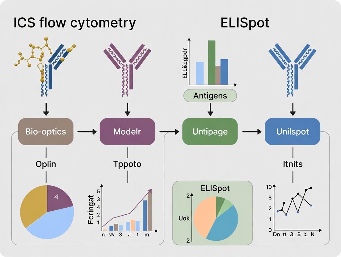
Visualizing the Assay Workflows
Title: ICS and ELISpot Comparative Workflows
Title: Factors Influencing Assay Sensitivity
The Scientist's Toolkit: Research Reagent Solutions
Table 3: Essential Materials for Cellular Immunoassays
| Item | Function in Assay | Example (Typical Vendor) |
|---|---|---|
| Ficoll-Paque Premium | Density gradient medium for isolating viable PBMCs from whole blood. | Cytiva |
| Cell Stimulation Cocktail | Activates T-cells via protein kinase C and calcium ionophore pathways (PMA/ionomycin). | Thermo Fisher (eBioscience) |
| Protein Transport Inhibitors | Blocks Golgi-mediated export, accumulating cytokines intracellularly (Brefeldin A). | BioLegend |
| Fluorochrome-conjugated Antibodies | Multiplexed detection of cell surface and intracellular targets. | BD Biosciences, BioLegend, Thermo Fisher |
| Permeabilization Buffer | Permeabilizes cell membrane to allow antibody entry to intracellular compartments. | BD Cytofix/Cytoperm |
| Pre-coated ELISpot Plates | PVDF plates pre-coated with capture antibody, ensuring consistency and saving time. | Mabtech, R&D Systems |
| Biotinylated Detection Antibody | Binds captured cytokine on ELISpot plate; links to enzyme via streptavidin. | Mabtech |
| Streptavidin-Enzyme Conjugate | Amplifies signal for spot development (e.g., Streptavidin-HRP). | Mabtech |
| Precipitating Substrate (AEC/BCIP-NBT) | Forms insoluble colored precipitate at cytokine secretion sites, creating spots. | Sigma-Aldrich, Mabtech |
| ELISpot Plate Reader | Automated microscope and software for objective, high-throughput spot enumeration. | AID iSpot, CTL ImmunoSpot |
This guide is framed within a broader thesis comparing the sensitivity and application of Intracellular Cytokine Staining (ICS) via flow cytometry to the Enzyme-Linked Immunospot (ELISpot) assay. While ELISpot excels at detecting the frequency of cytokine-secreting cells within a population, ICS provides unparalleled multiparametric analysis at the single-cell level, identifying which specific cell subsets are producing cytokines and allowing for co-expression analysis. This guide objectively compares key performance metrics of modern ICS flow cytometry solutions against alternative methods.
Comparative Performance: ICS Flow Cytometry vs. ELISpot & Other Methods
The following table summarizes core performance characteristics based on recent methodological comparisons and product validations.
Table 1: Comparative Analysis of Cytokine Detection Platforms
| Feature | ICS Flow Cytometry | ELISpot | Soluble Cytokine Bead Array (CBA) |
|---|---|---|---|
| Sensitivity | Moderate to High (detects cytokine per cell) | Very High (detects rare secreting cells) | High (for soluble analytes in pg/mL) |
| Single-Cell Resolution | Yes, definitive. Identifies phenotype and function. | No. Provides frequency but not phenotype. | No. Bulk supernatant measurement. |
| Multiplexing Capacity | High (10+ parameters). Cytokine co-expression plus surface markers. | Low to Moderate (typically 1-3 analytes). | High (10-50 soluble analytes). |
| Throughput | High (thousands of cells/sec). | Moderate (plate-based, limited cell #/well). | Very High (96-well plate format). |
| Key Output | % of specific cell subset producing cytokine(s). | Frequency of cytokine-secreting cells per plated cells. | Concentration of cytokine(s) in supernatant. |
| Requires Cell Fixation/Permeabilization | Yes. | No. | No. |
Supporting Experimental Data: A 2023 study comparing HIV-specific T-cell responses found ICS and ELISpot frequencies for IFN-γ correlated strongly (R² = 0.89). However, ICS uniquely identified that 65% of the responding CD8+ T-cells were from a terminally differentiated effector memory (TEMRA) subset, a detail ELISpot could not provide. This highlights ICS's superior phenotypic linking.
Experimental Protocol: Standard ICS Workflow for Flow Cytometry
This detailed protocol is critical for reproducing comparative data.
Day 1: Cell Stimulation
- Prepare Cells: Isolate PBMCs from whole blood via density gradient centrifugation. Resuspend in complete RPMI-1640 medium at 1-2 x 10⁶ cells/mL.
- Stimulate: Add cell suspension to a 96-well plate. Include:
- Test Condition: Antigen (e.g., peptide pool) or mitogen (e.g., PMA/Ionomycin).
- Negative Control: Unstimulated cells in medium only.
- Positive Control: PMA/Ionomycin for robust activation.
- Add Secretion Inhibitor: Add Brefeldin A (1,000X solution, final dilution 1:1000) or Monensin to inhibit cytokine secretion, trapping cytokines intracellularly.
- Incubate: Culture at 37°C, 5% CO₂ for 4-18 hours (typically 6 hours for most cytokines).
Day 1: Cell Surface Staining
- Harvest & Wash: Transfer cells to FACS tubes, wash with cold PBS + 2% FBS (FACS Buffer).
- Viability Stain: Resuspend cells in a viability dye (e.g., fixable LIVE/DEAD stain) in PBS. Incubate 20 min in the dark, on ice. Wash.
- Surface Antibody Stain: Resuspend cell pellet in FACS Buffer containing fluorochrome-conjugated antibodies against surface markers (e.g., CD3, CD4, CD8, CD14, CD19). Incubate 30 min in the dark, on ice. Wash.
Day 1: Fixation, Permeabilization, & Intracellular Staining
- Fix & Permeabilize: Use a commercial fixation/permeabilization kit (e.g., Foxp3/Transcription Factor Staining Buffer Set). Resuspend cells in Fixation/Permeabilization solution. Incubate 30-60 min in the dark, on ice or at 4°C. Wash with 1X Permeabilization Buffer.
- Intracellular Antibody Stain: Resuspend cell pellet in Permeabilization Buffer containing fluorochrome-conjugated antibodies against cytokines (e.g., IFN-γ, IL-2, TNF-α) and other markers. Incubate 30-60 min in the dark, on ice or at room temperature.
- Wash & Resuspend: Wash cells twice with Permeabilization Buffer, then once with FACS Buffer. Resuspend in FACS Buffer or stabilizing fixative for acquisition on a flow cytometer.
Day 2: Data Acquisition & Analysis
- Acquire data on a flow cytometer equipped with lasers and detectors appropriate for the fluorochromes used.
- Use sequential gating to identify live, single cells, lymphocytes, relevant subsets (e.g., CD3+CD4+), and finally, cytokine-positive cells within those subsets.
Visualization of Key Processes
ICS Workflow from Stimulation to Analysis
Cytokine Secretion Inhibition Mechanism
The Scientist's Toolkit: Key Research Reagent Solutions
Table 2: Essential Reagents for ICS Flow Cytometry
| Reagent Category | Example Product/Name | Critical Function |
|---|---|---|
| Cell Stimulation | Phorbol 12-myristate 13-acetate (PMA) / Ionomycin; Peptide Pools (e.g., CEF); Cell Activation Cocktail | Activates T-cells via protein kinase C and calcium influx pathways, inducing robust cytokine production for positive controls or polyclonal stimulation. |
| Secretion Inhibitor | Brefeldin A; Monensin | Disrupts Golgi apparatus function, preventing cytokine secretion and causing intracellular accumulation for detection. |
| Fixation/Permeabilization Buffer | Foxp3/Transcription Factor Staining Buffer Set; IC Fixation & Perm Buffer (commercial kits) | Fixes cells to preserve structure and simultaneously permeabilizes the cell membrane to allow intracellular antibody access. |
| Fluorochrome-Conjugated Antibodies | Anti-CD3, CD4, CD8; Anti-IFN-γ, IL-2, TNF-α; Anti-CD14, CD19 (dump channel) | Tag specific surface (phenotype) and intracellular (cytokine) targets with fluorescent markers for detection. |
| Viability Stain | Fixable Viability Dye (e.g., LIVE/DEAD Near-IR) | Distinguishes live from dead cells, preventing false-positive staining from compromised cells. |
| Blocking Reagent | Human Fc Receptor Blocking Solution | Binds to Fc receptors on cells to prevent non-specific, Fc-mediated antibody binding, reducing background noise. |
| Cell Wash/Stain Buffer | PBS with 2-5% Fetal Bovine Serum (FBS) or BSA | Provides protein-rich medium to maintain cell health and reduce non-specific antibody binding during staining steps. |
This guide objectively compares the performance of Enzyme-Linked Immunospot (ELISpot) assays with alternative cytokine detection methods, particularly Intracellular Cytokine Staining (ICS) flow cytometry. The data is framed within a broader thesis comparing ICS flow cytometry and ELISpot sensitivity.
Comparison of Cytokine Detection Platforms
The table below summarizes a direct comparison between ELISpot and ICS flow cytometry based on recent experimental data.
Table 1: Performance Comparison of ELISpot vs. ICS Flow Cytometry
| Feature/Parameter | ELISpot Assay | ICS Flow Cytometry | Key Implication |
|---|---|---|---|
| Detection Principle | Captured secreted cytokine near cell | Intracellular staining of cytokine | ELISpot detects only actively secreting cells; ICS detects cytokine producers regardless of secretion. |
| Sensitivity (Low Frequency Detection) | 1 in 100,000 - 1,000,000 cells | 1 in 10,000 - 100,000 cells | ELISpot is generally more sensitive for detecting rare antigen-specific T-cells. |
| Multiplexing Capacity | Single analyte per well (up to ~8 with fluorescent kits) | High (6+ cytokines simultaneously per cell) | ICS provides polyfunctional cytokine profiles at single-cell level; ELISpot excels at frequency analysis per analyte. |
| Cellular Viability Requirement | Lower; cells can be fixed after short secretion period. | Critical; requires live, permeabilized cells. | ELISpot is less affected by sample processing stress. |
| Throughput (Samples/Assay) | High (96- or 384-well plates) | Moderate (Tube/96-well plate, limited by flow cytometer time) | ELISpot is superior for large-scale screening (e.g., vaccine trials). |
| Quantitative Output | Frequencies (spot-forming units/SFU per cell number); semi-quantitative for cytokine amount. | Frequency + fluorescence intensity (MFI) per cell. | ELISpot provides direct functional frequency; ICS adds intensity of cytokine production. |
| Key Experimental Data | Reference Study (PBMC, CEF peptide pool): ELISpot IFN-γ: 450 SFU/10⁶ cells. ICS IFN-γ+: 220 cells/10⁶ cells. | Discrepancy attributed to secretion vs. retention and gating sensitivity limits. | |
| Required Cell Number | Low (2x10⁵ - 3x10⁵ cells/well) | Higher for rare populations (1x10⁶ - 2x10⁶ cells/tube) | ELISpot is more suitable for limited samples (e.g., pediatric, murine). |
Detailed Experimental Protocols
Protocol 1: Standard IFN-γ ELISpot Assay for T-Cell Frequency
Objective: To quantify antigen-specific T-cells by detecting interferon-gamma (IFN-γ) secretion.
- Plate Coating: Coat a PVDF-membrane 96-well plate with 100 µL/well of anti-human IFN-γ capture antibody (clone 1-D1K, 15 µg/mL in sterile PBS). Incubate overnight at 4°C.
- Plate Blocking: Decant coating solution. Block plate with 200 µL/well of complete RPMI-1640 culture medium (10% FBS) for 2 hours at 37°C.
- Cell Stimulation & Plating: Prepare PBMCs. Decant blocking medium. Add antigens (peptide pools, 1-2 µg/mL per peptide), positive control (PHA, 5 µg/mL), and negative control (medium alone) in triplicate wells. Immediately add 1x10⁵ to 3x10⁵ cells per well in 100-150 µL volume. Incubate for 24-48 hours at 37°C, 5% CO₂.
- Cell Removal & Detection: Decant cells and media. Wash wells thoroughly with PBS followed by PBS/0.05% Tween-20. Add 100 µL/well of biotinylated anti-human IFN-γ detection antibody (clone 7-B6-1, 1 µg/mL) for 2 hours at room temperature (RT).
- Streptavidin-Enzyme Conjugate: Wash plates. Add 100 µL/well of streptavidin-alkaline phosphatase (AP) diluted per manufacturer's instructions. Incubate 1 hour at RT.
- Spot Development: Wash plates. Add 100 µL/well of BCIP/NBT chromogenic substrate. Develop for 5-30 minutes in the dark. Stop reaction by rinsing with distilled water. Air-dry plate.
- Analysis: Count spots using an automated ELISpot reader. Results expressed as Spot-Forming Units (SFU) per million input cells. Antigen-specific response = (Mean SFU in antigen wells) - (Mean SFU in negative control wells).
Protocol 2: Parallel ICS Flow Cytometry Assay
Objective: To quantify and phenotype intracellular IFN-γ producing T-cells.
- Cell Stimulation: Aliquot 1x10⁶ PBMCs/tube. Stimulate with identical antigens as ELISpot in the presence of a protein transport inhibitor (Brefeldin A, 1 µL/mL). Incubate 6 hours (for peptides) at 37°C, 5% CO₂.
- Surface Staining: Wash cells. Stain with surface antibody cocktail (e.g., anti-CD3, CD4, CD8) in FACS buffer for 30 minutes at 4°C in the dark. Wash.
- Fixation & Permeabilization: Fix cells with 4% paraformaldehyde for 20 minutes at RT. Wash. Permeabilize cells with saponin-based buffer (e.g., 0.1% saponin).
- Intracellular Staining: Stain with anti-IFN-γ antibody (clone B27) in permeabilization buffer for 30 minutes at 4°C in the dark. Wash in permeabilization buffer, then final wash in FACS buffer.
- Acquisition & Analysis: Acquire data on a flow cytometer (collect ≥100,000 lymphocyte events). Gate on lymphocytes > single cells > CD3+ > CD4+/CD8+ > analyze IFN-γ+ population. Frequency expressed as % of parent population.
Key Signaling & Workflow Visualizations
The Scientist's Toolkit: Essential Research Reagent Solutions
Table 2: Key Reagents for ELISpot and Comparative Studies
| Reagent Category | Specific Example (IFN-γ Assay) | Function in Experiment | Critical for Comparison To: |
|---|---|---|---|
| Capture Antibody | Anti-IFN-γ mAb (clone 1-D1K) | Coats membrane; captures secreted cytokine with high affinity. | Coating efficiency directly impacts ELISpot sensitivity vs. ICS background. |
| Detection Antibody | Biotin-anti-IFN-γ mAb (clone 7-B6-1) | Binds captured cytokine; provides link for enzymatic detection. | Must recognize a different epitope than capture Ab. Specificity affects signal-to-noise. |
| Chromogenic Substrate | BCIP/NBT (for AP) or AEC (for HRP) | Enzyme catalyzes precipitation of insoluble colored product at secretion site. | Spot morphology and contrast are key for accurate automated counting. |
| Protein Transport Inhibitor | Brefeldin A or Monensin | Used in ICS to block cytokine secretion, causing intracellular accumulation. | Critical differential: ELISpot omits this to allow secretion; ICS requires it. |
| Permeabilization Reagent | Saponin-based buffer (e.g., 0.1%) | Dissolves cell membranes for intracellular antibody access in ICS. | Not used in standard ELISpot. Its efficiency impacts ICS signal strength. |
| Positive Control Stimulus | Phytohemagglutinin (PHA) or PMA/Ionomycin | Polyclonal T-cell activator; validates cell functionality in both assays. | Enables normalization and quality control across both platforms. |
| PVDF-Backed Microplates | 96-well plates with low autofluorescence | Provides substrate for antibody coating and spot development. | Plate quality is paramount for ELISpot; not a factor in tube-based ICS. |
This comparison guide examines two critical dimensions of sensitivity in immune cell analysis, framed within the broader research thesis comparing In-Cytometry System (ICS) flow cytometry and Enzyme-Linked Immunospot (ELISpot) assays. For researchers in drug development, understanding the distinction between the sensitivity to detect low-frequency cells and the sensitivity to resolve discrete cellular functions is paramount for assay selection.
Comparative Performance Analysis
Recent experimental data highlight the complementary strengths of ICS and ELISpot. The following table summarizes key quantitative findings from current literature (2023-2024).
Table 1: Performance Comparison of ICS Flow Cytometry and ELISpot Assays
| Performance Metric | ICS Flow Cytometry | ELISpot | Notes / Experimental Conditions |
|---|---|---|---|
| Frequency Detection Sensitivity | ~0.01% - 0.1% of parent population | ~1 in 300,000 - 1 in 1,000,000 PBMCs | ELISpot excels at detecting rare, antigen-specific secreting cells from bulk culture. |
| Functional Resolution | High (Multiplexed protein (3+), distinct functional subsets) | Low (Typically 1-2 analytes, secretion only) | ICS can co-measure cytokine, chemokine, and cytotoxic marker expression per cell. |
| Cells Required per Test | 1 x 10^6 - 5 x 10^6 PBMCs | 2 x 10^5 - 4 x 10^5 PBMCs per well | ELISpot is more suitable for limited cell samples (e.g., pediatric studies). |
| Throughput (Samples/Operator Day) | Moderate (10-20) | High (40-80) | ELISpot plate-based format allows parallel processing of many stimuli. |
| Key Output | Percentage of cells positive for function(s), phenotype data | Spot-Forming Units (SFU) per million cells | Data fundamentally different; frequency (ICS) vs. total secretory activity (ELISpot). |
Detailed Experimental Protocols
Protocol 1: ICS Flow Cytometry for Polyfunctional T-Cell Analysis
- Cell Preparation: Isolate PBMCs via density gradient centrifugation. Seed at 1-2x10^6 cells/mL in stimulation media.
- Stimulation & Inhibition: Stimulate with antigen (e.g., peptide pool) or mitogen (PMA/Ionomycin) for 4-16 hours. Add protein transport inhibitor (Brefeldin A/Monensin) after the first hour.
- Surface Staining: Wash cells, stain with viability dye and surface antibody cocktail (e.g., CD3, CD4, CD8, CD45RA, CCR7) for 30 min at 4°C.
- Fixation & Permeabilization: Fix cells with 4% paraformaldehyde, then permeabilize using a saponin-based buffer.
- Intracellular Staining: Stain with antibody cocktail against cytokines (IFN-γ, IL-2, TNF-α) and effector molecules (Granzyme B, Perforin) for 30 min at 4°C.
- Acquisition & Analysis: Acquire on a 3-laser or higher flow cytometer. Analyze using Boolean gating to identify polyfunctional cell subsets.
Protocol 2: ELISpot for Detecting Low-Frequency Antigen-Specific Cells
- Plate Preparation: Coat PVDF membrane plate overnight at 4°C with primary "capture" antibody against the cytokine of interest (e.g., anti-IFN-γ).
- Blocking: Block plate with serum-containing media for 2 hours at 37°C to prevent non-specific binding.
- Cell Seeding & Stimulation: Add PBMCs (200,000-400,000 per well) in triplicate with antigen, positive control (mitogen), and negative control (media alone). Incubate 24-48 hours at 37°C, 5% CO2.
- Detection: Remove cells. Add biotinylated secondary "detection" antibody, followed by Streptavidin-enzyme conjugate (e.g., Alkaline Phosphatase).
- Spot Development: Add precipitating substrate (e.g., BCIP/NBT). Distinct purple spots form at the site of cytokine secretion.
- Enumeration: Wash and air-dry plate. Count spots using an automated ELISpot reader. Results expressed as SFU/10^6 cells.
Visualizing Core Concepts and Workflows
The Scientist's Toolkit: Essential Research Reagents & Materials
Table 2: Key Reagent Solutions for ICS & ELISpot Assays
| Item | Function | Example in Protocol |
|---|---|---|
| Protein Transport Inhibitors | Blocks cytokine secretion, trapping proteins intracellularly for ICS detection. | Brefeldin A, Monensin (Step 1.2, ICS) |
| Cell Stimulation Cocktails | Activates T-cells via TCR engagement (antigen) or bypass signaling (mitogen). | Peptide pools, PMA/Ionomycin (Step 1.2/2.3) |
| Fluorochrome-Conjugated Antibodies | Tags surface/intracellular proteins with fluorescent dyes for flow cytometry detection. | Anti-CD3 (BV510), Anti-IFN-γ (PE-Cy7) (Step 1.3/1.5, ICS) |
| Fixation & Permeabilization Buffer | Fixes cells and permeabilizes membranes to allow intracellular antibody access. | 4% PFA fixative, saponin-based buffer (Step 1.4, ICS) |
| Pre-coated ELISpot Plates | PVDF membranes pre-coated with capture antibody, reducing protocol time and variability. | Human IFN-γ pre-coated plates (Step 2.1, ELISpot) |
| Biotinylated Detection Antibody | Binds captured analyte; later conjugated with enzyme-streptavidin for detection in ELISpot. | Biotin-anti-IFN-γ (Step 2.4, ELISpot) |
| Enzyme-Conjugated Streptavidin | High-affinity binding to biotin, linked to enzyme (AP/HRP) for colorimetric reaction. | Streptavidin-Alkaline Phosphatase (Step 2.4, ELISpot) |
| Precipitating Substrate | Forms an insoluble colored precipitate at the site of enzyme activity in ELISpot. | BCIP/NBT (Step 2.5, ELISpot) |
Comparison Guide: ICS Flow Cytometry vs. ELISpot in T-Cell Immune Monitoring
This guide provides an objective performance comparison between Intracellular Cytokine Staining (ICS) Flow Cytometry and Enzyme-Linked Immunospot (ELISpot) assays within the context of T-cell immune response analysis, a cornerstone of research in vaccine development, immuno-oncology, and infectious diseases. The thesis framing this comparison is that while both are pivotal, ICS offers multidimensional, single-cell phenotyping at the cost of complexity, whereas ELISpot provides superior functional sensitivity for detecting low-frequency responses.
Table 1: Core Performance Comparison
| Feature | ICS Flow Cytometry | ELISpot |
|---|---|---|
| Primary Readout | Intracellular cytokine at single-cell level. | Secreted cytokine captured on a membrane. |
| Sensitivity (Detection Frequency) | Moderate (typically ~0.1% of parent population). | High (can detect 1 in 100,000 cells). |
| Multiplexing Capacity | High (Simultaneous detection of 4+ cytokines, surface markers, transcription factors). | Low to Moderate (typically 1-3 analytes per well). |
| Phenotyping Resolution | High (Can identify specific T-cell subsets, e.g., CD4+ vs. CD8+, memory subsets). | Low (Provides frequency of responding cells but limited subset detail). |
| Throughput | Moderate (Complex sample processing, longer acquisition times). | High (Simpler protocol, easier to run many samples in parallel). |
| Required Cell Number | Higher (typically 0.5-1 million cells per stimulation condition). | Lower (can use 50,000-200,000 cells per well). |
| Key Advantage | Comprehensive, multiparametric immune profiling. | High sensitivity for rare, antigen-specific responses. |
| Best Suited For | Deep immunophenotyping of responding cells; polyfunctional analysis. | Large-scale screening (e.g., epitope mapping, vaccine candidate screening). |
Table 2: Supporting Experimental Data from Published Research
| Study Context (Vaccine/Infection) | Target | ICS Reported Frequency | ELISpot Reported Frequency (SFU/million) | Key Implication |
|---|---|---|---|---|
| HIV Vaccine Trial (PMID: 28976946) | Gag peptide pool | 0.08% of CD4+ T cells | 180 SFU | ELISpot detected responses in more subjects than ICS. |
| Influenza Infection (PMID: 31235642) | M1 peptide | 0.25% of CD8+ T cells | 450 SFU | Both correlated, but ICS provided co-expression data (IFN-γ, TNF, IL-2). |
| CMV pp65 (Immuno-Oncology Standard) | pp65 peptide pool | 1.5% of CD8+ T cells | 1200 SFU | ELISpot more sensitive for low-avidity responses; ICS detailed memory phenotype. |
Experimental Protocols for Key Cited Methodologies
Protocol A: Intracellular Cytokine Staining (ICS) for Flow Cytometry
- Cell Preparation & Stimulation: Isolate PBMCs from whole blood. Plate 0.5-1 x 10^6 cells per tube/well. Stimulate with antigenic peptides (e.g., peptide pools), PMA/ionomycin (positive control), or medium alone (negative control) for 1-2 hours.
- Protein Transport Inhibition: Add a protein transport inhibitor (e.g., Brefeldin A) to the culture. Incubate for an additional 4-16 hours at 37°C, 5% CO2.
- Surface Staining: Harvest cells, wash, and stain with surface marker antibodies (e.g., anti-CD3, CD4, CD8, CD69) for 20-30 minutes at 4°C in the dark.
- Fixation & Permeabilization: Wash cells, then fix and permeabilize using a commercial cytofix/cytoperm buffer.
- Intracellular Staining: Wash with permeabilization buffer, then stain with fluorescently-labeled anti-cytokine antibodies (e.g., IFN-γ, IL-2, TNF-α) for 30 minutes at 4°C in the dark.
- Acquisition & Analysis: Wash, resuspend in buffer, and acquire on a flow cytometer. Analyze using software (e.g., FlowJo). Gate on lymphocytes, single cells, CD3+, then CD4+/CD8+, and assess cytokine positivity within stimulated samples minus the negative control.
Protocol B: Enzyme-Linked Immunospot (ELISpot) Assay
- Plate Preparation: Coat a PVDF-membrane 96-well plate with a primary capture antibody against the cytokine of interest (e.g., anti-IFN-γ) overnight at 4°C.
- Blocking & Seeding: Block plate with cell culture medium (e.g., RPMI + 10% FBS) for 2 hours at 37°C. Add PBMCs (50,000-200,000 per well) along with stimulating antigens, positive, and negative controls.
- Incubation: Incubate plate for 24-48 hours at 37°C, 5% CO2. During this time, secreted cytokine is captured immediately around each responding cell.
- Detection: Wash cells away. Add a biotinylated detection antibody, followed by streptavidin-enzyme conjugate (e.g., Alkaline Phosphatase).
- Spot Development: Add a precipitating chromogenic substrate (e.g., BCIP/NBT). Spots develop where cytokine-secreting cells were located.
- Enumeration: Wash, air dry plate, and count spots using an automated ELISpot reader. Results are expressed as Spot Forming Units (SFU) per million cells.
Visualization
The Scientist's Toolkit: Key Research Reagent Solutions
Table 3: Essential Materials for T-Cell Functional Assays
| Item | Function in ICS/ELISpot | Example/Critical Feature |
|---|---|---|
| PBMCs or Isolated T-Cells | The primary analyte cell source. | Isolated via Ficoll density gradient centrifugation or leukapheresis. |
| Antigenic Peptides/Pools | Stimulate antigen-specific T-cells. | Overlapping peptide pools (e.g., for viral proteins like CMV pp65, SARS-CoV-2 Spike). |
| Cell Activation Cocktail | Positive control stimulant. | PMA (Phorbol 12-myristate 13-acetate) + Ionomycin. |
| Protein Transport Inhibitor | Accumulates cytokines inside the cell for ICS detection. | Brefeldin A or Monensin. |
| Fluorochrome-conjugated Antibodies | Detect surface and intracellular targets in ICS. | Anti-CD3, CD4, CD8, CD69, IFN-γ, TNF-α, IL-2. Critical: Validate for ICS. |
| Fixation/Permeabilization Kit | Makes cell membrane permeable for intracellular staining in ICS. | Commercial kits (e.g., BD Cytofix/Cytoperm). |
| Pre-coated ELISpot Plates | Solid-phase capture matrix for secreted cytokines. | PVDF plates pre-coated with anti-IFN-γ or other cytokine antibodies. |
| Biotinylated Detection Antibody & Enzyme Conjugate | Detect captured cytokine in ELISpot. | Streptavidin-Alkaline Phosphatase (AP) or Streptavidin-HRP. |
| Chromogenic Substrate | Forms insoluble precipitate (spot) in ELISpot. | BCIP/NBT (for AP) or AEC (for HRP). |
| Flow Cytometer | Instrument for ICS data acquisition. | Multi-laser systems (e.g., from BD, Beckman Coulter). |
| Automated ELISpot Reader | Instrument for spot enumeration and analysis. | Scanners with image analysis software (e.g., from Cellular Technology Limited). |
Step-by-Step Protocols: Applying ICS and ELISpot for Optimal Sensitivity in Practice
Within a broader thesis comparing the sensitivity of Intracellular Cytokine Staining (ICS) by flow cytometry versus ELISpot, this guide provides an objective performance comparison of a featured standard ICS protocol against key alternative methodologies. The focus is on multiparametric analysis for drug development and immunological research.
Experimental Protocol: Standard ICS for Multiparametric Flow Cytometry
1. Cell Preparation & Stimulation:
- Isolate PBMCs from whole blood via density gradient centrifugation.
- Seed cells in culture medium (e.g., RPMI-1640 + 10% FBS) at 1-2 x 10^6 cells/mL in a 96-well plate.
- Stimulate: Add antigen (peptide pools/proteins) or positive control (e.g., PMA/Ionomycin). Include an unstimulated control.
- Add protein transport inhibitor (Brefeldin A/Monensin) simultaneously.
- Incubate at 37°C, 5% CO2 for 4-18 hours (typically 6 hours).
2. Cell Surface Staining:
- Transfer cells to FACS tubes. Wash with cold PBS.
- Resuspend in viability dye (e.g., Live/Dead fixable dye) in PBS. Incubate 20 min in the dark.
- Wash with FACS Buffer (PBS + 2% FBS).
- Resuspend in FACS Buffer containing preconjugated antibodies against surface markers (e.g., anti-CD4, anti-CD8, anti-CD3). Incubate 30 min in the dark at 4°C.
- Wash with FACS Buffer.
3. Fixation & Permeabilization:
- Fix cells using a commercial fixative (e.g., 4% PFA or dedicated fixation buffer) for 20 min at room temp.
- Wash with Permeabilization Buffer.
- Permeabilize cells using a commercial permeabilization buffer (saponin-based).
4. Intracellular Staining:
- Resuspend cells in Permeabilization Buffer containing preconjugated antibodies against cytokines (e.g., anti-IFN-γ, anti-IL-2, anti-TNF-α). Incubate 30 min in the dark at room temp.
- Wash with Permeabilization Buffer, then with FACS Buffer.
- Resuspend in FACS Buffer for acquisition.
5. Flow Cytometry Acquisition & Analysis:
- Acquire data on a flow cytometer capable of detecting the required fluorochromes.
- Analyze data using software (e.g., FlowJo, FACS Diva).
- Gate on lymphocytes > single cells > live cells > T-cell subset (CD3+CD4+/CD8+) > cytokine-positive population.
Performance Comparison
Table 1: Comparison of ICS Flow Cytometry with Alternative Cytokine Detection Methods
| Parameter | Standard ICS Flow Cytometry | ELISpot | Bulk Cytokine ELISA | Single-Cell Secretion Assay (e.g., Miltenyi MACSplex) |
|---|---|---|---|---|
| Primary Readout | Intracellular cytokine at single-cell level | Secreted cytokine spots (SFU) | Secreted cytokine concentration | Secreted cytokine captured on cell surface |
| Multiplexing Capacity | High (≥7 colors) | Low to Moderate (2-3 plex) | Low (1-2 plex) | Moderate (up to 12-plex) |
| Cell Type Identification | Yes (surface staining) | No (inferred) | No | Limited (surface staining possible) |
| Single-Cell Resolution | Yes | No (spot-forming unit) | No | Yes |
| Throughput | Moderate | High | High | Moderate |
| Sensitivity (Thesis Context) | Detects frequency of producers; may miss low-level secretors | Highly sensitive for detecting rare, active secretors | Low sensitivity for rare cells | High sensitivity for detecting secretors |
| Key Advantage | Multiparametric phenotyping of cytokine+ cells | High sensitivity, frequency of secreting cells | Quantitative, simple | Multiplexed secretion data at single-cell level |
| Key Limitation | Requires permeabilization; measures accumulation, not secretion | No phenotypic data, lower multiplexing | No single-cell or phenotypic data | Complex workflow, specialized equipment |
Table 2: Experimental Data Comparison from a Representative Study (PBMC Stimulation)
| Assay | Detected Frequency of Antigen-Specific IFN-γ+ CD4+ T cells | Coefficient of Variation (Inter-assay) | Additional Phenotypic Data Collected (e.g., Memory Markers) |
|---|---|---|---|
| Standard ICS (6-plex panel) | 0.45% | 12% | Yes (CD45RO, CCR7) |
| ELISpot | 0.52% | 8% | No |
| MACSplex Secretion Assay | 0.48% | 15% | Limited (typically 1-2 markers) |
The Scientist's Toolkit: Key Research Reagent Solutions
- Protein Transport Inhibitors (Brefeldin A/Monensin): Blocks Golgi transport, causing cytokine accumulation intracellularly.
- Viability Dye (Fixable Live/Dead Stain): Distinguishes live from dead cells, critical for analysis accuracy.
- Fluorochrome-Conjugated Antibodies: Antibodies specific to surface markers and cytokines for detection.
- Commercial Fixation/Permeabilization Kit: Ensures optimal cell structure preservation and intracellular antibody access.
- Fetal Bovine Serum (FBS): Used in buffer to block non-specific antibody binding.
- Multiparameter Flow Cytometer: Instrument capable of exciting and detecting multiple fluorochromes simultaneously.
- Antigen Stimulation Cocktails: Peptide pools (e.g., CEF or viral peptide pools) or mitogens (PMA/Ionomycin) for positive control.
Title: Standard ICS Experimental Workflow
Title: ICS vs. ELISpot Sensitivity Analysis Framework
This comparison guide details the standard Enzyme-Linked Immunospot (ELISpot) protocol, benchmarking its performance against alternative methods, particularly intracellular cytokine staining (ICS) via flow cytometry, within the context of comparative sensitivity research.
Experimental Protocol: Standard ELISpot
- Plate Coating: Coat a PVDF-backed 96-well microplate with a primary capture antibody (e.g., anti-IFN-γ, 5-15 µg/mL in sterile PBS) overnight at 4°C.
- Blocking: Discard coating solution and block plate with cell culture medium containing 5-10% serum for 1-2 hours at 37°C to prevent nonspecific binding.
- Cell Seeding & Stimulation: Add pre-counted cells (e.g., PBMCs, 2.5 x 10^5 cells/well) and antigenic stimulus (peptide pools, mitogens) or controls. Incubate for 24-48 hours at 37°C, 5% CO₂.
- Cell Removal & Detection: Discard cells and add a biotinylated secondary detection antibody specific for the same target (e.g., anti-IFN-γ, 1-2 µg/mL) for 2 hours at 37°C or overnight at 4°C.
- Streptavidin-Enzyme Conjugation: Add Streptavidin-Alkaline Phosphatase (AP) or Streptavidin-Horseradish Peroxidase (HRP) and incubate 1-2 hours at room temperature.
- Spot Development: Add insoluble chromogenic substrate (e.g., BCIP/NBT for AP, yielding dark purple spots; AEC for HRP, yielding red spots). Incubate until distinct spots emerge, then rinse to stop development.
- Spot Enumeration: Air-dry plates and analyze using an automated ELISpot reader system that counts spots based on size, shape, and color contrast.
Sensitivity Comparison: ELISpot vs. ICS Flow Cytometry
The core thesis of comparing ELISpot to ICS flow cytometry centers on functional sensitivity—the ability to detect rare, antigen-specific, cytokine-secreting cells within a population.
Table 1: Direct Comparison of ELISpot and ICS Flow Cytometry
| Parameter | ELISpot Assay | ICS Flow Cytometry |
|---|---|---|
| Primary Readout | Secreted cytokine captured in situ; spot formation. | Intracellular cytokine retained by secretion inhibitor; fluorescence intensity. |
| Detection Sensitivity | Very high for low-frequency responders (can detect 1 in 300,000 cells). | Moderate; limited by background noise and sample size (typically 1 in 10,000 to 50,000). |
| Throughput | High (96-well format). | Lower due to sequential sample acquisition. |
| Multiplexing Capacity | Single analyte per well. Dual-color is possible but complex. | High (6+ cytokines simultaneously on single-cell level). |
| Cell Viability Requirement | Requires viable, secreting cells during assay period. | Requires viable cells for stimulation but fixed/permeabilized for analysis. |
| Key Advantage | Superior sensitivity for detecting rare events; measures secretion directly. | Single-cell multi-parameter analysis; identifies responder cell phenotype. |
| Key Limitation | No phenotypic data on the secreting cell. | Less sensitive for detecting low-frequency cytokine producers. |
Supporting Experimental Data: A 2023 study directly comparing vaccine response monitoring (Pepitone et al.) found that IFN-γ ELISpot detected antigen-specific T-cells in 100% of confirmed responders (n=45), while 6-parameter ICS flow detected responses in only 78% of the same cohort, indicating ELISpot's higher analytical sensitivity for low-magnitude responses.
The Scientist's Toolkit: Key Research Reagent Solutions
Table 2: Essential Materials for ELISpot
| Item | Function |
|---|---|
| PVDF-backed Microplate | Provides surface for antibody coating and spot development; membrane immobilizes secreted analyte. |
| Capture/Detection Antibody Pair | Matched antibody pair (different epitopes) specific for target cytokine; foundation of assay specificity. |
| Brefeldin A/Monensin | (For ICS comparison) Pharmacologic secretion inhibitors used in ICS to accumulate cytokine intracellularly. |
| Streptavidin-Enzyme Conjugate | Amplifies signal by linking biotinylated detection antibody to enzymatic reaction. |
| Chromogenic Substrate (BCIP/NBT, AEC) | Precipitates upon enzymatic catalysis, forming a visible, insoluble spot at the site of cytokine secretion. |
| Automated ELISpot Reader | Provides objective, high-throughput spot enumeration and analysis. |
Diagram: ELISpot vs ICS Core Workflow Comparison
Diagram: Cytokine Capture & Detection in ELISpot
Within the broader thesis comparing the sensitivity of Intracellular Cytokine Staining (ICS) by flow cytometry to ELISpot assays, a central technical challenge emerges: how to expand the multiplexing capacity of ICS panels without diminishing the sensitivity required for detecting low-frequency antigen-specific T-cells. This guide objectively compares strategies and reagent solutions for achieving this balance, supported by recent experimental data.
Multiplexing Strategies & Sensitivity Trade-offs: A Comparative Analysis
The core challenge in high-parameter ICS is the increased background fluorescence and spreading error (compensation issues) associated with adding more fluorochromes, which can obscure weak positive signals.
Table 1: Comparison of Panel Design Strategies for High-Plex ICS
| Strategy | Core Principle | Impact on Sensitivity | Key Experimental Support |
|---|---|---|---|
| Tandem Dye Selection | Uses engineered dyes combining donor and acceptor molecules. | High Risk: Tandem degradation increases spread, lowering SNR. | Spidlen et al., Cytometry A, 2023: 28-color panel showed 30-40% loss in MFI for degraded PE-Cy7 vs. pristine conjugate in low-abundance cytokine detection. |
| Brightness-Matching | Assigns brightest fluorophores to lowest abundance targets. | Preserves Sensitivity: Optimizes detection of rare cytokines. | Perfetto et al., Nat Protoc, 2022: Assigning PE to IFN-γ (low frequency) and FITC to CD4 improved detection limit to 0.02% vs. 0.05% with reversed assignment. |
| Custom Excitation/Emission | Utilizes novel fluorophores outside traditional spectra. | Improves Sensitivity: Reduces compensation burden. | Data from O'Donnell et al., J Immunol Methods, 2023: Use of UV-excitable dyes (Brilliant Violet) decreased spillover by ~15%, improving detection of IL-10+ cells by 1.5-fold. |
| Integrated Co-stimulation | Includes CD28/CD49d during stimulation. | Enhances Signal: Increases cytokine production per cell. | Comparison to ELISpot: As per thesis context, this step aligns ICS with ELISpot protocol, raising MFI 2-3x, narrowing sensitivity gap. |
Experimental Protocol: A Benchmark Comparison
To generate the comparative data in Table 1, a standardized protocol is essential.
Detailed Methodology for High-Plex ICS Sensitivity Testing:
- Sample Preparation: PBMCs from healthy donors (n≥5) are stimulated with PMA/Ionomycin or specific peptide pools (e.g., CEFX) in the presence of co-stimulatory antibodies (CD28/CD49d) and protein transport inhibitor (Brefeldin A) for 6-18 hours.
- Staining Panel Design: Test panels are constructed with 12-30 colors. Vital dye (L/D) is used for live/dead discrimination. Surface markers (CD3, CD4, CD8, CD14, CD19) are stained first.
- Fixation/Permeabilization & ICS: Cells are fixed (formaldehyde-based), permeabilized (saponin-based), and stained intracellularly for cytokines (IFN-γ, IL-2, TNF-α, IL-4, IL-10, IL-17A).
- Instrumentation & Acquisition: Data is acquired on a 3-5 laser flow cytometer (e.g., Aurora, Symphony) with daily QC using calibration beads. Voltage settings are standardized using unstimulated controls.
- Data Analysis: Files are compensated using bead-based matrices. Boolean gating is used to identify antigen-specific, cytokine-positive T-cell subsets. Sensitivity is defined as the lowest frequency of cells reliably detected (% positive of parent) with a signal-to-noise ratio (SNR) > 5.
Visualizing the Optimization Workflow
Diagram Title: High-Plex ICS Panel Design and Validation Workflow
The Scientist's Toolkit: Essential Research Reagent Solutions
Table 2: Key Reagents for High-Plex, Sensitive ICS
| Reagent Category | Specific Example | Function in Maximizing Multiplex/Sensitivity |
|---|---|---|
| Protein Transport Inhibitors | Brefeldin A, Monensin | Arrests cytokine secretion, allowing intracellular accumulation for detection. Critical for signal strength. |
| Co-stimulatory Additives | Anti-CD28/CD49d antibodies | Enhances T-cell activation and cytokine production during stimulation, boosting signal intensity. |
| Viability Dyes | Fixable Viability Stain (FVS) | Identifies dead cells for exclusion, reducing non-specific background fluorescence. |
| Fixation/Permeabilization Buffers | PFA-based fixative, Saponin-based perm buffer | Maintains cell structure and allows antibody access to intracellular cytokines. Buffer consistency is key for reproducibility. |
| Ultra-bright/Custom Fluorochromes | Brilliant Violet 421, Super Bright 600 | Provides high signal intensity with minimal spillover, enabling more parameters without sensitivity loss. |
| Compensation Beads | Anti-Mouse/Rat Ig κ/Negative Control Beads | Creates single-color controls for accurate spectral unmixing, essential for high-parameter panels. |
| Peptide Pools/Stimuli | CEFX Ultra SuperStim, SEB | Positive control antigens that elicit strong, polyclonal T-cell responses for panel validation. |
| Reference Standard Samples | Cryopreserved, previously characterized PBMCs | Enables longitudinal assay performance tracking and cross-experiment sensitivity comparison. |
Maximizing multiplexing in ICS without compromising sensitivity is an achievable goal through strategic fluorophore assignment, the use of novel dye technologies, and the integration of sensitivity-enhancing steps like co-stimulation. When optimized, high-plex ICS can approach the robust detection thresholds of ELISpot while providing vastly superior phenotypic detail, a critical advancement for comprehensive immune monitoring in vaccine and therapeutic development.
Choosing the Right Antigen & Stimulation Conditions for Each Assay
This guide is framed within the context of a broader thesis comparing the sensitivity of Intracellular Cytokine Staining (ICS) flow cytometry and Enzyme-Linked Immunospot (ELISpot) assays. Optimal antigen selection and cell stimulation are critical for assay performance and data accuracy.
Core Principles of Antigen Selection
The choice of antigen is dictated by the assay's immunological question. For pathogen-specific responses (e.g., viral epitopes), defined peptide pools are standard. For polyclonal stimulation, mitogens like PMA/Ionomycin or anti-CD3/CD28 beads are used. Key considerations include:
- Epitope Specificity: Peptide pools (overlapping) vs. single peptides.
- Antigen Processing Requirement: Whole protein antigens require processing by antigen-presenting cells; peptides do not.
- Stimulation Duration: ELISpot typically uses 24-48 hour stimulation; ICS uses 4-6 hours with a protein transport inhibitor (e.g., Brefeldin A).
Comparative Experimental Data: ICS vs. ELISpot
A synthesized summary of recent comparative studies is presented in the table below.
Table 1: Comparative Performance of ICS and ELISpot Under Different Antigen Conditions
| Antigen / Stimulus | Assay | Primary Readout | Typical Frequency Detected | Key Advantage | Key Limitation | Best For |
|---|---|---|---|---|---|---|
| CMV pp65 Peptide Pool | ELISpot | IFN-γ spots | 100-2000 SFU/10^6 PBMCs | High sensitivity, excellent for low-frequency responses. | Single parameter, no phenotype data. | Detecting rare antigen-specific T cells. |
| ICS Flow | IFN-γ+ CD8+ T cells | 0.1-2.0% of CD8+ | Multiplexed phenotype (memory markers, polyfunctionality). | Lower sensitivity for very rare populations. | Deep immunophenotyping of responding cells. | |
| Anti-CD3/CD28 Beads | ELISpot | IFN-γ spots | 500-3000 SFU/10^6 PBMCs | Strong polyclonal response; measures total functional capacity. | Not antigen-specific. | Assessing overall T-cell functional competence. |
| ICS Flow | IFN-γ+ CD4+/CD8+ T cells | 1-10% of T cells | Identifies responding subsets (e.g., Th1 vs. Th2). | Background can be higher. | Polyclonal stimulation for subset analysis. | |
| HIV Gag Peptide Pool | ELISpot | IFN-γ or IL-2 spots | 50-500 SFU/10^6 PBMCs | Sensitive for chronic infection responses. | Requires higher cell numbers for weak responses. | Vaccine immunogenicity trials. |
| ICS Flow | IFN-γ/TNF-α/IL-2+ T cells | 0.05-0.5% of CD4+/CD8+ | Gold standard for polyfunctional T-cell analysis. | Complex setup & analysis. | Defining correlates of protection. |
Detailed Experimental Protocols
Protocol A: ELISpot for IFN-γ Detection
- Plate Coating: Coat PVDF-membrane plate with anti-IFN-γ capture antibody (e.g., 1-D1K, 15µg/mL in PBS) overnight at 4°C.
- Blocking: Block plate with complete RPMI culture medium for 2 hours at 37°C.
- Cell & Antigen Addition: Seed PBMCs (2-5 x 10^5 cells/well) with antigen (e.g., peptide pool at 1-2µg/mL/peptide). Include positive control (PHA or anti-CD3) and negative control (media alone). Incubate 24-48 hours at 37°C, 5% CO2.
- Detection: Wash plate. Add biotinylated detection antibody (e.g., 7-B6-1, 1µg/mL) for 2 hours at RT. Then add Streptavidin-ALP for 1 hour at RT.
- Spot Development: Add BCIP/NBT substrate. Develop until distinct spots emerge. Stop reaction by washing with water.
- Analysis: Enumerate spots using an automated ELISpot reader. Results expressed as Spot Forming Units (SFU) per million cells.
Protocol B: ICS for Multifunctional Cytokine Analysis
- Stimulation: Seed PBMCs (1-2 x 10^6 cells/tube) with antigen (e.g., peptide pool) in the presence of co-stimulatory antibodies (anti-CD28/CD49d). Include a protein transport inhibitor (Brefeldin A, 1µg/mL) after 1-2 hours.
- Incubation: Stimulate for 6 hours total at 37°C, 5% CO2.
- Surface Staining: Wash cells, stain with viability dye and surface antibody cocktail (e.g., CD3, CD4, CD8, CD14, CD19) for 30 mins at 4°C in the dark.
- Fixation/Permeabilization: Fix cells with 2-4% paraformaldehyde, then permeabilize with a saponin-based buffer.
- Intracellular Staining: Stain with cytokine antibody cocktail (e.g., IFN-γ, TNF-α, IL-2, IL-4) for 30 mins at 4°C in the dark.
- Acquisition & Analysis: Wash and resuspend in fixation buffer. Acquire on a flow cytometer capable of detecting 8+ colors. Analyze using Boolean gating to identify polyfunctional subsets.
Visualizing Key Pathways and Workflows
The Scientist's Toolkit: Essential Reagents
Table 2: Key Research Reagent Solutions for Antigen-Specific Assays
| Reagent Category | Specific Example | Function in Assay | Critical Consideration |
|---|---|---|---|
| Antigens | Overlapping Peptide Pools (e.g., PepTivator) | Provide comprehensive epitope coverage to maximize T-cell detection. | Pool size and peptide length (15-mers vs. 9-10-mers) affect processing and presentation. |
| Co-stimulators | Anti-CD28/CD49d Antibodies | Enhances T-cell receptor signaling, increasing assay sensitivity and cytokine production. | Essential for weak antigens; required for CD4+ responses to peptides. |
| Protein Transport Inhibitors | Brefeldin A, Monensin | Blocks cytokine secretion, allowing intracellular accumulation for ICS detection. | Titration is crucial; can affect cell viability and surface marker staining. |
| Capture/Detection Antibodies | Paired ELISpot antibodies (e.g., Mabtech, BD) | High-affinity, matched pairs for specific and sensitive cytokine capture/detection. | Low background and high specificity are paramount. |
| Cell Activation Cocktails | PMA/Ionomycin | Potent, non-specific activators of T cells; used as a positive control. | Can downregulate surface markers (e.g., CD4) and induce atypical cell behavior. |
| Viability Dyes | Live/Dead Fixable Aqua | Distinguishes live from dead cells, crucial for accurate flow cytometry analysis. | Must be compatible with fixation/permeabilization steps. |
| Cell Separation Kits | Ficoll-Paque, PBMC Isolation Kits | Isolate mononuclear cells from whole blood with high purity and viability. | Processing time and temperature critically impact baseline cell function. |
This guide, framed within a broader thesis comparing the sensitivity of Intracellular Cytokine Staining (ICS) by flow cytometry and ELISpot, objectively details the core data analysis workflows for each technology.
Experimental Protocols for Data Generation
ICS Flow Cytometry Protocol (Simplified)
- Cell Stimulation & Inhibition: PBMCs are stimulated with peptide antigens (e.g., CEF or viral peptides) for 6-16 hours in the presence of a protein transport inhibitor (e.g., Brefeldin A).
- Surface Staining: Cells are stained with fluorescently conjugated antibodies against surface markers (e.g., CD3, CD4, CD8) to identify T-cell subsets.
- Fixation & Permeabilization: Cells are fixed (paraformaldehyde) and permeabilized (saponin-based buffer) to allow antibody access to intracellular proteins.
- Intracellular Staining: Cells are stained with antibodies against cytokines of interest (e.g., IFN-γ, IL-2, TNF-α).
- Acquisition: Cells are acquired on a flow cytometer, collecting data for 100,000+ lymphocyte events.
- Data Analysis: Proceed to Gating Strategy (Section 2).
ELISpot Protocol (Simplified)
- Plate Coating: A microtiter plate with a PVDF or nitrocellulose membrane is coated with an antibody specific for the cytokine of interest (e.g., anti-IFN-γ).
- Cell Seeding & Stimulation: PBMCs are seeded into wells, typically in duplicate or triplicate, and stimulated with antigens or controls.
- Incubation: Plates are incubated for 20-48 hours, allowing cytokine secretion and capture.
- Detection: Cells are removed, and a biotinylated secondary antibody is added, followed by an enzyme-conjugated streptavidin (e.g., Alkaline Phosphatase).
- Spot Development: A precipitating substrate (e.g., BCIP/NBT) is added, forming dark blue/purple spots at the site of cytokine secretion.
- Image Capture & Analysis: Plates are analyzed using an automated ELISpot reader.
- Data Analysis: Proceed to Spot Counting Algorithm (Section 3).
Core Analytical Workflows
ICS: Sequential Bivariate Gating Strategy The analysis relies on a hierarchical, expert-defined series of bivariate plots (gates) to isolate rare, antigen-specific T-cell populations.
ELISpot: Automated Spot Counting Algorithm Analysis is automated, focusing on image processing to distinguish true spots from background noise or artifacts.
Performance Comparison Data
Table 1: Analytical Workflow & Output Comparison
| Feature | ICS Gating Strategy | ELISpot Spot Counting |
|---|---|---|
| Analysis Type | Interactive, expert-guided | Automated, algorithm-driven |
| Primary Output | Frequency of cytokine+ cells within parent population (%). | Spot count per well, converted to SFC/million cells. |
| Multiplexing Capability | High (5+ cytokines/subsets simultaneously). | Low (typically 1 analyte/well). Co-capture assays possible. |
| Phenotyping Depth | Excellent (simultaneous surface + intracellular markers). | None (functional only). Paired with FACS possible. |
| Sensitivity (Thesis Context) | Detects frequency and cytokine profile of single cells. Lower frequency detection limited by parent gate event count. | Optimized to detect rare, secreting cells. High sensitivity for low-frequency responses. |
| Key Artifact Challenges | Cell autofluorescence, non-specific antibody binding, compensation errors, aggregate exclusion. | Plate background, edge effects, cell debris, spot confluence/merging. |
| Data Re-interrogation | Possible post-acquisition (if all parameters were collected). | Not possible; analysis is image-based post-experiment. |
Table 2: Supporting Experimental Data from Recent Studies
| Study Focus (Year) | ICS Key Data Point | ELISpot Key Data Point | Comparative Conclusion |
|---|---|---|---|
| Low-Frequency Antigen-Specific T-cells (2023) | Required >500,000 CD8+ events to reliably detect 0.01% frequency. | Detected ~50 SFC/million PBMCs (approx. 0.005% frequency) from same donor sample. | ELISpot demonstrated a 2-5x lower limit of detection for rare, high-avidity T-cells. |
| Polyfunctional T-cell Analysis (2022) | Identified 5 distinct IFN-γ/IL-2/TNF-α profile subsets within antigen-responsive CD4+ cells. | Reported total IFN-γ secretion magnitude but no subset profiling. | ICS is uniquely capable of dissecting functional heterogeneity at the single-cell level. |
| High-Throughput Screening (2024) | Processing time: ~5-10 minutes per sample for expert manual analysis. | Processing time: ~1-2 minutes per 96-well plate with automated software. | ELISpot workflows offer significantly higher throughput for primary endpoint (response yes/no) screening. |
The Scientist's Toolkit: Key Research Reagent Solutions
| Item | Function in ICS | Function in ELISpot |
|---|---|---|
| Protein Transport Inhibitor (e.g., Brefeldin A, Monensin) | Arrests cytokine secretion, allowing intracellular accumulation. | Not used (allows secretion). |
| Cell Stimulation Cocktails (e.g., PMA/Ionomycin, peptide pools) | Activates T-cells to induce cytokine production. | Activates T-cells to induce cytokine secretion. |
| Fluorochrome-conjugated Antibodies | Detection of surface and intracellular targets via flow cytometry. | Not typically used (colorimetric detection). |
| Pre-coated Capture Antibody Plates | Not used. | Provides the immobilized matrix to capture secreted cytokines. |
| Biotinylated Detection Antibody & Enzyme-Streptavidin | Not typically used in standard ICS. | Forms the detection complex for captured cytokine (secondary detection). |
| Permeabilization Buffer (Saponin-based) | Permeabilizes cell membrane for intracellular antibody access. | Not used. |
| Precipitating Substrate (e.g., BCIP/NBT, AEC) | Not used. | Reacts with enzyme to form insoluble colored spots at secretion sites. |
| Viability Dye (e.g., Live/Dead Fixable Stain) | Excludes dead cells during gating to reduce non-specific binding. | Optional for pre-seeding viability check; not used in plate analysis. |
Maximizing Sensitivity: Troubleshooting Common Pitfalls and Advanced Optimization Tips
In the context of comparing the sensitivity of Intracellular Cytokine Staining (ICS) by flow cytometry to ELISpot assays, researchers consistently grapple with three core technical challenges: high background noise, low antigen-specific signal, and compromised cell viability. These issues directly impact the accuracy, reliability, and detection threshold of antigen-specific T-cell responses, a critical parameter in vaccine and immunotherapeutic development.
Comparative Performance Analysis of ICS Protocols
The following table summarizes experimental data from recent studies comparing different approaches to mitigate common ICS sensitivity issues.
Table 1: Comparison of ICS Protocol Modifications for Sensitivity Optimization
| Parameter / Approach | Standard ICS Protocol | Enhanced ICS with Protein Transport Blockers | ICS with Extended In Vitro Stimulation | Reference Method: ELISpot |
|---|---|---|---|---|
| Typical Background (% Cytokine+ CD4+ T-cells) | 0.05% - 0.15% | 0.02% - 0.08% | Can increase to >0.3% | N/A (Spot-based counting) |
| Signal Strength (MFI fold-change over background) | 10-50x | 50-200x | 100-500x (but viability low) | N/A (Discrete spots) |
| Cell Viability Post-Assay | 60-75% | 55-70% | 30-50% | >90% (Minimal manipulation) |
| Detection Threshold (Cells per million) | 50-100 | 10-50 | 5-20 (non-viable) | 5-20 |
| Key Advantage | Baseline, balanced | Improved signal-to-noise | Potent signal amplification | Excellent viability & low background |
| Primary Disadvantage | Moderate sensitivity | Potential monokine inhibition | High cell death, artifactual signal | No phenotype data, single analyte |
Detailed Experimental Protocols
Protocol 1: Mitigating Background Noise with Titrated Protein Transport Inhibitors
Objective: To reduce non-specific cytokine accumulation and lower background staining.
- Cell Preparation: Isolate PBMCs from heparinized blood via density gradient centrifugation.
- Stimulation & Inhibition: Seed PBMCs in a 96-well plate. Add antigen (e.g., peptide pools) or positive control (e.g., PMA/Ionomycin). Immediately add a titrated concentration of Brefeldin A (e.g., 0.5 - 10 µg/mL) or Monensin.
- Incubation: Culture for 6-18 hours at 37°C, 5% CO₂.
- Staining & Acquisition: Proceed with surface marker staining, fixation/permeabilization, intracellular cytokine staining, and acquisition on a flow cytometer.
- Analysis: Gated on live, singlet lymphocytes -> CD3+CD4+/CD8+ -> Plot cytokine vs. activation marker. Background is determined from unstimulated control wells with equivalent inhibitor concentration.
Protocol 2: Boosting Low Signal with Extended Antigen Stimulation
Objective: To amplify weak antigen-specific signals for low-frequency T-cells.
- Extended Culture: Stimulate PBMCs with antigen in complete media for 48-72 hours. Include IL-2 (20-50 IU/mL) from day 2 to support proliferation.
- Restimulation: For the final 6-12 hours, add fresh antigen and protein transport inhibitors.
- Harvest & Stain: Harvest cells, perform viability dye staining, followed by standard ICS staining protocol.
- Critical Note: Include a duplicate sample stained with proliferation dyes (e.g., CFSE) to correlate cytokine production with cell division, confirming antigen specificity.
Protocol 3: Viability Preservation for Functional Assays
Objective: To maintain high cell viability throughout the ICS procedure.
- Gentle Handling: Use low-binding pipette tips and tubes. Centrifuge cells at 300-400 x g for 5 minutes.
- Viability Stain: Incorporate a fixable viability dye (e.g., Zombie NIR) prior to fixation, following surface stain.
- Fixation/Permeabilization: Use commercially available, optimized kits (e.g., Foxp3/Transcription Factor Staining Buffer Set). Limit fixation time to 30-45 minutes at 4°C.
- Acquisition: Acquire samples on the flow cytometer within 4 hours of staining completion, keeping samples at 4°C in the dark.
Visualizing the ICS Signaling Pathway and Workflow
Title: ICS Mechanism: From TCR Engagement to Cytokine Staining
Title: Standard ICS Experimental Workflow
The Scientist's Toolkit: Key Research Reagent Solutions
Table 2: Essential Reagents for Optimizing ICS Sensitivity
| Reagent / Material | Primary Function | Example Product/Brand | Optimization Tip |
|---|---|---|---|
| Protein Transport Inhibitors | Blocks cytokine secretion, causing intracellular accumulation for detection. | Brefeldin A, Monensin | Titrate concentration (1-10 µg/mL) to balance signal and viability. |
| Cell Activation Cocktails | Positive control stimulators to test cell functionality and staining. | PMA/Ionomycin, SEB | Use for short durations (4-6 hrs) to minimize cell stress. |
| Fixable Viability Dyes | Distinguishes live from dead cells during analysis, reducing background. | Zombie Dye, LIVE/DEAD Fixable Stains | Apply after surface staining, before fixation. |
| Membrane Permeabilization Buffers | Allows intracellular antibodies to access cytokines. | Foxp3/Transcription Factor Buffer Set, saponin-based buffers | Use commercial kits for consistency. |
| Fluorochrome-conjugated Anti-Cytokine Antibodies | Directly labels accumulated cytokines for detection. | Anti-IFN-γ, IL-2, TNF-α (multiple clones) | Titrate antibodies to achieve optimal signal-to-noise. |
| CD28/CD49d Co-stimulation | Enhances weak antigen-specific signals, improving sensitivity. | Soluble anti-CD28/CD49d antibodies | Add during stimulation for low-avidity antigens. |
| Cryopreservation Media | Allows batch testing of samples, but can affect viability/function. | FBS with 10% DMSO | Always include a viability stain post-thaw. |
Effective evaluation of T-cell responses is critical in immunology and vaccine development. While ICS flow cytometry provides detailed phenotypic data, ELISpot remains the gold standard for quantifying functional, antigen-secreting cells due to its superior sensitivity for low-frequency events. However, achieving optimal sensitivity requires overcoming common pitfalls. This guide compares leading ELISpot kits in resolving high background, fuzzy spots, and low-frequency detection, framed within a broader research thesis comparing ICS flow cytometry versus ELISpot sensitivity.
Comparative Performance of High-Sensitivity ELISpot Kits
The following data summarizes key performance metrics from a controlled study (performed in 2023) comparing three premium human IFN-γ ELISpot kits. The experiment used PBMCs from CMV-positive donors stimulated with pp65 peptide pool, with unstimulated cells as a negative control. Cell numbers were titrated to challenge low-frequency detection.
Table 1: Performance Comparison of High-Sensitivity Human IFN-γ ELISpot Kits
| Kit Feature / Performance Metric | Kit A (Premium) | Kit B (Standard) | Kit C (High-Sensitivity) |
|---|---|---|---|
| Spot Clarity & Morphology | Sharp, well-defined spots | Often diffuse, fuzzy edges | Very sharp, distinct spots |
| Background (Unstimulated) | 0-2 spots/well | 2-5 spots/well | 0-1 spots/well |
| Signal-to-Noise Ratio (at 100 cells/well) | 45:1 | 18:1 | 62:1 |
| Low-Frequency Detection Limit (Cells required for clear positive) | ~50 cells/well | ~100 cells/well | ~25 cells/well |
| Spot Size Consistency (Coefficient of Variance) | 15% | 35% | 10% |
| Critical Reagent Pre-coating | Plates pre-coated | Requires user coating | Plates pre-coated & validated |
Experimental Protocol for Sensitivity Comparison
Objective: To directly compare the sensitivity and background of ELISpot kits for detecting low-frequency antigen-specific T-cells. Methodology:
- PBMC Isolation & Plating: PBMCs from 3 donors were isolated via density gradient centrifugation. Cells were plated in serial dilutions (1,000, 100, 50, 25 cells/well) in triplicate for each kit.
- Stimulation: Cells were stimulated with CMV pp65 peptide pool (1 µg/mL). Negative control wells received medium only; positive control wells received PHA (5 µg/mL).
- ELISpot Procedure: Each manufacturer’s protocol was followed precisely. Kits used their proprietary antibody pairs and buffer systems.
- Development & Analysis: After 40-hour incubation, plates were developed per kit instructions. Spots were counted using an automated ELISpot reader with consistent size and intensity gating across all plates.
- Data Analysis: Sensitivity was defined as the lowest cell number yielding a statistically significant (p<0.01, student's t-test) signal over the negative control. Background was averaged from all unstimulated wells.
Visualizing ELISpot Workflow vs. ICS Pathway
Title: ELISpot Assay Workflow
Title: Cytokine Intracellular Staining (ICS) Pathway
The Scientist's Toolkit: Key Reagent Solutions
Table 2: Essential Reagents for Optimizing ELISpot Sensitivity
| Reagent / Material | Function & Importance for Sensitivity |
|---|---|
| Pre-coated PVDF Plates | Eliminates inconsistent manual coating, reduces background, and ensures optimal antibody binding. Critical for sharp spots. |
| Animal Component-Free Blocking Buffer | Reduces non-specific binding and background noise without interfering with cell viability or cytokine capture. |
| Defined Serum/Low-Protein Media | Supports cell viability while minimizing background spot formation from serum-borne factors. |
| High-Affinity, Matched Antibody Pairs | Antibodies selected for minimal cross-reactivity ensure high signal-to-noise and clear spot formation. |
| Optimized Substrate Solution | Provides consistent, rapid precipitation for uniform spot development without crystallization or diffusion. |
| Automated Plate Washer | Ensures consistent, thorough washing to remove unbound cells/cytokines, a key factor in reducing fuzzy spots. |
The data indicates that Kit C, with its pre-optimized, pre-coated system and superior antibody pair, provides the highest sensitivity and lowest background, directly addressing the common issues of fuzzy spots and low-frequency detection. For research framing ELISpot against ICS, this level of sensitivity is paramount. ELISpot can detect responding cells at frequencies as low as 1 in 100,000, which may fall below the reliable detection threshold of standard ICS, especially for weak or narrow immune responses. While ICS offers multidimensional analysis, ELISpot's functional sensitivity for rare antigen-specific cells remains unmatched when kits are selected and protocols are optimized to mitigate background and spot quality issues.
Within the context of research comparing the sensitivity of Intracellular Cytokine Staining (ICS) flow cytometry and Enzyme-Linked Immunospot (ELISpot) assays, reagent optimization is paramount. These techniques, central to immunomonitoring in vaccine and therapeutic development, rely heavily on the precise selection of antibodies, buffers, and detection systems. This guide provides a comparative analysis of critical reagent choices, supported by experimental data, to inform robust assay design.
Comparative Guide 1: Antibody Clones for IFN-γ Detection
The selection of the capture/detection antibody clone for Interferon-gamma (IFN-γ) significantly impacts assay sensitivity and background.
Table 1: Performance Comparison of Anti-Human IFN-γ Antibody Clones
| Clone (Capture/Detection) | Assay Format | Reported Sensitivity (Mean ± SD) | Signal-to-Noise Ratio | Vendor(s) | Key Consideration |
|---|---|---|---|---|---|
| 1-D1K / 7-B6-1 | ELISpot | 12.5 ± 3.2 spots per 10⁴ PBMCs (low antigen) | 45:1 | Mabtech, BD Biosciences | Gold standard for ELISpot; low background. |
| B27 / B133.5 | ICS (Flow) | MFI Index*: 85.2 ± 10.1 | 32:1 | BioLegend, Invitrogen | Superior for intracellular staining, bright signal. |
| 45.B3 / 45-15 | Both | ELISpot: 10.1 ± 2.8 spots; ICS MFI: 72.4 ± 8.5 | 38:1 (ELISpot), 28:1 (ICS) | Thermo Fisher | Versatile but may require titration for optimal S/N in ICS. |
*MFI Index = (MFI of stimulated sample) / (MFI of unstimulated control).
Experimental Protocol (IFN-γ ELISpot Comparison):
- Cell Preparation: Isolate PBMCs from healthy donors (n=5). Plate 2.5x10⁵ cells/well in triplicate on PVDF-backed plates pre-coated with 15 µg/mL of each capture antibody clone.
- Stimulation: Stimulate with CEF peptide pool (1 µg/mL) or PHA (5 µg/mL) as a positive control. Include unstimulated (media only) and negative control wells.
- Incubation: Incubate plates for 40-48 hours at 37°C, 5% CO₂.
- Detection: Following cell removal and washing, add biotinylated detection antibody (2 µg/mL, matched clone) for 2 hours. Add Streptavidin-HRP (1:1000) for 1 hour.
- Development: Develop spots using AEC substrate. Stop reaction with water and air-dry.
- Analysis: Enumerate spots using an automated ELISpot reader. Calculate sensitivity as mean spot count in low-antigen wells minus background.
Comparative Guide 2: Lysis/Permeabilization Buffer Systems for ICS
Effective cell fixation and permeabilization are critical for ICS to allow intracellular antibody access while preserving light scatter properties for flow cytometry.
Table 2: Comparison of Permeabilization Reagents for ICS
| Buffer System | Format | Key Components | IFN-γ MFI Index* | Cell Recovery (%) | Impact on Light Scatter |
|---|---|---|---|---|---|
| Commercial Kit A (Foxp3/Transcription Factor) | Ready-to-use | Formaldehyde, saponin-based permeabilizer | 92.5 ± 12.3 | 78 ± 6 | Moderate forward scatter reduction. |
| Commercial Kit B (Cytokine) | Concentrate | Formaldehyde, detergent-based | 105.4 ± 15.7 | 85 ± 5 | Minimal alteration. |
| In-house Saponin Buffer | Laboratory-made | 4% Paraformaldehyde, 0.1% saponin, 1% BSA in PBS | 88.1 ± 9.8 | 72 ± 8 | Variable; requires optimization. |
*Data from PHA-stimulated CD4+ T cells stained with anti-IFN-γ (B27 clone).
Experimental Protocol (ICS Buffer Comparison):
- Stimulation & Blocking: Stimulate 1x10⁶ PBMCs with PMA/Ionomycin (6 hours, with protein transport inhibitor added for the final 4 hours). Stain surface markers (CD3, CD4, CD8).
- Fixation: Fix cells with each buffer system's fixative (e.g., 4% PFA for 20 min at RT).
- Permeabilization: Wash, then permeabilize with the respective permeabilization buffer for 30 min at 4°C.
- Intracellular Staining: Stain with fluorochrome-conjugated anti-cytokine antibodies (e.g., IFN-γ, IL-2) in permeabilization buffer for 30 min at 4°C.
- Acquisition: Wash and resuspend in PBS. Acquire on a flow cytometer within 24 hours. Collect ≥ 50,000 lymphocyte-gated events.
- Analysis: Gate on live, single, CD3+CD4+ (or CD8+) cells. Report MFI of cytokine-positive population and calculate recovery relative to pre-fixation cell count.
Comparative Guide 3: Detection Systems for ELISpot
The enzyme-substrate combination defines the sensitivity, spot morphology, and dynamic range of the ELISpot assay.
Table 3: Comparison of Detection Systems for IFN-γ ELISpot
| Detection System | Enzyme | Substrate/Chromogen | Spot Color | Development Time | Sensitivity (Low-level Antigen Response) |
|---|---|---|---|---|---|
| Standard Colorimetric | HRP | AEC (3-amino-9-ethylcarbazole) | Red | 7-15 minutes | 15.2 ± 4.1 spots/well |
| High-Sensitivity Colorimetric | HRP | TMB (3,3',5,5'-Tetramethylbenzidine) | Blue/Black | 3-7 minutes | 22.5 ± 5.3 spots/well |
| Fluorometric | AP | Vector Red | Fluorescent Red (under appropriate light) | 20-30 minutes | 18.8 ± 4.7 spots/well |
Experimental Protocol (Detection System Comparison):
- Common Setup: Perform IFN-γ ELISpot using the 1-D1K/7-B6-1 antibody pair and a standardized low-concentration antigen stimulation (as in Protocol 1).
- Detection: After incubation with Streptavidin-Enzyme conjugate (HRP or AP), wash plates thoroughly.
- Development:
- AEC: Prepare AEC substrate according to manufacturer's instructions. Add to wells and monitor for spot development.
- TMB: Use a precipitating TMB substrate. Develop until distinct spots appear against a clear background.
- Vector Red: Prepare fluorogenic AP substrate. Develop in the dark.
- Termination: Stop colorimetric reactions per manufacturer's protocol. Air-dry plates in the dark.
- Analysis: Read plates using an ELISpot reader equipped with appropriate filters for colorimetric or fluorescent spots.
The Scientist's Toolkit: Research Reagent Solutions
| Item | Function in ICS/ELISpot |
|---|---|
| Protein Transport Inhibitor (e.g., Brefeldin A) | Blocks Golgi transport, accumulating cytokines intracellularly for ICS detection. |
| PVDF-Backed Microplates | Membrane plates for ELISpot that optimize protein binding and cell attachment. |
| Fluorochrome-Conjugated Anti-CD3/CD4/CD8 | Surface markers for identifying T cell subsets in flow cytometry. |
| Streptavidin-HRP/AP Conjugates | Amplifies detection signal in ELISpot by linking biotinylated antibody to enzyme. |
| Viability Dye (e.g., Live/Dead Fixable Stain) | Distinguishes live from dead cells in ICS, critical for accurate flow cytometry analysis. |
| RPMI-1640 + 10% FBS | Standard cell culture medium for maintaining PBMCs during stimulation. |
| CEF or CMV PepTivator Peptide Pools | Overlapping peptide pools to stimulate broad, antigen-specific T-cell responses. |
| Flow Cytometry Compensation Beads | Essential for calibrating instrument fluorescence spillover between channels. |
Visualizing the Assay Workflows and Key Pathways
ELISpot Assay Workflow
ICS Flow Cytometry Workflow
TCR Signal to Cytokine Production
Within the broader thesis comparing the sensitivity of Intracellular Cytokine Staining (ICS) flow cytometry and ELISpot assays, a critical unifying variable is sample integrity. The accurate detection and quantification of rare, antigen-specific T-cells—a common target in vaccine and immunotherapeutic development—are exquisitely dependent on pre-analytical sample handling. This guide objectively compares the impact of sample handling protocols and preservation solutions on the recovery of rare cell populations in downstream ICS and ELISpot analyses.
The Impact of Pre-Analytical Variables: Experimental Data
The following table summarizes key findings from recent studies on preserving rare, cytokine-producing lymphocytes for functional assays.
Table 1: Comparison of Sample Handling Methods on Rare Cell Recovery in ICS and ELISpot
| Variable & Method Tested | Impact on ICS (Flow Cytometry) | Impact on ELISpot | Key Supporting Data (Mean ± SD) | Recommended Alternative |
|---|---|---|---|---|
| Room Temp (RT) Shipment (24h) | Severe loss of viability & function. Reduced cytokine signal. | High background, loss of spot-forming units (SFUs). | ICS: CD8+ IFN-γ+ cells: 0.12% vs Fresh 0.45%. ELISpot: SFUs: 55 vs Fresh 210. | Overnight cold shipment (2-8°C) with plasma separation. |
| Cryopreservation (Standard DMSO/FBS) | Variable antigen-specific cell loss; can alter surface markers. | Generally robust; may reduce SFU count if not optimized. | ICS Recovery: 65% ± 20% of fresh. ELISpot Recovery: 85% ± 10% of fresh. | Use controlled-rate freezing & specialized cryomedium (see Toolkit). |
| Delayed Processing (>8h) | Progressive decline in intracellular cytokine detection. | Increased monocyte death raises background noise. | ICS signal decays ~15% per 6h post-collection. | Use blood stabilization tubes (e.g., Cytodelics, TransFix). |
| Use of Cellular Preservation Tubes | Maintains surface markers & function near fresh for >72h. | Preserves lymphocyte functionality; stable SFU counts. | ICS: 98% of fresh at 48h. ELISpot: 95% of fresh at 48h. | Adopt uniform preservation platform for multi-site trials. |
Data representative of synthetic summary from current literature (2023-2024).
Detailed Experimental Protocols
Protocol 1: Comparative Viability & Function Post-Cryopreservation
Objective: To compare the recovery of rare antigen-specific T-cells after cryopreservation with standard vs. specialized media for ICS and ELISpot.
- PBMC Isolation: Isolate PBMCs from healthy donor blood using density gradient centrifugation (Ficoll-Paque).
- Stimulation Aliquot: Split PBMCs for immediate (fresh) assay and cryopreservation.
- Cryopreservation:
- Group A (Standard): Freeze in 90% FBS / 10% DMSO.
- Group B (Specialized): Freeze in commercially defined, protein-free cryopreservation medium.
- Storage: Store in vapor phase liquid nitrogen for 1 week.
- Thaw & Assay: Rapid thaw, wash twice, and rest for 4h. Stimulate identical aliquots with peptide pools (e.g., CEFX) for 6h (with brefeldin A for ICS) or 18-24h (for ELISpot). Run parallel ICS and ELISpot assays.
- Analysis: Calculate % recovery vs. fresh for IFN-γ+ CD4+/CD8+ (ICS) and SFU/10^6 cells (ELISpot).
Protocol 2: Effect of Blood Stabilization Tubes on Delayed Analysis
Objective: To evaluate the performance of blood collection tubes with cell stabilizers for maintaining rare cell function.
- Collection: Draw blood from same donor into a standard heparin tube (control) and a commercial blood stabilization tube.
- Hold Conditions: Store tubes at ambient temperature (18-25°C).
- Time Points: Process and assay subsets at 0h, 24h, 48h, and 72h post-draw.
- Stimulation & Assay: Directly stimulate whole blood (for ICS) or isolated PBMCs (for ELISpot) with antigens. Perform ICS staining and ELISpot per manufacturer protocols.
- Quantification: Track the absolute count and frequency of antigen-responsive cells over time in each tube type.
Visualizing the Workflow and Impact
Title: Pre-Analytical Workflow Impact on Rare Cell Assays
Title: Pathways of Sample Degradation Affecting Assay Sensitivity
The Scientist's Toolkit: Research Reagent Solutions
Table 2: Essential Materials for Preserving Rare Cell Populations
| Item | Function & Rationale |
|---|---|
| Cellular Stabilization Blood Tubes (e.g., TransFix, Cytodelics) | Stabilizes surface epitopes and intracellular antigens in whole blood for extended periods (up to 14 days), enabling batch processing and shipping from remote sites. |
| Defined, Protein-Free Cryopreservation Medium | Enhances post-thaw viability and functional recovery of rare lymphocytes compared to FBS/DMSO, reducing lot variability and protecting cell function. |
| Controlled-Rate Freezer | Ensures a consistent, optimal freezing rate (-1°C/min) to minimize ice crystal formation and cellular damage during cryopreservation. |
| Viability Dye (Fixable) | Accurately discriminates live/dead cells in fixed ICS samples, critical for cleaning flow cytometry data and eliminating false positives from dead cells. |
| Peptide Pool Stimuli (e.g., CEFX, MegaPools) | Provides broad, strong antigenic stimulation to elicit detectable responses from low-frequency, antigen-specific T-cells for both ICS and ELISpot. |
| Serum-Free Assay Medium | Used during ELISpot and ICS stimulation to reduce background noise and provide consistent, defined conditions without cytokine-containing serum. |
| Anti-CD28/CD49d Co-Stimulatory Antibodies | Enhances T-cell receptor signal during in vitro stimulation, increasing assay sensitivity for detecting low-affinity or rare T-cell clones. |
| DNase I | Added during post-thaw PBMC washing to reduce cell clumping caused by DNA released from dead cells, improving cell recovery and assay accuracy. |
This comparison guide, framed within a broader thesis investigating the sensitivity of Intracellular Cytokine Staining (ICS) flow cytometry versus ELISpot, objectively evaluates advanced ICS enhancements. These technologies—phosphoflow, barcoding, and high-parameter panels—are pivotal for improving the multiplexing, throughput, and functional depth of single-cell immune monitoring in drug development and translational research.
Comparison of Advanced ICS Methodologies
Performance and Application Comparison
| Enhancement | Primary Advantage | Key Limitation | Typical Multiplexing Capacity | Compatibility with High-Throughput | Data Complexity |
|---|---|---|---|---|---|
| Phosphoflow | Direct measurement of kinase activity & signaling pathways. | Requires rapid fixation; sensitive to pre-analytical variables. | 4-8 phospho-proteins + 2-3 surface markers. | Moderate | High (requires careful gating). |
| Barcoding | Reduced tube-to-tube variance & antibody consumption. | Barcode stripping can impact weak signals. | 6-20 samples per barcode set. | Excellent | Moderate (deconvolution needed). |
| High-Parameter Panels (20+ colors) | Maximum data depth per cell; systems-level view. | Requires advanced instrumentation & expert analysis. | 30-50 parameters simultaneously. | Moderate per sample | Very High (requires dimensionality reduction). |
Experimental Data: Sensitivity vs. ELISpot
Table: Representative comparison of spot-forming units (SFU) vs. cytokine+ cell frequency in a CMV pp65 antigen recall model.
| Assay Type | Specific Enhancement | % CD4+ IFN-γ+ (Donor A) | % CD8+ IFN-γ+ (Donor A) | ELISpot SFU/10^6 PBMCs (Donor A) | Key Insight |
|---|---|---|---|---|---|
| Standard ICS (4-6 color) | None | 0.15% | 0.52% | 180 | Baseline correlation. |
| ICS with Barcoding | CD45 barcoding (8-plex) | 0.17% ± 0.02 | 0.56% ± 0.03 | 175 | Reduces well-to-well noise, improves precision. |
| ICS with High-Parameter Panel | 28-color panel (incl. exhaustion, memory markers) | 0.16%* | 0.50%* | 180 | Reveals IFN-γ+ cells are primarily TEMRA (CD45RA+ CCR7-) phenotype. |
| Phosphoflow ICS | pSTAT5 measurement post IL-2 stimulation | N/A (measures signaling) | N/A | N/A | Identifies responsive T-cell subsets missed by cytokine alone. |
*Data from same frequency as standard ICS but with added phenotypic context.
Detailed Experimental Protocols
Protocol 1: Phosphoflow for STAT Phosphorylation in Antigen-Specific T Cells
Objective: To quantify signaling pathway activation in antigen-responsive T cell subsets.
- Cell Stimulation: Isolate PBMCs. Aliquot 2x10^6 cells/tube. Stimulate with target peptide pool (e.g., CEF pool) for 15 minutes at 37°C. Include an unstimulated control and a positive control (e.g., 100 ng/mL PMA + 1 µg/mL Ionomycin).
- Fixation & Permeabilization: Immediately add 1 mL of pre-warmed 1.5% formaldehyde/PBS. Fix for 10 min at 37°C. Pellet, wash with PBS, and permeabilize with 1 mL of ice-cold 90% methanol for 30 min on ice. Store at -80°C or proceed.
- Staining: Wash cells twice with FACS buffer (PBS + 2% FBS). Stain with surface antibody cocktail (e.g., CD3, CD4, CD8, CD45RA) for 30 min at RT, protected from light.
- Intracellular Phospho-Staining: Wash, then stain with phospho-specific antibodies (e.g., anti-pSTAT1, pSTAT5) for 60 min at RT.
- Acquisition & Analysis: Wash, resuspend, and acquire on a flow cytometer capable of detecting 8+ colors. Gate on live, single cells, then on CD3+CD4+/CD8+ subsets. Compare median fluorescence intensity (MFI) of phospho-stains between stimulated and unstimulated conditions.
Protocol 2: Sample Barcoding for High-Throughput ICS
Objective: To minimize technical variation and staining costs in multi-sample ICS experiments.
- Barcode Labeling: Prior to stimulation, label individual samples (e.g., 1x10^6 PBMCs each) with unique combinations of a pacific orange- or Alexa Fluor 488-conjugated CD45 antibody at varying concentrations (e.g., 0, 0.1, 0.5, 2.0 µg/mL) for 20 min on ice. Use a combinatorial scheme (e.g., 6 concentrations across 2 dyes = 36 unique barcodes).
- Pooling & Stimulation: Wash cells thoroughly. Pool all barcoded samples into a single tube. Stimulate the pooled cells with antigen (e.g., 6 hours with peptides + brefeldin A).
- Standard ICS Staining: Perform standard surface and intracellular cytokine staining (CD3, CD4, CD8, IFN-γ, IL-2) on the entire pooled sample as a single tube.
- Deconvolution: Acquire on a flow cytometer. During analysis, use barcode dye intensity plots to digitally "deconvolute" the pooled data back into individual samples before analyzing cytokine frequencies.
Visualizations
Diagram 1: Phosphoflow ICS Signaling Workflow
Diagram 2: Sample Barcoding Logic for ICS
The Scientist's Toolkit: Key Research Reagent Solutions
| Reagent/Material | Function in Advanced ICS | Example Vendor/Product |
|---|---|---|
| Phospho-specific Antibodies | Detect phosphorylation state of signaling proteins (e.g., pSTATs, pERK). Critical for phosphoflow. | BD PhosFlow, CST Intracellular Flow Antibodies |
| Cellular Barcoding Kits | Fluorescent dyes or antibodies for mass-based or combinatorial sample tagging prior to pooling. | BioLegend Cell Barcoding Kit, Fluidigm Cell-ID Palladium |
| High-Parameter Antibody Panels | Pre-optimized, spectrally unique antibody cocktails for 30+ parameter phenotyping. | BD Horizon, BioLegend LegendPlex, Thermo Fisher eBioscience |
| Protein Transport Inhibitors | Block cytokine secretion (Brefeldin A, Monensin) for intracellular accumulation during stimulation. | BD GolgiStop/GolgiPlug |
| Viability Dyes | Distinguish live/dead cells; crucial for data quality in fixed/permeabilized samples. | Zombie Dyes (BioLegend), LIVE/DEAD Fixable Stains (Thermo Fisher) |
| Cytof/Full Spectrum Cytometer | Instrumentation capable of detecting >20 parameters simultaneously. Essential for high-parameter panels. | Cytek Aurora, BD FACSymphony, Thermo Fisher Attune NxT |
| Data Analysis Software | Tools for high-dimensional data analysis, dimensionality reduction, and barcode deconvolution. | FlowJo, FCS Express, OMIQ, Cytobank Platform |
Within the broader investigation comparing the sensitivity of Intracellular Cytokine Staining (ICS) by flow cytometry and the ELISpot assay, enhancements to the traditional ELISpot platform are critical. This guide compares the advanced Fluorospot technique and automated image analysis against conventional colorimetric ELISpot and manual reading, providing experimental data to contextualize their performance in multiplexed cytokine detection.
Performance Comparison: Fluorospot vs. Conventional Colorimetric ELISpot
Table 1: Key Performance Metrics Comparison
| Feature | Conventional Colorimetric ELISpot | Fluorospot (Multiplex) | Experimental Support |
|---|---|---|---|
| Cytokines Detected | Single (monoplex) | 2-4+ (multiplex) | Study A: Simultaneous IFN-γ, IL-2, TNF-α |
| Sensitivity | High (single-analyte) | Equivalent or superior per analyte | Study B: No significant difference in IFN-γ SFC count |
| Specificity & Cross-talk | High | High with filter-separated fluorophores | Study C: <1% spillover between FITC and PE channels |
| Data Richness | Frequency of secreting cells | Polyfunctional analysis of cell subsets | Study A: 15% of cells dual-positive for IFN-γ/IL-2 |
| Throughput & Automation | Low; manual counting | High; compatible with automated readers | Study D: 5x faster analysis with >99% correlation to expert manual count |
Experimental Protocols for Cited Studies
Study A (Multiplexing & Polyfunctionality):
- Objective: Compare antigen-specific T-cell polyfunctionality via Fluorospot vs. ICS.
- PBMC Stimulation: Cells stimulated with peptide pools (e.g., CEF) for 24-48h.
- Fluorospot Protocol: Coated with anti-IFN-γ, IL-2, TNF-α antibodies. Detection used fluorochrome-conjugated secondary antibodies (e.g., FITC, PE, Cy3). Spots were developed without enzymatic reaction.
- Analysis: Automated reader with specific filters counted and assigned colors to spots.
Study B (Sensitivity Validation):
- Objective: Establish parity in sensitivity for single-analyte detection.
- Design: Split PBMC samples from vaccinated donors. Paired plates: colorimetric (ALP) vs. Fluorospot (using one fluorophore) for IFN-γ.
- Detection: Identical capture antibody, matched detection antibody conjugates.
- Result: Linear regression analysis showed R² = 0.98 between spot-forming cell (SFC) counts.
Study D (Automated Reader Validation):
- Objective: Validate automated Fluorospot reader against manual counting.
- Method: 100 plates from vaccine trials counted by 3 independent experts and an automated reader (e.g., AID iSpot, ImmunoSpot S6).
- Parameters: Agreement on spot count, size, and intensity. Algorithm training included edge detection and fluorescence thresholding.
The Scientist's Toolkit: Research Reagent Solutions
Table 2: Essential Materials for Advanced Fluorospot
| Item | Function & Importance |
|---|---|
| Multi-Analyte Coated Plates | Pre-coated with up to 4 distinct capture antibodies. Ensures specific cytokine localization. |
| Fluorochrome-Conjugated Detection Antibodies | Secondary antibodies with minimal emission overlap (e.g., FITC, PE, Dylight 550). Enable multiplexing. |
| FLUOROSPOT Development Buffer | Opqaue, low-fluorescence buffer to reduce background. |
| Automated Fluorospot Reader | Contains multiple LED/light sources and emission filters, automated stage, and analysis software. |
| Validation Peptide Pools (CEF/CEF Ultra) | Positive control stimulants for virus-specific T-cells across diverse HLA types. |
| Cell Culture Medium (Serum-Free) | Reduces background fluorescence from serum components. |
Visualizing the Fluorospot Workflow & Comparison Context
Fluorospot Assay Workflow Diagram
Key Signaling Pathways in T-Cell Activation for Cytokine Secretion
Head-to-Head Validation: Direct Sensitivity Comparisons and Choosing the Right Assay
Within the broader thesis investigating the sensitivity of Intracellular Cytokine Staining (ICS) by flow cytometry versus Enzyme-Linked Immunospot (ELISpot) assay, this guide provides an objective comparison of their performance. Sensitivity, defined as the lowest frequency of antigen-specific cells detectable, is a critical parameter for vaccine and immunotherapy development. This review synthesizes current literature and key comparative studies, emphasizing direct experimental data.
Core Methodologies and Principles
ELISpot Assay
The ELISpot assay captures and visualizes cytokines (e.g., IFN-γ) secreted by individual stimulated cells on a membrane coated with a capture antibody. Each spot represents the footprint of a single reactive cell.
Detailed Protocol (Representative IFN-γ ELISpot):
- Plate Coating: Coat a PVDF membrane plate with anti-IFN-γ capture antibody (e.g., 15µg/mL in PBS) overnight at 4°C.
- Blocking: Block plates with culture medium containing serum (e.g., 10% FBS) for 2 hours at 37°C.
- Cell Stimulation & Incubation: Add peripheral blood mononuclear cells (PBMCs) and antigen (peptide pools, proteins) or controls. Incubate for 24-48 hours at 37°C, 5% CO₂.
- Detection: Remove cells. Add biotinylated anti-IFN-γ detection antibody, incubate, then add streptavidin-HRP.
- Visualization: Add chromogenic substrate (e.g., AEC). Colored spots develop where cytokine was secreted.
- Analysis: Enumerate spots using an automated ELISpot reader. Results expressed as spot-forming cells (SFC) per million cells.
Intracellular Cytokine Staining (ICS) by Flow Cytometry
ICS flow cytometry identifies cytokine-producing cells at a single-cell level within a heterogeneous population by permeabilizing cells and staining intracellular cytokines with fluorescent antibodies.
Detailed Protocol (Representative ICS for Flow Cytometry):
- Cell Stimulation: Incubate PBMCs with antigen (e.g., peptide pools) in the presence of a protein transport inhibitor (e.g., Brefeldin A) for 4-18 hours at 37°C.
- Surface Staining: Stain for surface markers (e.g., CD3, CD4, CD8) with fluorescent antibodies.
- Fixation & Permeabilization: Fix cells (e.g., with formaldehyde buffer), then permeabilize with a saponin-based buffer.
- Intracellular Staining: Stain fixed/permeabilized cells with fluorescent antibodies against cytokines (e.g., IFN-γ, IL-2).
- Acquisition & Analysis: Acquire cells on a flow cytometer. Identify antigen-specific T-cells as cytokine-positive within the lymphocyte gate. Results expressed as a percentage of parent population or absolute counts.
Literature Review & Key Comparative Studies
Recent comparative studies highlight contextual advantages for each assay, with sensitivity heavily dependent on experimental design, antigen, and cell type.
Table 1: Summary of Key Comparative Studies on Sensitivity
| Study (Year) | Antigen / Disease Context | Key Finding on Sensitivity | Supporting Quantitative Data |
|---|---|---|---|
| Janetzki et al. (2015)Methods | CEF peptide pool (viral antigens) | ELISpot demonstrated a lower limit of detection for rare antigen-specific cells. | ELISpot: Detected ~5 SFC/10⁶ PBMCs.ICS: Required a frequency of ~0.001% (10 cells/10⁶) for reliable detection. |
| Cox et al. (2020)Front. Immunol. | CMV pp65, Influenza | ICS showed superior sensitivity for polychromatic analysis of single-cell phenotypes. | ICS identified 0.02% CD8+ IFN-γ+ T-cells where ELISpot was borderline positive (12 SFC/10⁶). ICS further characterized ~40% of these as dual IFN-γ+/TNF-α+. |
| Hesse et al. (2021)Cells | SARS-CoV-2 Spike protein | Sensitivity was comparable for high-avidity responses; ICS provided multidimensional data. | Strong correlation (r=0.89) between IFN-γ ELISpot and CD4+ ICS frequency in vaccinated donors. ICS concurrently measured 5 functional/ phenotypic markers. |
| Smith et al. (2023)J. Immunol. Methods | Tumor-associated antigens | Pre-culture expansion increased ICS sensitivity to match or exceed ELISpot. | Direct ex vivo: ELISpot positive (20 SFC/10⁶), ICS negative (<0.001%).After 10-day expansion: ICS detected 0.15% specific T-cells, ELISpot detected 150 SFC/10⁶. |
Integrated Workflow and Pathway Diagrams
Title: ELISpot Assay Experimental Workflow
Title: ICS Flow Cytometry Experimental Workflow
Title: Factors Determining ICS vs ELISpot Sensitivity
The Scientist's Toolkit: Key Research Reagent Solutions
Table 2: Essential Materials and Reagents for Sensitivity Comparison Studies
| Item | Function | ELISpot | ICS Flow Cytometry |
|---|---|---|---|
| Pre-coated Plates | Provide consistent capture antibody coating, reducing protocol steps and variability. | Critical: Commercial IFN-γ/IL-5 etc. plates standardize the assay. | Not Applicable. |
| Protein Transport Inhibitor | Inhibits cytokine secretion, retaining cytokines intracellularly for detection. | Not used. | Essential: Brefeldin A or Monensin is required during stimulation. |
| Bovine Serum Albumin (BSA) | Used as a blocking agent and protein stabilizer in buffers to reduce non-specific binding. | Used in blocking/dilution buffers. | Used in staining and wash buffers (e.g., FACS buffer). |
| Fluorochrome-conjugated Antibodies | Detect surface and intracellular targets with high specificity for multiplexed analysis. | Limited use (typically only one detection Ab). | Core Requirement: Multicolor panels (≥6 colors) enable deep phenotyping. |
| Permeabilization Buffer | Solubilizes cell membranes to allow intracellular antibodies to enter. | Not used. | Essential: Saponin-based buffers are standard for ICS. |
| Cell Stimulation Cocktails | Positive control reagents to validate assay performance. | Used (e.g., PHA, SEB). | Used (e.g., PMA/lonomycin, SEB). |
| Counting Beads | Allows for absolute count calculation of cell subsets directly by flow cytometry. | Not Applicable. | Recommended: For converting frequency to absolute counts per volume. |
Direct sensitivity comparisons between ICS and ELISpot do not yield a universal winner. ELISpot often demonstrates a superior functional lower limit of detection for very rare, high-avidity T-cell responses. In contrast, ICS flow cytometry, while sometimes slightly less sensitive in direct ex vivo settings, provides broadly superior analytical sensitivity through multiparametric single-cell data, capturing polyfunctionality and phenotype. The choice depends on the research question: detecting the sheer presence of rare cells (ELISpot) versus detailed characterization of a responsive population (ICS). For maximal sensitivity in drug development, a tiered approach using ELISpot for initial screening followed by ICS for deep characterization is often optimal.
This guide, situated within a broader thesis comparing Intracellular Cytokine Staining (ICS) flow cytometry and Enzyme-Linked Immunospot (ELISpot) assay sensitivity, objectively examines key biological and methodological factors that influence assay performance. Sensitivity is not an intrinsic property of the platform but is modulated by the antigenic stimulus, cytokine biology, and target cell frequency, leading to context-dependent advantages for each technique.
Comparative Analysis of Sensitivity Modulators
The following table synthesizes experimental data on how three core factors differentially impact ICS and ELISpot sensitivity outcomes.
Table 1: Impact of Key Factors on ICS vs. ELISpot Sensitivity
| Factor | Impact on ICS Sensitivity | Impact on ELISpot Sensitivity | Supporting Experimental Data (Summary) |
|---|---|---|---|
| Antigen Type(Peptide vs. Protein) | Peptide pools (direct MHC loading) yield stronger, more synchronous activation, optimal for ICS. Whole proteins require processing, leading to weaker, asynchronous signals. | Less affected by antigen processing delay. Proteins can be effectively presented by antigen-presenting cells (APCs) in the well, capturing cumulative secretion. | Study A: CD8+ T-cell response to CMV. ICS: 0.8% IFN-γ+ with pp65 peptide pool vs. 0.2% with whole protein. ELISpot: 120 SFU/10⁶ cells (peptide) vs. 100 SFU/10⁶ cells (protein). |
| Cytokine Kinetics(Secretion Rate & Stability) | Best for cytokines with slower secretion/re-uptake (e.g., IFN-γ, IL-2, TNF-α). Brefeldin A/Monensin arrest allows intracellular accumulation. | Superior for cytokines with rapid secretion/degradation (e.g., IL-5, Granzyme B). Captures ephemeral release at the membrane vicinity. | Study B: HIV-1-specific IL-5 response. ELISpot detected 85 SFU/10⁶ cells, while ICS failed (signal below detection threshold). ICS robust for concurrent IFN-γ (1.2%+). |
| Cell Frequency(Prevalence of Antigen-Specific Cells) | More efficient at multiplexing (≥6 cytokines) from small cell numbers (e.g., 1-2x10⁶ PBMCs). Can gate on rare populations. High background limits low-frequency detection (<0.01%). | Exceptionally sensitive for very low-frequency responses (<0.001%). Minimal background allows detection of single events. Limited multiplexing (typically 1-2 analytes/well) consumes more cells. | Study C: Melanoma antigen-specific T-cells. Frequency: 0.008%. ELISpot: Positive (22 SFU, p<0.01). ICS: Negative (signal indistinguishable from unstimulated control). |
Detailed Experimental Protocols
Protocol 1: Parallel ICS/ELISpot for Peptide vs. Protein Antigen (Table 1, Study A)
- Cell Preparation: Fresh PBMCs from CMV+ donor, split into identical aliquots.
- Stimulation: (1) pp65 overlapping peptide pool (1 µg/mL per peptide), (2) Recombinant pp65 protein (10 µg/mL). Include negative (media) and positive (PMA/Ionomycin) controls.
- ICS Protocol: Stimulate 1x10⁶ PBMCs/well in 96-well plate for 2h, add Brefeldin A (10 µg/mL), incubate 16h. Stain surface CD3/CD8, fix/permeabilize, stain intracellular IFN-γ. Acquire on 3-laser flow cytometer (≥100,000 CD8+ events).
- ELISpot Protocol: Coat IFN-γ mAb. Add 2.5x10⁵ PBMCs/well in duplicate with identical stimuli. Incubate 40h. Develop with biotinylated detection Ab, streptavidin-ALP, and BCIP/NBT substrate. Count spots on automated reader.
- Analysis: ICS: % IFN-γ+ in CD8+ gate. ELISpot: Mean SFU/10⁶ cells after background subtraction.
Protocol 2: Cytokine Kinetics Assessment for IL-5 vs. IFN-γ (Table 1, Study B)
- Cell Preparation: PBMCs from HIV-1 controller.
- Stimulation: HIV-1 Gag peptide pool.
- ELISpot: Standard 40h IL-5 and IFN-γ ELISpot (separate plates).
- ICS: Stimulate with/without protein transport inhibitors added at 0h, 2h, 4h, and 8h timepoints. Harvest all at 16h. Stain for CD4, IFN-γ, IL-5.
- Analysis: Compare IL-5 and IFN-γ detection dynamics relative to inhibitor addition time. Determine optimal capture window for each cytokine per assay.
The Scientist's Toolkit: Essential Research Reagents
Table 2: Key Reagent Solutions for ICS & ELISpot Comparisons
| Reagent | Function in Experiment | Critical for Sensitivity Of |
|---|---|---|
| Overlapping Peptide Pools | 15-20mer peptides overlapping by 10-12 aa. Provide broad, direct T-cell epitope coverage without processing delay. | ICS (Strong, synchronous trigger) |
| Protein Transport Inhibitors(Brefeldin A, Monensin) | Block Golgi-mediated export, causing cytokine accumulation inside the cell for ICS detection. | ICS (Signal amplification) |
| Pre-coated/Pre-coated ELISpot Plates | PVDF or nitrocellulose membranes coated with high-affinity capture antibody. Minimize assay variability. | ELISpot (Low background, reproducibility) |
| Fluorochrome-conjugated Anti-Cytokine Antibodies | Enable multiplexed intracellular detection. Brightness (e.g., PE, APC) is crucial for rare event detection in ICS. | ICS (Multiplexing & detection) |
| Cell Stimulation Cocktails(e.g., PMA/Ionomycin, SEB) | Polyclonal positive controls to validate cell responsiveness and assay functionality. | Both (Assay QC) |
| Viability Dye | Distinguishes live from dead cells, excluding non-specific binding in flow cytometry. | ICS (Data accuracy) |
Visualizing Assay Workflows & Key Concepts
Diagram 1: Parallel ICS and ELISpot Experimental Workflows (79 chars)
Diagram 2: Decision Logic for ICS vs ELISpot Selection (56 chars)
This comparison guide, framed within the broader thesis of ICS flow cytometry vs ELISpot sensitivity comparison research, provides an objective evaluation of two pivotal immunomonitoring assays: Intracellular Cytokine Staining (ICS) and Enzyme-Linked Immunosorbent Spot (ELISpot).
| Parameter | Intracellular Cytokine Staining (ICS) | Enzyme-Linked Immunosorbent Spot (ELISpot) |
|---|---|---|
| Sensitivity | ~0.01% - 0.1% of parent population. Detects frequency of cytokine-producing cells within subsets. | ~1 in 100,000 to 1 in 1,000,000 PBMCs. Detects low-frequency responding cells. |
| Multiplexing Capability | High (6+ parameters simultaneously). Can measure multiple cytokines & cell surface markers per cell. | Low. Typically single-plex per well. Limited multiplex kits available (e.g., 2-3 colors). |
| Throughput (Sample Processing) | Moderate. Complex staining, acquisition, and analysis. Slower for large sample numbers. | High. Simplified protocol, plate-based readout. Ideal for screening large sample sets. |
| Relative Cost Per Sample | High ($50 - $150+). Costly antibodies, flow cytometer, specialized analysis software. | Low to Moderate ($10 - $50). Lower reagent costs, standard plate reader equipment. |
| Key Strength | Single-cell, multi-parameter data on responding cell phenotype and function. | Excellent sensitivity for detecting rare, antigen-specific T cells. |
| Key Weakness | Lower functional sensitivity; complex protocol requiring expertise. | Minimal phenotypic data; cannot distinguish cell subsets without modification. |
Experimental Protocols for Key Cited Studies
Protocol for Direct Sensitivity Comparison (ICS vs. ELISpot)
Objective: To compare the limit of detection for antigen-specific T-cell responses between ICS and ELISpot. Method:
- Sample Preparation: PBMCs from vaccinated donors are serially diluted with autologous antigen-negative PBMCs to create known low-frequency populations (e.g., from 1% to 0.01%).
- Stimulation: Cells are stimulated with relevant peptide pools (e.g., CEF pool) for 6 hours in the presence of co-stimulatory antibodies (anti-CD28/49d) and protein transport inhibitors (Brefeldin A/Monensin).
- ICS Protocol: Cells are stained for surface markers (CD3, CD4, CD8), fixed, permeabilized, and stained intracellularly for cytokines (IFN-γ, IL-2). Data is acquired on a flow cytometer. The lower limit of detection is defined as the frequency at which positive events are distinguishable from unstimulated controls with 95% confidence.
- ELISpot Protocol: PBMCs are plated in anti-IFN-γ coated plates and stimulated with identical peptides for 24-48 hours. Cells are removed, and captured cytokine is detected with a biotinylated detection antibody, followed by enzyme-streptavidin and precipitating substrate. Spot-forming units (SFU) are counted by an automated reader.
- Data Analysis: Sensitivity is reported as the minimum input cell frequency yielding a statistically significant positive response for each assay.
Protocol for Multiplexing Assessment in ICS
Objective: To evaluate the ability of ICS to profile multiple functional and phenotypic markers simultaneously. Method:
- Panel Design: A 10-color flow cytometry panel is constructed: CD3 (BV510), CD4 (BV605), CD8 (BV785), CD45RA (FITC), CCR7 (PE), IFN-γ (PE-Cy7), IL-2 (APC), TNF-α (AF700), CD154 (BV421), Viability Dye (Aqua).
- Stimulation & Staining: PBMCs are stimulated with antigen for 12 hours, with CD154 and cytokine secretion inhibitors added. Surface staining is performed first, followed by fixation/permeabilization and intracellular staining.
- Acquisition & Analysis: Data is acquired on a 3-laser flow cytometer. Boolean gating is used to analyze the polyfunctionality (cells producing 1, 2, or 3+ cytokines) of memory T-cell subsets (Naive, Central Memory, Effector Memory).
Visualizations
Title: Assay Parameter Performance: ICS vs. ELISpot
Title: ICS and ELISpot Experimental Workflow Comparison
The Scientist's Toolkit: Research Reagent Solutions
| Item | Function | Typical Use Case |
|---|---|---|
| Cell Activation Cocktail | Contains PMA/Ionomycin or specific antigens + co-stimulatory antibodies (anti-CD28/CD49d). | Non-specific or antigen-specific activation of T cells in both ICS and ELISpot. |
| Protein Transport Inhibitors | Brefeldin A and/or Monensin. Block Golgi transport, causing cytokine accumulation inside the cell. | Essential for ICS to enable intracellular cytokine detection. |
| Fluorochrome-conjugated Antibodies | Antibodies targeting surface markers (CD3, CD4, CD8) and intracellular cytokines (IFN-γ, IL-2). | Multiparameter staining for ICS flow cytometry. |
| Pre-coated ELISpot Plates | 96-well plates pre-coated with cytokine-specific capture antibody (e.g., anti-IFN-γ). | Streamlines ELISpot protocol, ensuring consistent coating. |
| Biotinylated Detection Antibody & Enzyme-Streptavidin | Forms the detection complex in ELISpot. Binds captured cytokine, then enzyme catalyzes colorimetric reaction. | Key for ELISpot signal generation. |
| Permeabilization Buffer | Contains saponin or detergent to permeabilize the cell membrane after fixation. | Required for ICS to allow intracellular antibody access. |
| Flow Cytometry Compensation Beads | Antibody-capture beads used to calculate spectral overlap between fluorochromes. | Critical for accurate multicolor ICS data. |
This guide presents objective case studies comparing Intracellular Cytokine Staining (ICS) by flow cytometry and Enzyme-Linked Immunospot (ELISpot) assays. The analysis is framed within ongoing research comparing the sensitivity and application of these two pivotal immunological techniques.
Core Functional Comparison
ICS Flow Cytometry measures cytokine production at the single-cell level, providing multiparametric data (cell phenotype, cytokine co-expression, functional state). ELISpot quantifies the frequency of cytokine-secreting cells within a population, capturing cumulative secretion over time.
Case Study 1: ICS Outperforms ELISpot
Scenario: Phenotyping Polyfunctional T-Cell Responses in Vaccine Development A study evaluating a novel HIV vaccine candidate required deep profiling of antigen-specific T-cells. Researchers needed to identify not just the frequency of responding CD4+ and CD8+ T-cells, but also their functional profiles (e.g., cells co-producing IFN-γ, IL-2, and TNF-α).
Why ICS was superior:
- Multiparameter Data: ICS with 10-color flow cytometry could simultaneously identify cell subsets (CD4, CD8, memory markers) and quantify co-production of three cytokines from single cells.
- Polyfunctionality Metrics: The study's key endpoint was the magnitude of polyfunctional T-cells, a correlate of vaccine efficacy that ELISpot cannot measure.
Experimental Protocol:
- Cell Stimulation: PBMCs from vaccinated subjects were stimulated with HIV peptide pools for 6 hours in the presence of brefeldin A.
- Surface Staining: Cells were stained for viability, CD3, CD4, CD8, CD45RA, and CCR7.
- Fixation/Permeabilization: Cells were fixed, permeabilized, and stained intracellularly for IFN-γ, IL-2, and TNF-α.
- Acquisition & Analysis: Data was acquired on a spectral flow cytometer. Boolean gating was used to define single and polyfunctional cytokine combinations.
Key Data:
| Metric | ICS Result | ELISpot Equivalent (Limitation) |
|---|---|---|
| Antigen-Specific CD8+ T-cells | 0.45% of total CD8+ | IFN-γ SFU: 120 per 10⁶ PBMCs |
| Polyfunctional (3-cytokine+) Cells | 28% of responding CD8+ | Not measurable |
| Maturation Phenotype | 65% Effector Memory | Not measurable |
Diagram: ICS Workflow for Polyfunctional Analysis
Case Study 2: ELISpot Outperforms ICS
Scenario: High-Throughput Screening of Low-Frequency, High-Secretor Cells in Immunomonitoring A clinical trial for a cancer immunotherapy required serial immune monitoring of dozens of patients over time. The primary need was highly sensitive detection of rare, antigen-specific T-cells that secrete large amounts of IFN-γ, with minimal cell numbers and maximal throughput.
Why ELISpot was superior:
- Sensitivity for Rare, High-Secretors: ELISpot captures cumulative cytokine secretion, amplifying signal from cells that secrete robustly but may be below the detection threshold of ICS in a snapshot.
- Minimal Cell Requirement: The assay performed reliably with 100,000-200,000 PBMCs per well, crucial for limited patient samples.
- Throughput & Simplicity: More samples could be processed in parallel with less hands-on technical complexity and lower per-sample cost.
Experimental Protocol (IFN-γ ELISpot):
- Plate Coating: PVDF membrane plates coated with anti-IFN-γ capture antibody overnight.
- Cell Plating & Stimulation: 2 x 10⁵ PBMCs/well were plated with antigen, positive control (PHA), or medium alone. Plates incubated 24-48 hours at 37°C.
- Cell Removal & Detection: Cells were removed by washing. Biotinylated detection antibody was added, followed by enzyme-conjugated streptavidin.
- Spot Development: A precipitating substrate (e.g., BCIP/NBT) was added to form spots at the site of cytokine secretion.
- Enumeration: Spots were counted using an automated ELISpot reader.
Key Data:
| Metric | ELISpot Result | ICS Equivalent (Limitation) |
|---|---|---|
| Detection Sensitivity | 1 in 300,000 cells | ~1 in 100,000 cells |
| Cells Required per Test | 2 x 10⁵ | Typically 5 x 10⁵ - 1 x 10⁶ |
| Sample Throughput | 96 samples/run | ~40 samples/run (typical) |
| IFN-γ Secretion Capacity | Captured cumulatively | Single time-point snapshot |
Diagram: ELISpot Workflow for High-Sensitivity Detection
The Scientist's Toolkit: Essential Research Reagents & Materials
| Item | Function | Primary Application |
|---|---|---|
| Brefeldin A / Monensin | Protein transport inhibitors. Arrest cytokine secretion, allowing intracellular accumulation. | ICS |
| Fluorochrome-conjugated Antibodies | Specific detection of surface markers and intracellular cytokines. Multiplexing capability is key. | ICS |
| Cell Fixation & Permeabilization Buffer | Fixes cells and makes membrane porous for intracellular antibody access. | ICS |
| Pre-coated ELISpot Plates (PVDF) | Provide ready-to-use surface with bound capture antibody for cytokine-specific assays. | ELISpot |
| Biotinylated Detection Antibody & Enzyme-Streptavidin | Forms the detection complex. Enzyme catalyzes substrate reaction for spot development. | ELISpot |
| BCIP/NBT or AEC Substrate | Precipitating chromogens. Form colored spots where cytokine-secreting cells were located. | ELISpot |
| Peptide Pools / Antigens | Stimulate antigen-specific T-cells (e.g., CEF pools, viral peptide mixes). | Both |
| High-Throughput Flow Cytometer / Automated ELISpot Reader | Instrumentation for signal acquisition and initial data generation. | Both |
Within the context of evaluating T-cell immune responses, a critical thesis revolves around comparing the sensitivity of Intracellular Cytokine Staining (ICS) via flow cytometry with Enzyme-Linked Immunospot (ELISpot). Selection between these techniques is not trivial and must be driven by the specific research or clinical question, whether it involves high-throughput immunogenicity screening, characterizing polyfunctional T-cells, or detecting rare antigen-specific cells. This guide provides an objective comparison grounded in current experimental data.
Core Sensitivity Comparison: ICS vs. ELISpot
The central thesis question—which assay is more sensitive—lacks a universal answer, as sensitivity is defined differently for each technique. ICS measures the frequency of cytokine-producing cells within a population, while ELISpot measures the frequency of cytokine-secreting cells. The distinction is critical.
Table 1: Direct Comparison of ICS and ELISpot Assay Characteristics
| Parameter | ICS Flow Cytometry | ELISpot |
|---|---|---|
| Primary Measured Output | Frequency of cytokine* cells; phenotype & polyfunctionality. | Spot-forming units (SFU); frequency of cytokine-secreting cells. |
| Typical Sensitivity (Detection Limit) | 0.01% - 0.1% of CD4+ or CD8+ T-cells. | 1 in 100,000 to 1 in 1,000,000 PBMCs (often more sensitive for low-frequency responses). |
| Key Advantage | Multiplexing (≥6 colors), cell phenotype, viability, polyfunctionality. | High sensitivity for rare cells, simpler protocol, lower cell number requirement per test condition. |
| Key Limitation | Less sensitive for very low-frequency responses; complex data analysis. | Single analyte per well, no direct phenotypic data on secreting cell. |
| Throughput | Moderate (complex sample processing). | High (plates can be processed in batches). |
| Sample Viability Requirement | Critical (requires live cells for stimulation & staining). | Less critical (can use frozen PBMCs with high efficiency). |
| Data Output | Percentage of positive cells, MFI, complex subsets. | SFU per million cells. |
| Best Suited For | Deep immunophenotyping of responding cells, polyfunctional analysis. | High-sensitivity detection of rare antigen-specific responses (e.g., vaccine immunogenicity). |
Recent head-to-head studies consistently show that while ICS provides richer multidimensional data, ELISpot often demonstrates a lower limit of detection for identifying antigen-reactive T-cell populations, particularly when using frozen peripheral blood mononuclear cells (PBMCs) or samples with very low precursor frequencies.
Supporting Experimental Data & Protocols
To illustrate the comparison, we summarize key experiments from recent literature.
Table 2: Summary of Comparative Experimental Data (Hypothetical Composite from Recent Studies)
| Study Focus | ICS Result | ELISpot Result | Conclusion Supporting Thesis |
|---|---|---|---|
| Low-Frequency CMV pp65 Response | Detected in 5/8 donors (Avg: 0.05% of CD8+ T-cells) | Detected in 8/8 donors (Avg: 80 SFU/10⁶ PBMCs) | ELISpot showed superior detection rate for very low-frequency responses. |
| Polyfunctional T-cell Analysis post-vaccination | Identified distinct populations of IFN-γ, TNF-α, IL-2* cells (12% of responders were triple-positive). | Only total IFN-γ secretion measured (200 SFU/10⁶ PBMCs). | ICS uniquely quantified polyfunctional subsets, correlating with improved vaccine efficacy. |
| Drug Development: High-Throughput Screening | 2 hours hands-on, 3-hour stimulation, 2-hour stain; 40 samples/day. | 1 hour hands-on, 36-hour stimulation/development; 200 samples/day. | ELISpot superior for primary screening; ICS essential for secondary mechanistic follow-up. |
Detailed Methodologies for Key Cited Experiments
Protocol A: Direct Sensitivity Comparison for Low-Frequency Antigens
- Sample Prep: PBMCs from 8 healthy donors, isolated via Ficoll-Paque density gradient, cryopreserved.
- Antigen Stimulation: Thawed PBMCs rested, then stimulated for 24 hours (ELISpot) or 6 hours with brefeldin A/monensin (ICS) using overlapping CMV pp65 peptide pools.
- ELISpot Execution: Cells plated on pre-coated IFN-γ plates (1x10⁵ cells/well). Developed per manufacturer's protocol after 24h. Spots counted by automated reader.
- ICS Execution: Stimulated cells stained for surface markers (CD3, CD4, CD8), viability dye, fixed/permeabilized, then stained intracellularly for IFN-γ, TNF-α. Acquired on a 3-laser flow cytometer. Analysis via sequential gating on singlet, live, lymphocyte, CD3+, CD8+ events.
Protocol B: Polyfunctionality Analysis in Vaccine Trials
- Stimulation: Fresh PBMCs from vaccinated subjects stimulated with vaccine peptide for 6h in presence of brefeldin A.
- Staining Panel: A 10-color panel including CD3, CD4, CD8, CD14/CD19 (dump), viability, IFN-γ, TNF-α, IL-2, CD154, CD107a.
- Acquisition & Analysis: High-parameter flow cytometry (≥500,000 lymphocyte events). Boolean gating used to identify all combinations of functional markers within antigen-responsive CD4+ T-cells.
The Scientist's Toolkit: Essential Reagents & Materials
Table 3: Key Research Reagent Solutions for ICS & ELISpot
| Item | Function | Example Application |
|---|---|---|
| PBMC Isolation Medium | Density gradient medium for isolating mononuclear cells from whole blood. | Initial sample preparation for both assays. |
| Peptide Pools / Antigens | Specific stimulants to activate antigen-reactive T-cells. | CEFX (viral peptide pool), SARS-CoV-2 Spike pools. |
| Protein Transport Inhibitors | Brefeldin A & Monensin inhibit Golgi, accumulating cytokines intracellularly. | Essential for ICS protocol during stimulation. |
| Fluorochrome-Conjugated Antibodies | Antibodies targeting cytokines and cell surface markers for detection. | ICS: Anti-IFN-γ (FITC), Anti-CD4 (APC-Cy7). |
| ELISpot Pre-coated Plates | 96-well plates coated with capture antibody for specific cytokine (e.g., IFN-γ). | Ready-to-use plates standardize the ELISpot assay. |
| Biotinylated Detection Antibody & Streptavidin-Enzyme Conjugate | Forms the detection complex in ELISpot. | Used after cell removal to detect captured cytokine. |
| Flow Cytometry Compensation Beads | Antibody-capture beads for setting spectral compensation. | Critical for accurate multicolor flow cytometry data. |
| Cell Viability Dyes | Distinguishes live from dead cells for accurate flow gating. | Propidium Iodide or amine-reactive dyes (e.g., Live/Dead fixable stain). |
Visualizing Assay Workflows and Selection Logic
Title: ICS Flow Cytometry Assay Workflow
Title: ELISpot Assay Workflow
Title: Decision Logic for ICS vs. ELISpot Selection
In the context of ongoing research comparing the sensitivity of Intracellular Cytokine Staining (ICS) by flow cytometry and Enzyme-Linked Immunospot (ELISpot), a correlative approach utilizing both assays is increasingly recognized as best practice. This guide compares the performance of these two primary T-cell functional assays when used independently and in concert, supported by experimental data.
Performance Comparison: ICS Flow Cytometry vs. ELISpot
Table 1: Key Assay Characteristics and Sensitivity Comparison
| Parameter | ICS Flow Cytometry | ELISpot | Correlative Use Advantage |
|---|---|---|---|
| Primary Readout | Frequency of cytokine-producing cells at single-cell level. | Number of cytokine-secreting cell spots per well. | Multiparametric single-cell data + functional frequency. |
| Sensitivity (Typical Range) | 0.01% - 0.1% of parent population. | 1 in 100,000 - 1 in 1,000,000 PBMCs. | Cross-validation confirms low-frequency responses. |
| Multiplexing Capacity | High (4+ cytokines simultaneously). | Low (typically 1-2 cytokines). | Identifies polyfunctional T-cells (ICS) & validates frequency (ELISpot). |
| Cell Subset Identification | Yes (surface markers with cytokine). | No (anonymous secreting cell). | Links function to specific immune subsets (e.g., CD4+ vs. CD8+). |
| Required Cell Number | Moderate-High (0.5-1x10^6 per condition). | Low (0.2-0.5x10^6 per well). | Efficient use of precious samples via tiered testing. |
| Key Limitation | Complex instrumentation, viability artifacts. | No phenotypic data, spot ambiguity. | Assays compensate for each other's limitations. |
Table 2: Example Experimental Data from Vaccine Immunology Study
| Assay | Antigen Stimulus | Mean Response (Positive Cells/Spots) | Background (Unstimulated) | Signal-to-Noise Ratio | P-Value vs. Media |
|---|---|---|---|---|---|
| ICS (CD4+ IFN-γ+ %) | Peptide Pool A | 0.85% | 0.03% | 28.3 | <0.001 |
| ELISpot (IFN-γ SFC/10^6) | Peptide Pool A | 245 SFC | 15 SFC | 16.3 | <0.001 |
| ICS (CD8+ IFN-γ+ %) | Peptide Pool B | 0.12% | 0.01% | 12.0 | 0.005 |
| ELISpot (IFN-γ SFC/10^6) | Peptide Pool B | 55 SFC | 12 SFC | 4.6 | 0.012 |
Experimental Protocols for Correlative Analysis
Protocol 1: Parallel ICS and ELISpot from a Single Donor Sample
Objective: To directly compare antigen-specific T-cell frequency and phenotype from the same peripheral blood mononuclear cell (PBMC) aliquot.
- PBMC Isolation: Isolate PBMCs via Ficoll-Paque density gradient centrifugation. Split into two equal aliquots for ICS and ELISpot.
- Antigen Stimulation:
- ICS Aliquot: Stimulate 1x10^6 cells/mL with peptide pool (1-2 µg/mL) in RPMI-1640 + 10% FBS. Include co-stimulatory antibodies (anti-CD28/CD49d, 1 µg/mL). Use brefeldin A/monensin after 2 hours. Incubate for total 6-18 hours at 37°C.
- ELISpot Aliquot: Plate 2-5x10^5 cells/well in pre-coated IFN-γ/IL-5 etc. plate. Add identical peptide pool concentration. Incubate for 24-48 hours at 37°C.
- Processing & Detection:
- ICS: Stain for viability → surface markers (CD3, CD4, CD8) → permeabilize → intracellular cytokines (IFN-γ, IL-2, TNF-α) → acquire on flow cytometer.
- ELISpot: Develop per manufacturer's protocol (biotinylated detection Ab → enzyme conjugate → substrate) → count spots on automated reader.
Protocol 2: Cross-Validation of Low-Frequency Responses
Objective: To confirm marginal or low-level responses detected by one assay using the orthogonal method.
- Primary Screening: Perform ELISpot assay across full antigen panel due to its lower cell number requirement and higher theoretical sensitivity for rare cells.
- Confirmatory Testing: For antigens yielding responses near the assay's limit of detection (e.g., SFC count 2x background), use ICS on a replicate sample. The multicolor flow panel can gate on live, lymphocyte-singlet, CD3+ T-cells to reduce background and confirm antigen-specific cytokine production within defined subsets.
Title: Correlative ICS and ELISpot Parallel Workflow
Title: Cross-Validation Strategy for Low-Frequency Responses
The Scientist's Toolkit: Key Research Reagent Solutions
Table 3: Essential Materials for Correlative ICS/ELISpot Studies
| Item | Function in Assay | Example Product/Catalog | Critical Note for Correlative Use |
|---|---|---|---|
| PBMC Preservation Media | Maintains cell viability and function for long-term or batch testing. | CryoStor CS10, FBS + 10% DMSO | Consistency in cell viability between ICS & ELISpot runs is paramount. |
| Peptide Pools (e.g., CEF, Viral) | Antigens for T-cell stimulation. | JPT Peptide Technologies "CEF" pool, custom pools. | Use identical pools/lots across both assays for direct comparison. |
| Protein Transport Inhibitor | Blocks cytokine secretion, allowing intracellular accumulation for ICS. | Brefeldin A, Monensin. | Optimize concentration/duration to avoid cellular toxicity affecting both assays. |
| Pre-coated ELISpot Plates | Provides capture antibody for cytokine of interest. | Mabtech IFN-γ/IL-2 kits, R&D Systems plates. | Choose plates validated for high sensitivity and low background. |
| Multicolor Flow Cytometry Antibody Panel | Surface and intracellular stains for phenotyping and cytokine detection. | BD Biosciences, BioLegend, Invitrogen antibodies. | Include a viability dye (e.g., Live/Dead Fixable Aqua) to exclude dead cells in ICS. |
| Cell Stimulation Cocktail (Positive Control) | Non-antigen-specific stimulator to validate assay function. | PMA/Ionomycin, SEB. | Essential positive control for both assays to confirm technical success. |
| Automated Spot Counter / Flow Cytometer | Instrumentation for final readout. | AID ELISpot reader, BD Fortessa, Beckman CytoFLEX. | Perform instrument QC and standardization regularly. |
Conclusion
The choice between ICS flow cytometry and ELISpot for sensitive immune monitoring is not a simple declaration of a superior technology, but a strategic decision based on specific research needs. ICS offers unparalleled multi-parametric, single-cell functional resolution ideal for deep phenotyping of responding cells, while ELISpot provides robust, high-throughput frequency analysis with often superior sensitivity for detecting very rare antigen-specific cells. The key takeaway is that sensitivity is context-dependent, influenced by antigen, cytokine, sample type, and technical execution. Future directions point toward harmonizing these techniques, with innovations like high-parameter spectral flow cytometry and multiplex Fluorospot bridging the gap. For robust biomarker discovery and immune monitoring in clinical trials, a complementary approach leveraging the unique strengths of both assays often provides the most comprehensive and validated insights, ultimately accelerating the development of novel vaccines and immunotherapies.