From Theory to Treatment: Mastering the Arrhenius Equation for Predictive Thermal Damage Modeling in Biological Tissues
This comprehensive guide explores the application of the Arrhenius equation for modeling thermal damage in biological tissues, a cornerstone of modern thermal therapy research and development.
From Theory to Treatment: Mastering the Arrhenius Equation for Predictive Thermal Damage Modeling in Biological Tissues
Abstract
This comprehensive guide explores the application of the Arrhenius equation for modeling thermal damage in biological tissues, a cornerstone of modern thermal therapy research and development. We first establish the fundamental chemical kinetics principles underlying the model, explaining the critical parameters of activation energy (Ea) and frequency factor (A). We then detail practical methodologies for implementing the model, from experimental data acquisition to computational integration for procedural planning. The article addresses common challenges in parameter determination and model calibration, offering optimization strategies for enhanced predictive accuracy. Finally, we critically evaluate the model's performance against experimental data and alternative modeling approaches, assessing its validity and limitations across different tissue types and thermal modalities. Aimed at researchers, scientists, and drug development professionals, this resource provides a rigorous framework for leveraging the Arrhenius model to advance hyperthermia treatments, thermal ablation technologies, and safety protocols for medical devices.
The Kinetic Foundation: Understanding the Arrhenius Equation in Biothermal Contexts
This guide establishes the foundational principles of chemical kinetics and their direct application to modeling thermal damage in biological tissues, a cornerstone of modern therapeutic and diagnostic research. The central thesis frames reaction rate theory, particularly the Arrhenius formalism, as the critical bridge connecting in vitro enzyme studies to the prediction of macroscopic tissue phenomena like coagulation and denaturation. This mechanistic understanding is vital for advancing surgical lasers, radiofrequency ablation, and thermal therapy protocols in oncology.
Fundamental Kinetics: The Arrhenius Formalism
The rate constant ( k ) for a chemical reaction exhibits an exponential dependence on absolute temperature ( T ), as described by the Arrhenius equation: [ k = A \exp\left(-\frac{E_a}{RT}\right) ] where:
- ( A ): Pre-exponential factor (frequency factor), representing the collision frequency.
- ( E_a ): Activation energy (J mol⁻¹), the energy barrier for the reaction.
- ( R ): Universal gas constant (8.314 J mol⁻¹ K⁻¹).
- ( T ): Absolute temperature (K).
In thermal damage modeling, the rate of tissue damage (( d\Omega/dt )) is assumed to follow first-order kinetics relative to the concentration of native tissue (( C )), with ( k ) being the temperature-dependent damage rate constant: [ \frac{d\Omega}{dt} = k(1 - \Omega) \quad \text{where} \quad \Omega = 1 - \frac{C}{C0} ] The extent of damage (( \Omega ), ranging from 0 to 1) is calculated by integrating the rate constant over time at a given temperature profile ( T(t) ): [ \Omega(t) = 1 - \exp\left[ -\int0^t A \exp\left(-\frac{E_a}{RT(\tau)}\right) d\tau \right] ]
Table 1: Representative Arrhenius Parameters for Tissue Damage
| Tissue Type / Process | ( A ) (s⁻¹) | ( E_a ) (kJ mol⁻¹) | Reference Temperature for k=1x10⁻³ s⁻¹ | Primary Application |
|---|---|---|---|---|
| Skin Collagen Denaturation | ~1.6 x 10⁴⁴ | ~280 | ~62 °C | Laser Surgery |
| Liver Protein Coagulation | ~7.4 x 10³⁹ | ~257 | ~67 °C | Tumor Ablation |
| Myocardial Cell Death | ~3.1 x 10⁹⁸ | ~627 | ~49 °C* | Cardiac Ablation |
| Albunex Microbubble Rupture | ~3.7 x 10⁵¹ | ~321 | ~72 °C | Ultrasound Contrast |
Note: High ( E_a ) indicates extreme temperature sensitivity.
Experimental Protocol: Determining ( E_a ) and ( A ) for Tissue
A standard protocol for determining kinetic parameters via isothermal testing:
Materials: Precision-controlled water or metal block bath (±0.1°C), thin tissue samples (<1mm thickness), histological staining (e.g., H&E, picrosirius red for collagen), spectrophotometer or polarized light microscope for quantitative analysis.
Procedure:
- Sample Preparation: Cut uniform tissue sections (e.g., liver, skin) to ensure consistent thermal diffusion times.
- Isothermal Exposure: Immerse samples in the bath at a fixed target temperature (e.g., 55, 60, 65, 70°C) for varying durations (t₁, t₂, ..., tₙ).
- Damage Quantification: Fix exposed tissue, stain, and quantify native structure. For collagen, birefringence loss measured via polarized microscopy provides a precise metric for ( \Omega ).
- Rate Constant Calculation: For each temperature ( Ti ), plot ( \ln(1 - \Omega) ) vs. time ( t ). The slope of the linear fit is ( -k(Ti) ).
- Arrhenius Plot: Plot ( \ln(k) ) vs. ( 1/T ) for all temperatures. Perform a linear regression: Slope = ( -E_a/R ), Intercept = ( \ln(A) ).
From Molecular Denaturation to Macroscopic Damage
The kinetic model maps directly to cellular and extracellular events. Protein denaturation unfolds tertiary structures, leading to:
- Enzyme Inactivation: Loss of catalytic function.
- Membrane Disruption: Increased permeability, cell death.
- Collagen Shrinkage: Hyalinization and loss of birefringence.
- Nucleic Acid Damage: Helix denaturation.
These molecular-scale events, when integrated spatially and temporally, manifest as the clinically observed zones of coagulation, necrosis, and hyperthermia.
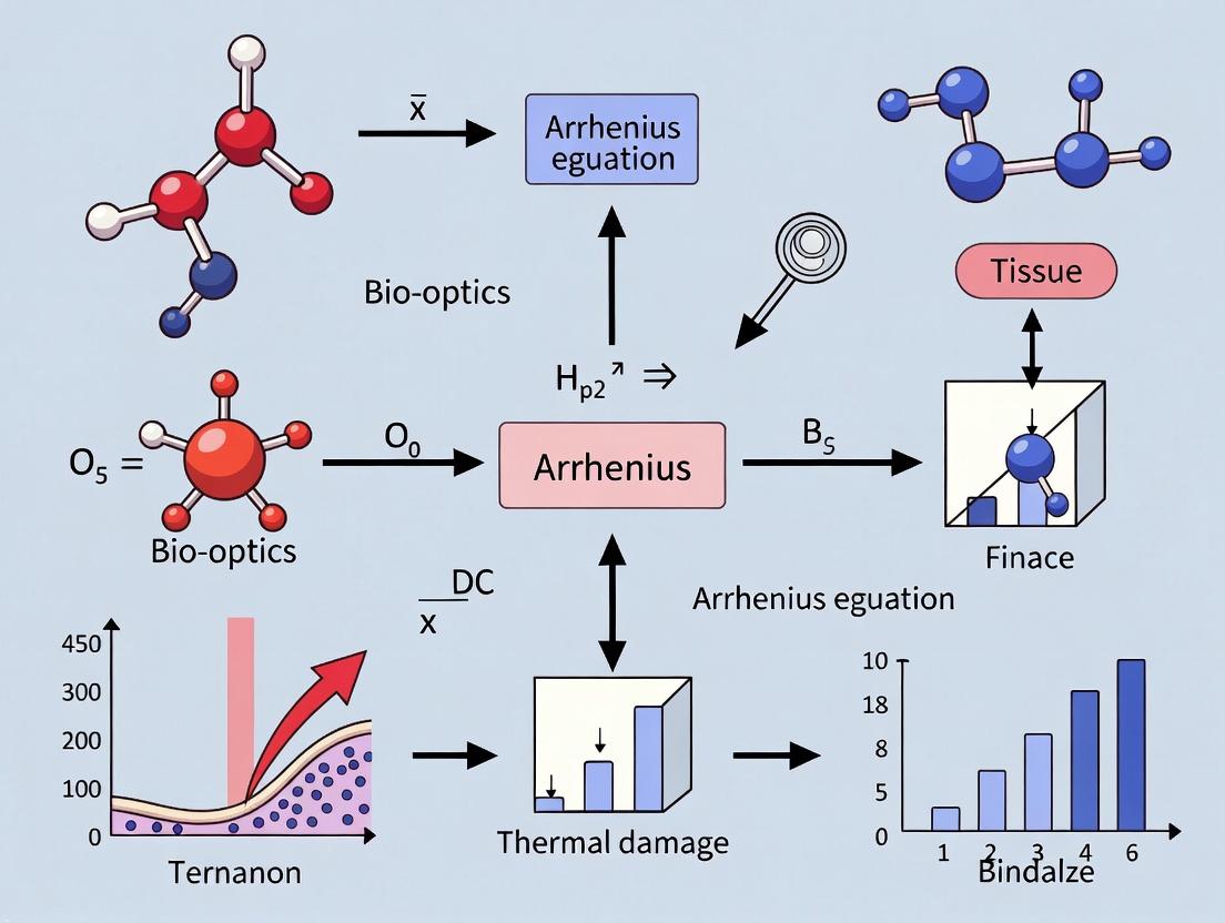
Title: Logical Flow from Thermal Input to Tissue Damage
The Scientist's Toolkit: Key Research Reagents & Materials
Table 2: Essential Materials for Kinetics & Thermal Damage Studies
| Item/Category | Function/Application in Research |
|---|---|
| Precision Thermocouples | Real-time, localized temperature measurement during thermal exposure (<0.1°C accuracy). |
| Isothermal Bath | Provides stable, uniform temperature environment for kinetic parameter determination. |
| Histological Stains (H&E, Picrosirius Red) | H&E for general cell morphology; Picrosirius Red with polarized light for specific, quantitative collagen damage. |
| Differential Scanning Calorimetry (DSC) | Directly measures heat flow during protein denaturation, providing thermodynamic data (ΔH, Tm). |
| Fluorescent Viability Probes (Propidium Iodide, Calcein-AM) | Distinguish live/dead cells post-thermal insult in in vitro models. |
| Recombinant Enzymes | Purified protein systems for studying fundamental kinetics of thermal inactivation without tissue complexity. |
| Mathematical Software (MATLAB, Python SciPy) | For numerical integration of the Arrhenius integral and fitting of experimental damage data. |
Title: Experimental Workflow for Kinetic Parameter Extraction
Advanced Context: Non-Isothermal Protocols & Validation
Clinical thermal therapies (laser interstitial thermal therapy, focused ultrasound) involve complex, dynamic temperature profiles ( T(t) ). Validation requires:
- Real-Time Thermometry: MR thermometry or distributed fiber optic sensors.
- Endpoint Histology: Precise spatial correlation of ( \Omega(x,y,z) ) with modeled damage zones.
- Multi-Process Models: Incorporating simultaneous damage processes (e.g., separate rate equations for collagen and cell viability) for higher fidelity in heterogeneous tissues.
The Arrhenius model remains a powerful, parsimonious framework, though contemporary research explores its limits at very high heating rates and addresses tissue-specific variations in ( A ) and ( E_a ).
The Arrhenius equation, ( k = A \exp(-E_a / RT) ), is a cornerstone of chemical kinetics, modeling the temperature dependence of reaction rates. In the context of biological tissue research, it provides a fundamental framework for quantifying thermal damage. This whitepaper deconstructs the formula's components within a thesis focused on modeling thermally induced protein denaturation, cell death, and drug efficacy degradation. Accurate application is critical for developing therapeutic hyperthermia protocols, optimizing drug storage, and ensuring biomedical device safety.
Theoretical Deconstruction
The equation's parameters are biophysically interpretable in a tissue context:
- k: The rate constant for the damage process (e.g., s⁻¹).
- A (Pre-exponential factor): Represents the frequency of collisions or attempts at the reaction transition state. In tissue, this correlates with the inherent frequency of molecular configurations leading to denaturation.
- E_a (Activation Energy): The energy barrier (J mol⁻¹ or kJ mol⁻¹) that must be overcome for the damage event (e.g., protein unfolding) to occur. It is the most critical tissue-specific parameter.
- R (Universal Gas Constant): 8.314 J mol⁻¹ K⁻¹.
- T (Absolute Temperature): In Kelvin.
The model assumes a single, rate-limiting step for damage accumulation, often characterized by first-order kinetics.
Application in Thermal Damage Modeling for Biological Tissues
Thermal damage (Ω) is modeled as a first-order rate process: [ \Omega(\tau) = \ln\left(\frac{C0}{C(\tau)}\right) = \int0^{\tau} A \exp\left(-\frac{Ea}{RT(t)}\right) dt ] where ( C0 ) and ( C(\tau) ) are the concentrations of native and damaged tissue constituents, and ( \tau ) is the total heating time. A damage threshold (often Ω = 1) signifies a visible, irreversible effect.
Key Tissue Damage Parameters
Quantitative parameters for various tissue components, derived from recent studies, are summarized below.
Table 1: Arrhenius Parameters for Biological Tissue Damage Models
| Tissue / Process | Activation Energy, E_a (kJ mol⁻¹) | Pre-exponential Factor, A (s⁻¹) | Reference & Notes |
|---|---|---|---|
| General Protein Denaturation | 300 - 700 | 1.0e45 - 1.0e110 | Classic range; highly variable |
| Collagen Denaturation (Type I) | 450 - 550 | 5.0e70 - 1.0e90 | Key for connective tissue shrinkage |
| Cell Viability Loss (HeLa cells) | 280 - 350 | 3.1e45 - 2.0e55 | In vitro hyperthermia models |
| Enzyme Inactivation (LDH) | 200 - 300 | 5.0e30 - 1.0e45 | Model for therapeutic protein decay |
| Skin Burn Injury (Dermal) | 425 - 625 | 1.8e66 - 3.1e98 | Basis for many clinical thermal safety standards |
Experimental Protocols for Parameter Determination
Protocol: Determining Ea and A via Isothermal Testing
Objective: To derive Arrhenius parameters by measuring the rate of damage at constant temperatures. Materials: See "The Scientist's Toolkit" (Section 6). Methodology:
- Sample Preparation: Prepare uniform samples of the target tissue or biomolecule in a controlled buffer solution.
- Isothermal Exposure: Expose replicates to a series of precise, constant temperatures (e.g., 45°C, 50°C, 55°C, 60°C) for varying durations in a calibrated thermal cycler or water bath.
- Damage Assay: Quantify remaining native state post-exposure. For collagen, use differential scanning calorimetry (DSC) to measure enthalpy loss. For cells, use a viability assay (e.g., live/dead staining with calcein-AM/propidium iodide). For proteins, use fluorescence spectroscopy or activity assays.
- Rate Constant Extraction: For each temperature (T), fit the damage progression data to a first-order kinetic model to extract the rate constant ( k_T ).
- Arrhenius Plot: Plot ( \ln(k_T) ) vs. ( 1/T ) (where T is in Kelvin). Perform linear regression.
- Parameter Calculation: The slope of the line is ( -E_a/R ). The y-intercept is ( \ln(A) ).
Protocol: Validation via Non-Isothermal (Linear Ramp) DSC
Objective: To independently validate Ea using Differential Scanning Calorimetry. Methodology:
- DSC Run: Subject a small tissue sample (~5 mg) to a constant temperature ramp (e.g., 1-5°C/min) in a DSC instrument.
- Peak Analysis: Identify the endothermic peak corresponding to protein denaturation. Record the peak temperature (T_p).
- Kinetics by ASTM E2070: Using the Borchardt and Daniels method, the software analyzes the shape of the heat flow curve to directly compute Ea and A, assuming nth-order kinetics. This provides a comparison to isothermal results.
Diagram 1: Isothermal Parameter Determination Workflow
Advanced Considerations & Pathway Integration
Thermal damage in cells is not a single event but a cascade. The classical Arrhenius model can be linked to pathways of programmed cell death triggered by heat stress.
Diagram 2: Thermal Stress to Cell Death Signaling Pathway
The Scientist's Toolkit
Table 2: Essential Research Reagents & Materials for Arrhenius-Based Tissue Studies
| Item | Function/Application |
|---|---|
| Differential Scanning Calorimeter (DSC) | Gold-standard for measuring thermal transitions (denaturation enthalpy, Tm) and extracting kinetic parameters via non-isothermal methods. |
| Real-Time PCR Thermocycler with High-Precision Blocks | Provides accurate isothermal exposure for small sample volumes, essential for generating k(T) data points. |
| Calcein-AM / Propidium Iodide (PI) Viability Kit | Fluorescent live/dead assay. Calcein-AM (live, green) and PI (dead, red) allow quantification of cell viability loss rate after thermal insult. |
| Collagenase Activity Assay Kit | Measures enzymatic activity decay of collagenases or other enzymes as a model for protein inactivation kinetics at elevated temperatures. |
| Thermocouple Data Logger (Microprobe) | For direct, real-time temperature measurement within tissue samples during heating protocols, critical for accurate T(t) history. |
| Phosphate-Buffered Saline (PBS) & Stabilizing Buffers | Maintain physiological pH and ionic strength during experiments to prevent non-thermal degradation artifacts. |
| Matlab or Python (SciPy) with Custom Scripts | For numerical integration of the damage integral and nonlinear regression fitting of Arrhenius parameters from experimental data. |
The Biological Meaning of Activation Energy (Ea) and Frequency Factor (A) for Proteins and Cells.
Within the framework of Arrhenius equation-based thermal damage modeling of biological tissue, the kinetic parameters of the Arrhenius equation—activation energy (Ea) and the pre-exponential frequency factor (A)—are traditionally treated as empirical constants for predicting macroscopic tissue coagulation. However, these parameters have profound and distinct biological meanings at the molecular and cellular levels. Ea quantifies the energy barrier for specific biomolecular events, such as protein denaturation or enzyme inactivation, while A relates to the frequency of attempts to overcome that barrier, reflecting the system's configurational entropy. This whitepaper delineates their biological interpretations, providing researchers and drug development professionals with a foundational guide for applying kinetic models beyond phenomenological damage prediction to mechanistic insights into cellular stress response and therapeutic targeting.
Fundamental Principles: The Arrhenius Equation in Biology
The Arrhenius equation describes the temperature dependence of reaction rates: [ k = A e^{-E_a/(RT)} ] Where:
- k is the rate constant (e.g., for protein denaturation, cell death rate).
- A is the frequency factor (s⁻¹), interpreted as the attempt frequency for the reaction.
- Ea is the activation energy (J mol⁻¹), the minimum energy required for the reaction to proceed.
- R is the universal gas constant (8.314 J mol⁻¹ K⁻¹).
- T is the absolute temperature (K).
In biological thermal damage models, the rate constant k is used to compute a damage integral (Ω), predicting the extent of irreversible change. The biological fidelity of such models hinges on accurate, context-specific Ea and A values.
Biological Interpretation of Activation Energy (Ea)
Ea is not an abstract fitting parameter but corresponds directly to the energy required to disrupt critical stabilizing forces within a biological macromolecule or cellular system.
Table 1: Biological Correlates of Activation Energy (Ea)
| Biological Process | Typical Ea Range (kJ mol⁻¹) | Molecular/Cellular Meaning | Key Stabilizing Forces Overcome |
|---|---|---|---|
| Protein Denaturation | 150 - 600 | Energy to disrupt native fold, leading to loss of function. | Hydrogen bonds, hydrophobic packing, van der Waals interactions. |
| Enzyme Inactivation | 200 - 500 | Energy to alter active site geometry or global structure. | Specific active site interactions, cofactor binding energy. |
| Membrane Permeabilization | 150 - 300 | Energy for lipid phase transition (gel to liquid-crystalline) or pore formation. | Lipid bilayer cohesion, lipid-protein interactions. |
| Cell Clonogenic Death | 300 - 700 | Energy required for cumulative, lethal damage to critical targets (proteins, DNA, membranes). | Integrated cellular homeostasis and repair systems. |
| Collagen Shrinkage | 400 - 600 | Energy to break heat-labile crosslinks and unravel the triple helix. | Intermolecular crosslinks, hydrogen bonding network. |
Biological Interpretation of the Frequency Factor (A)
A reflects the probability of the reacting system being in the correct configuration to attempt barrier crossing. A higher A indicates a larger number of accessible transitional states.
- For a Single Protein: A is related to the entropy change (ΔS‡) upon reaching the transition state: ( A = (k_B T / h) e^{ΔS‡/R} ). A large, flexible protein may have a higher A (more conformational pathways to denaturation) than a small, rigid one.
- For a Cell Population: A can reflect phenotypic heterogeneity. A population with diverse starting states may exhibit a higher effective A for death, as more cells are nearer to a lethal configuration.
Experimental Protocols for Determination
Determining Ea and A requires measuring the rate constant k at multiple temperatures.
Protocol 1: Differential Scanning Calorimetry (DSC) for Protein Denaturation
- Sample Prep: Prepare protein solution in appropriate buffer. Dialyze extensively against buffer. Degas sample to prevent artifacts.
- Instrumentation: Use a high-sensitivity DSC. Load reference (buffer) and sample cells.
- Temperature Ramp: Scan from low (e.g., 20°C) to high temperature (e.g., 110°C) at a constant rate (e.g., 1°C/min).
- Data Analysis: Obtain heat capacity (Cp) vs. temperature (T) curve. The peak corresponds to the denaturation transition.
- Kinetic Analysis (for irreversible scans): For each scan rate (β = dT/dt), record the peak denaturation temperature (Tm). Plot ln(β/Tm²) vs. 1/T_m. The slope yields Ea/R and the intercept relates to A.
Protocol 2: Clonogenic Survival Assay for Cellular Thermal Damage
- Cell Culture: Seed cells at low density in culture flasks.
- Heating: Immerse flasks in a precision-controlled water bath at a specific temperature (e.g., 44, 46, 48, 50°C) for varying durations (t).
- Post-treatment: Return cells to normal culture conditions for 7-14 days to form colonies.
- Analysis: Fix, stain colonies (>50 cells), and count. Determine surviving fraction (SF) for each time point.
- Kinetic Fitting: For each temperature, fit SF vs. time to a first-order kinetic model: SF = exp(-kt). Plot ln(k) vs. 1/T (Arrhenius plot). The slope is -Ea/R* and the intercept is ln(A).
Pathway and Conceptual Visualization
Thermal Damage Molecular Pathway
Experimental Determination of Ea and A
The Scientist's Toolkit: Research Reagent & Material Solutions
Table 2: Essential Materials for Kinetic Studies of Thermal Damage
| Item | Function / Rationale |
|---|---|
| Precision Recirculating Water Bath | Provides stable, uniform, and accurate (±0.1°C) heating for cell or protein samples in sealed vials or flasks. |
| High-Sensitivity Differential Scanning Calorimeter (DSC) | Directly measures heat flow associated with protein denaturation, enabling calculation of thermodynamic and kinetic parameters. |
| Clonogenic Assay Kit | Typically includes crystal violet or methylene blue stain for colony visualization and quantification post-thermal stress. |
| Recombinant, Lyophilized Protein | A well-characterized protein standard (e.g., lysozyme, RNase A) for calibrating DSC protocols and validating kinetic models. |
| Phase-Change Cells or Beads | Calibration standards with known melting points for temperature verification of heating devices. |
| Insulin-like Growth Factor-1 (IGF-1) | A critical reagent in cell stress studies; used in post-heat treatment media to assess/modulate survival pathways and repair kinetics. |
| Thermostable DNA Polymerase (e.g., Taq) | Serves as a positive control for protein thermal stability in functional assays, with known high Ea for inactivation. |
| Annexin V / Propidium Iodide Apoptosis Kit | Distinguishes modes of cell death (apoptosis vs. necrosis) induced by thermal stress, informing on the mechanism behind the observed k. |
In the context of modeling thermal damage to biological tissues, the Arrhenius equation provides a kinetic framework for describing the rate of damage accumulation from a single, isothermal reaction. However, real-world thermal therapies (e.g., radiofrequency ablation, laser surgery) involve complex, time-temperature histories. The Damage Integral (Ω) is the critical mathematical construct that extends the Arrhenius model from single reactions to cumulative damage, enabling the prediction of total tissue damage from variable thermal exposures. This whitepaper defines Ω, details its derivation, and provides protocols for its experimental validation, framing it as the cornerstone of modern thermal damage assessment in biophysical research and therapeutic device development.
Theoretical Foundation: From Arrhenius Rate to Cumulative Ω
The classical Arrhenius model for a single, irreversible damage process is:
k(T) = A exp(-E_a/(RT))
where k(T) is the damage rate coefficient (s⁻¹), A is the frequency factor (s⁻¹), E_a is the activation energy (J mol⁻¹), R is the universal gas constant (8.314 J mol⁻¹ K⁻¹), and T is the absolute temperature (K).
For a constant temperature exposure over time t, the fraction of native tissue transformed into damaged tissue (α) is: α = 1 - exp(-k(T)*t).
The Damage Integral (Ω) generalizes this for a time-varying temperature profile, T(τ):
Ω(t) = ∫_0^t A exp(-E_a/(R T(τ))) dτ
The total accumulated damage is then: α(t) = 1 - exp(-Ω(t)). Thus, Ω serves as a dimensionless measure of total effective "dose," where Ω = 1 corresponds to α ≈ 0.632, or 63.2% damage, analogous to one "reaction event" per initial target.
Title: Logical Progression from Arrhenius to Damage Integral
Quantitative Kinetic Parameters for Biological Tissues
The accuracy of Ω hinges on precise, tissue-specific Arrhenius parameters (A and E_a). These are determined via controlled isothermal experiments. The table below summarizes canonical values from literature.
Table 1: Arrhenius Parameters for Thermal Damage in Selected Tissues
| Tissue / Process | Frequency Factor (A) [s⁻¹] | Activation Energy (E_a) [J mol⁻¹] | Reference Temperature for k | Method |
|---|---|---|---|---|
| Myocardial Tissue (Coagulation) | 3.1 x 10⁹⁸ | 6.28 x 10⁵ | k(60°C) ≈ 0.1 | Histology, Enzyme denaturation |
| Collagen Denaturation (Type I) | 1.606 x 10⁴⁰ | 2.577 x 10⁵ | k(60°C) ≈ 0.02 | Birefringence loss, DSC |
| Skin Epidermis (Necrosis) | 3.1 x 10⁹⁹ | 6.27 x 10⁵ | k(54°C) ≈ 0.001 | Vital stain (Propidium Iodide) |
| Liver Parenchyma (Ablation) | 7.39 x 10⁴² | 2.80 x 10⁵ | k(70°C) ≈ 1.0 | NADH diaphorase assay |
| Protein (Albumin) Denaturation | 7.95 x 10⁴⁴ | 3.06 x 10⁵ | k(65°C) ≈ 0.5 | Fluorescence (Sypro Orange) |
Note: Values exhibit wide range; validation for specific experimental context is critical. DSC = Differential Scanning Calorimetry.
Experimental Protocol: Determining Arrhenius Parameters
This protocol details the core experiment required to define Ω for a new tissue or damage endpoint.
Title: Isothermal Kinetic Analysis for A and E_a Determination.
Objective: To measure the rate of damage accumulation at multiple constant temperatures to calculate the Arrhenius parameters.
Materials: See "The Scientist's Toolkit" below.
Procedure:
- Tissue Preparation: Prepare uniform samples (e.g., 3mm thick slices, 5mm diameter) in phosphate-buffered saline (PBS) to prevent desiccation.
- Isothermal Bath Exposure: Place samples in a precisely controlled thermal bath (±0.2°C) set to target temperatures (e.g., 50, 55, 60, 65, 70°C).
- Time-Series Sampling: Remove replicate samples (n≥5) at logarithmically spaced time points (e.g., 1, 3, 10, 30, 100, 300 s).
- Immediate Cooling: Quench samples in ice-cold PBS to halt thermal processes.
- Damage Quantification: Assay each sample using a calibrated damage endpoint metric (see Step 6).
- Data Analysis:
- For each temperature
T, plot the damage metric vs. time. Fit to a first-order kinetic model:α(t) = 1 - exp(-k * t)to extract the rate constantk(T). - Perform an Arrhenius Plot:
ln(k) vs. 1/(RT). The slope is-E_a, and the y-intercept isln(A).
- For each temperature
Title: Experimental Workflow for Arrhenius Parameter Determination
The Scientist's Toolkit: Key Research Reagent Solutions
Table 2: Essential Materials for Thermal Damage Kinetics Experiments
| Item | Function / Rationale |
|---|---|
| Precision Thermostatic Bath | Provides stable, uniform isothermal environment (±0.1°C) for kinetic studies. |
| NADH Diaphorase Assay Kit | Gold-standard histochemical stain for quantifying viable vs. non-viable cells in liver/heart; measures enzyme activity loss. |
| Propidium Iodide (PI) / Fluorescein Diacetate (FDA) | Vital stains for cell viability. PI enters dead cells (red), FDA metabolized by live cells (green). |
| Differential Scanning Calorimeter (DSC) | Directly measures heat flow associated with protein denaturation, providing E_a and ΔH. |
| Sypro Orange Protein Gel Stain | Fluorescent dye that binds hydrophobic patches exposed during protein denaturation; usable in real-time PCR machines for kinetics. |
| Polarized Light Microscope | Quantifies birefringence loss in collagen as a direct measure of structural denaturation. |
| Custom MATLAB/Python Scripts | For numerical integration of Ω from complex T(τ) data and nonlinear curve fitting of kinetic models. |
Protocol: Validating Ω with a Time-Varying Thermal Protocol
Once A and E_a are known, Ω's predictive power must be validated against a non-isothermal exposure.
Title: Predictive Validation of the Damage Integral.
Objective: To compare predicted damage (calculated from T(τ) and Ω) to experimentally measured damage following a controlled, time-varying heat exposure.
Procedure:
- Design Thermal Protocol: Define a heating profile with a known time-temperature path,
T(τ)(e.g., linear ramp, simulated ablation profile). - Instrumentation: Use a thermocouple or infrared thermal camera to record the actual
T(τ)at the sample site with high temporal resolution. - Apply Protocol: Subject tissue samples (n≥10) to the designed heating profile.
- Compute Predicted Ω: Numerically integrate the recorded
T(τ)data using the equation forΩ(t)and the previously determinedAandE_a. Calculate predictedα_pred = 1 - exp(-Ω). - Measure Actual Damage: Quantify the actual damage fraction
α_measin the samples using the same assay from Protocol 1. - Validation: Perform a linear regression between
α_predandα_meas. A slope of 1 and high R² value validates the Ω model for that tissue and protocol.
Title: Workflow for Validating the Damage Integral Model
Advanced Application: Ω in Therapeutic Dose Planning
In clinical thermal ablation, Ω is used to define the "lethal dose" boundary (typically Ω ≥ 1). Treatment planning software integrates real-time T(τ) from imaging to compute and display a cumulative Ω field, predicting the final ablation zone.
Table 3: Typical Damage Integral Thresholds for Clinical Endpoints
| Clinical Endpoint | Damage Integral (Ω) Threshold | Corresponding α |
|---|---|---|
| Immediate Cell Necrosis | 0.53 | 0.41 |
| Complete Coagulation (Ablation) | 1.0 | 0.63 |
| Microvascular Damage | 4.6 | 0.99 |
| Collagen Shrinkage | ≥ 30* | ~1.00 |
*Note: Collagen shrinkage involves very high apparent Ω, suggesting a multi-step process not fully captured by a single Arrhenius model.
The Damage Integral (Ω) is the essential mathematical bridge linking the fundamental Arrhenius kinetics of a single reaction to the cumulative, irreversible damage observed in complex biological systems under thermal stress. Its rigorous definition, grounded in experimentally derived tissue-specific parameters, transforms thermal therapy from an empirical art into a predictive science. For researchers and drug developers, Ω provides a quantitative framework for optimizing therapeutic protocols, evaluating device safety, and modeling tissue response, ultimately enabling more precise and effective thermal interventions.
The development of precise thermal surgical tools, such as radiofrequency and ultrasonic devices, is fundamentally grounded in the quantitative modeling of thermal damage in biological tissue. This modeling paradigm originates not in medicine, but in food science. The seminal work of food chemists and engineers in the 19th and 20th centuries to predict nutrient degradation and bacterial spore inactivation during thermal processing provided the kinetic framework—specifically, the Arrhenius equation—that was later adapted to model collagen denaturation and cell necrosis. This whitepaper explores this historical continuum, detailing the core kinetic models and their experimental validation, framed within a thesis on Arrhenius-based thermal damage modeling for modern surgical tool development.
Theoretical Foundation: The Arrhenius Equation and Damage Integration
The core model describes the rate of damage accumulation (k) as a function of absolute temperature (T):
k = A * exp(-Ea/(R*T))
where A is the frequency factor (s⁻¹), Ea is the activation energy (J/mol), and R is the universal gas constant (8.314 J/mol·K).
For a time-varying temperature history T(t), the total damage (Ω) is expressed as the integral of the rate:
Ω = ∫₀ᵗ A * exp(-Ea/(R*T(τ))) dτ
A value of Ω = 1.0 typically represents a threshold for irreversible damage (e.g, 63% protein denaturation). This formulation is directly analogous to the "C-value" or "sterilizing value" (F₀) used in food canning.
Table 1: Kinetic Parameters for Thermal Damage Across Fields
| Material/System | A (s⁻¹) | Ea (kJ/mol) | Ω Threshold | Reference Context |
|---|---|---|---|---|
| C. botulinum Spore Inactivation | 1.0 × 10³⁶ | 283.0 | F₀=3 min (Ω=1) | Food Sterilization (Low-Acid Canned Foods) |
| Vitamin C Degradation | 1.0 × 10¹⁹ | 125.0 | 50% Loss (Ω=0.69) | Food Nutrient Retention |
| Collagen Denaturation (Bovine Tendon) | 1.0 × 10⁸⁴ | 550.0 | Ω=1.0 (Shrinkage) | Surgical Tool Target (Historic) |
| Myocardial Cell Necrosis (Porcine) | 2.8 × 10⁶⁴ | 430.0 | Ω=1.0 (Irreversible) | Radiofrequency Ablation |
| Pancreatic Tissue Ablation (Ex Vivo) | 5.6 × 10⁶² | 415.0 | Ω=4.6 (Complete Lesion) | High-Intensity Focused Ultrasound (HIFU) |
Experimental Protocols for Parameter Determination
Determining kinetic parameters (A, Ea) for a specific tissue requires controlled isothermal experiments.
Protocol 3.1: Isothermal Tube Heating for Kinetic Analysis
- Sample Preparation: Prepare uniform tissue samples (e.g., 1mm thick slices, 5mm diameter) in phosphate-buffered saline.
- Isothermal Bath: Use a precision-controlled mineral oil or water bath stabilized at target temperatures (e.g., 50°C, 55°C, 60°C, 65°C).
- Heat Exposure: Seal samples in thin-walled glass tubes. Immerse tubes in the bath for varying, precisely timed durations (seconds to minutes).
- Damage Assessment: Quantify damage post-exposure. Methods include:
- Histology: H&E staining for coagulation necrosis; measure fraction of damaged cells via image analysis.
- Birefringence Loss: Use polarized light microscopy on collagenous tissues; loss of birefringence correlates with denaturation.
- Electrical Impedance: Measure drop in tissue impedance, which increases with protein coagulation.
- Data Fitting: For each temperature, plot log(1 - Fraction Undamaged) vs. time. The slope is the rate constant k. Plot ln(k) vs. 1/T (Arrhenius plot); the slope is -Ea/R and the y-intercept is ln(A).
Protocol 3.2: Calorimetric Validation (Differential Scanning Calorimetry - DSC)
- Equipment: Use a power-compensated DSC with high-pressure capsules.
- Method: Load a small tissue sample (5-10 mg) and seal. Run a controlled temperature ramp (e.g., 1-10°C/min).
- Analysis: The endothermic peak corresponds to protein denaturation. The peak temperature (Tₚ) and shape provide validation for the energy barrier (Ea) derived from kinetic experiments.
The Scientist's Toolkit: Key Research Reagent Solutions
Table 2: Essential Materials for Thermal Damage Kinetics Research
| Item | Function & Rationale |
|---|---|
| Ex Vivo Tissue Model (e.g., Porcine Liver/Myocardium) | Provides a reproducible, ethically-sourced biological substrate with properties similar to human tissue for initial tool validation. |
| Precision Temperature-Controlled Bath | Enables accurate isothermal exposure for fundamental kinetic parameter determination (A, Ea). |
| Thermocouple Microprobes (Type T or K, <0.5mm) | For direct spatial and temporal temperature measurement during energy delivery; critical for validating predictive models. |
| Radiofrequency Ablation Generator & Needle Electrode | Standardized energy delivery system for creating controlled thermal lesions; allows correlation of power/time/damage volume. |
| High-Intensity Focused Ultrasound (HIFU) Transducer | Non-contact energy delivery system for studying thermal damage in deep tissues without conductive interference. |
| Triphenyltetrazolium Chloride (TTC) Stain | Vital stain for macroscopic visualization of necrotic tissue (unstained) vs. viable tissue (red) in immediate post-ablation analysis. |
| H&E Staining Kit | Gold standard for histological assessment of coagulation necrosis, cell morphology, and collagen structure post-thermal exposure. |
| MATLAB/Python with PDE Toolbox/NumPy | Software for implementing finite-element models that solve the Bioheat Equation coupled with the Arrhenius damage integral. |
Signaling Pathways in Thermal Damage Response
Thermal insult activates complex cellular stress response pathways that determine survival or death.
Cellular Fate Post Thermal Insult
Modern Surgical Workflow Integration
The historical kinetic models are now embedded in treatment planning software for thermal surgery.
Computational Planning for Thermal Surgery
The journey from predicting spoilage in canned goods to planning tumor ablations exemplifies interdisciplinary translation. The Arrhenius equation remains the universal kinetic bridge. Future development of surgical tools—particularly in pulsed regimes and combined electrothermal therapies—relies on refining these models with tissue-specific parameters and real-time feedback, a direct legacy of the rigorous quantification pioneered in food science.
Key Assumptions and Theoretical Limitations of the Classic Model
The classic Arrhenius damage integral model is a cornerstone for predicting thermal damage kinetics in biological tissues. It serves as the principal theoretical framework for a wide range of applications, from laser surgery and radiofrequency ablation to thermal therapy planning. Within broader thesis research, this model's assumptions directly impact the fidelity of predicting protein denaturation, cell death, and tissue necrosis. This whitepaper critically examines the foundational assumptions and inherent limitations of this classic model, providing a technical guide for researchers aiming to refine thermal damage predictions in drug development and therapeutic interventions.
Core Assumptions of the Classic Arrhenius Model
The classic model is built upon several key assumptions that simplify complex biophysical processes.
Formal Assumptions
- First-Order Kinetics: Damage accumulation is assumed to follow an irreversible, first-order rate process. The rate of transformation of native tissue to damaged tissue is directly proportional to the concentration of native state.
- Single-Step Process: The damage mechanism is represented as a single, irreversible step from a native state (N) to a damaged state (D), ignoring intermediate states.
- Arrhenius Dependence: The rate constant k for the damage process follows the Arrhenius law, dependent solely on absolute temperature T: k(T) = A exp(-Eₐ/RT) where A is the frequency factor (s⁻¹), Eₐ is the activation energy (J mol⁻¹), and R is the universal gas constant.
- Damage Integral (Ω) Independence: The total damage integral Ω is calculated by integrating the rate constant over time, assuming the kinetics are independent of prior damage history (i.e., no rate dependence on the current state of damage). Ω(τ) = ∫₀ᵗ A exp(-Eₐ/RT(t)) dt
- Spatial Homogeneity: The pre-exponential factor A and activation energy Eₐ are treated as constants for a given tissue type, assuming tissue is homogeneous and isotropic.
- Neglect of Thermo-Mechanical Effects: The model disregards mechanical stress induced by thermal expansion and phase changes (e.g., water vaporization) that can accelerate damage.
Quantitative Parameter Assumptions
Table 1: Commonly Used Classic Arrhenius Parameters for Selected Tissues
| Tissue / Protein | Activation Energy, Eₐ (J mol⁻¹) | Frequency Factor, A (s⁻¹) | Reference Temperature for Validation | Typical Application |
|---|---|---|---|---|
| Skin Collagen | ~6.0 x 10⁵ | ~1.0 x 10⁸⁴ | 50-70°C | Laser skin resurfacing |
| Myocardium | ~5.8 x 10⁵ | ~1.0 x 10⁸³ | 50-80°C | Cardiac ablation |
| Liver Parenchyma | ~6.7 x 10⁵ | ~7.4 x 10¹⁰⁷ | 60-100°C | Tumor ablation |
| Egg Albumin | ~5.5 x 10⁵ | ~3.1 x 10⁷¹ | 55-90°C | In vitro benchmark |
Detailed Theoretical Limitations and Modern Critiques
Kinetic Limitations
The assumption of first-order, single-step kinetics is a significant simplification. Real thermal damage involves multiple parallel and sequential reactions (e.g., protein unfolding, aggregation, membrane disruption). Intermediate states can have different activation energies, making the effective Eₐ temperature-dependent.
Experimental Protocol for Validating Kinetics: Isothermal Calorimetry & Spectroscopy
- Sample Preparation: Prepare homogeneous samples of purified tissue protein (e.g., collagen) or cell suspensions in a physiologically relevant buffer.
- Isothermal Hold: Expose samples to precise, constant temperatures (e.g., 55, 60, 65°C) in a differential scanning calorimeter (DSC) or a temperature-controlled spectrophotometer.
- Real-Time Measurement:
- In DSC, measure the heat flow associated with protein denaturation over time.
- In spectroscopy, measure changes in optical properties (turbidity at 400 nm for aggregation, fluorescence for conformational change) over time.
- Rate Constant Calculation: For each temperature, fit the time-dependent damage progress curve to various kinetic models (e.g., first-order, nth-order, multi-step) to extract an apparent rate constant k(T).
- Arrhenius Plot: Plot ln(k) versus 1/T to check for linearity. Non-linearity indicates a breakdown of the single-activation-energy assumption.
Spatial and Temporal Limitations
The homogeneity assumption fails at microscale and macroscale. Tissue is hierarchically structured, with cells, extracellular matrix, and vasculature each having distinct thermal and kinetic properties. Blood perfusion causes significant convective cooling, creating steep thermal gradients not accounted for in the basic integral.
Experimental Protocol for Spatial Validation: Multi-Photon Microscopy
- Labeling: Label live tissue explants with fluorescent viability probes (e.g., Calcein-AM for live cells, Propidium Iodide for dead cells) and a structural label (e.g., second harmonic generation for collagen).
- Controlled Heating: Use a micro-heating stage or focused laser to apply a defined thermal dose to a specific region of interest.
- Time-Lapse Imaging: Use multi-photon microscopy to acquire 3D image stacks of the heated region over time (pre-heat, during heat, and post-heat recovery).
- Damage Mapping: Correlate the spatiotemporal evolution of fluorescence signals (indicating damage) with finite-element model predictions of temperature and damage integral (Ω) fields.
- Analysis: Quantify the discrepancy between the predicted Ω=1 contour (theoretical damage boundary) and the observed boundary of cell death or protein denaturation.
Visualization of Concepts and Workflows
Diagram Title: Classic vs. Complex Thermal Damage Kinetics (76 chars)
Diagram Title: Workflow for Validating Arrhenius Kinetics Assumption (76 chars)
The Scientist's Toolkit: Essential Research Reagents & Materials
Table 2: Key Research Reagent Solutions for Thermal Damage Studies
| Item / Reagent | Function in Experiment | Key Consideration for Model Validation |
|---|---|---|
| Recombinant Human Collagen I | Standardized protein substrate for foundational kinetic studies, free from tissue variability. | Allows isolation of pure protein denaturation kinetics without confounding cellular effects. |
| 3D Bioprinted Tissue Constructs | Provides a more physiologically relevant, yet controlled, heterogeneous tissue model. | Enables testing of spatial homogeneity assumption with defined cell-matrix architecture. |
| FLIR/IR Thermal Camera | Provides high-resolution, real-time 2D surface temperature mapping during heating. | Critical for accurate input T(t) for damage integral, especially at boundaries. |
| Fluorescent Viability Kit (Live/Dead Assay) | Dual-color fluorescence (Calcein-AM/PI) for immediate post-heat viability assessment. | Provides the experimental endpoint (Ω threshold) to correlate with calculated damage integral. |
| Thermally Responsive Nanoparticles (e.g., gold nanorods) | Act as localized nanoscale heat sources under NIR laser for micro-scale thermal challenge. | Used to probe damage kinetics at sub-cellular level, challenging homogeneity. |
| Differential Scanning Calorimeter (DSC) | Precisely measures heat flow associated with protein denaturation under controlled temperature ramps. | Gold standard for determining thermodynamic parameters (ΔH, Tm) and validating kinetic models. |
| MatLab/Python with PDE Toolkits | Software for implementing finite-element models of bioheat transfer coupled with damage integrals. | Essential for moving beyond the classic model to incorporate spatial effects (Pennes' Bioheat Equation). |
Implementing the Model: A Step-by-Step Guide to Calculating Thermal Damage
In the context of Arrhenius equation-based thermal damage modeling of biological tissue, the temperature-time history, T(t), is the foundational input. The Arrhenius damage integral, Ω, is expressed as:
Ω(τ) = ∫₀ᵗ A exp(-Eₐ/RT(t)) dt
where A is the frequency factor (s⁻¹), Eₐ is the activation energy (J mol⁻¹), R is the universal gas constant (8.314 J mol⁻¹ K⁻¹), and T(t) is the absolute temperature (K) as a function of time. The accuracy of the predicted damage fraction, often modeled as FD = 1 - exp(-Ω), is directly contingent upon the fidelity of the T(t) measurement. This guide details the methodologies, technologies, and protocols for acquiring this critical data.
Core Measurement Technologies & Comparative Analysis
The selection of a temperature measurement technique depends on required spatial and temporal resolution, invasiveness, tissue type, and the thermal therapy modality (e.g., radiofrequency ablation, laser irradiation, high-intensity focused ultrasound).
Decision Tree for T(t) Measurement Method Selection
Table 1: Quantitative Comparison of Primary Temperature Measurement Techniques
| Technique | Spatial Resolution | Temporal Resolution | Accuracy | Invasiveness | Key Limitation |
|---|---|---|---|---|---|
| Thermocouple (Type T) | ~0.5-1 mm | ~10-100 ms | ±0.5°C | Invasive (Penetrating) | Conduction artifacts, punctures tissue |
| Fiber Bragg Grating (FBG) | ~1-5 mm | ~1-10 ms | ±0.1°C | Minimally Invasive | Fragility, cost, limited multiplexing |
| Infrared Thermography | ~10-50 µm (lateral) | ~1-100 ms | ±1-2°C (surface) | Non-invasive | Surface measurement only |
| MR Thermometry (Proton Resonance Freq.) | ~1-3 mm | 1-5 seconds | ±1°C | Non-invasive | Slow, expensive, motion-sensitive |
| Ultrasound (Time-Shift of Echo) | ~1-2 mm | ~10-100 ms | ±1°C (in-vitro) | Non-invasive | Under development, tissue heterogeneity effects |
Detailed Experimental Protocols
Protocol: Ex-Vivo Tissue T(t) Measurement Using Multipoint Thermocouples During Laser Irradiation
Aim: To capture high-temporal-resolution thermal gradients in ex-vivo porcine liver during 1064 nm Nd:YAG laser exposure.
The Scientist's Toolkit:
| Item | Function |
|---|---|
| Nd:YAG Laser (1064 nm) | Provides controlled radiative heating source. |
| Type T (Copper-Constantan) Thermocouples | High accuracy, low noise for 0-350°C range. |
| Data Acquisition System (DAQ) | High-speed (>1 kHz) multichannel logger for simultaneous T(t) capture. |
| Thermal Gel (e.g., ultrasound gel) | Ensures acoustic/thermal coupling, reduces air gaps. |
| Polyimide Tape/Sheath | Electrically insulates thermocouples in conductive media. |
| Ex-Vivo Tissue Chamber | Maintains tissue hydration (e.g., with PBS-soaked gauze). |
Methodology:
- Tissue Preparation: Prepare uniform 20 mm x 20 mm x 10 mm blocks of fresh ex-vivo porcine liver. Place in chamber, maintaining 37°C with saline mist.
- Sensor Placement: Insert four bare-wire Type T thermocouple needles (75 µm diameter) at depths of 1 mm, 3 mm, 5 mm, and 7 mm directly below the planned laser irradiation spot. Secure with micro-positioners. Connect to DAQ.
- Laser Calibration: Measure laser output power with a calibrated power meter. Set laser to continuous wave (CW) mode at 3 W.
- Data Acquisition: Initiate DAQ recording at 2 kHz. Irradiate tissue surface for 30 seconds. Continue recording for 60 seconds post-irradiation to capture cooling phase.
- Data Processing: Apply moving average filter (50 ms window) to raw voltage data. Convert to temperature using NIST-standard polynomials. Align all T(t) traces to irradiation start time (t=0).
Protocol: In-Vivo MR Thermometry (MRT) for Focused Ultrasound (FUS)
Aim: To non-invasively map 3D T(t) during MR-guided focused ultrasound (MRgFUS) ablation of uterine fibroids.
Methodology:
- Subject/Phantom Positioning: Position patient or tissue-mimicking phantom in 3T MRI scanner integrated with FUS transducer.
- Sequence Selection: Use a 2D multi-slice gradient echo sequence sensitive to proton resonance frequency (PRF) shift. Typical parameters: TR/TE = 50/20 ms, flip angle = 30°, matrix = 128x128, FOV = 280 mm, slice thickness = 5 mm.
- Baseline Scan: Acquire 5-10 baseline phase images before heating to establish reference phase, φ₀.
- Heating & Acquisition: Initiate FUS sonication at therapeutic acoustic power. Simultaneously, acquire repeated phase images throughout the heating and cooling cycle.
- Temperature Calculation: Compute temperature change ΔT for each voxel and time point using: ΔT(t) = (φ(t) - φ₀) / (γ α B₀ TE) where γ is gyromagnetic ratio, α is PRF shift coefficient (~ -0.01 ppm/°C for aqueous tissue), B₀ is static field strength.
- Validation: Correlate predicted thermal dose (CEM43) from MRT-derived T(t) with post-procedure contrast-enhanced MRI ablation zone.
Data Processing & Integration into Arrhenius Models
Raw T(t) data requires conditioning before integration.
Table 2: Common Data Processing Steps for T(t)
| Step | Purpose | Typical Method |
|---|---|---|
| Noise Reduction | Remove electrical/thermal noise | Low-pass Butterworth filter (cutoff ~10 Hz) |
| Sensor Lag Correction | Compensate for finite thermal mass of sensor | Inverse filtering using sensor's known time constant |
| Spatial Interpolation | Create continuous T(x,y,z,t) field from discrete points | Kriging or linear interpolation on a 3D grid |
| Temporal Integration | Compute Arrhenius integral Ω(t) | Numerical integration (e.g., trapezoidal rule) with high frequency (≥100 Hz) data |
Workflow from T(t) Measurement to Damage Prediction
Emerging Technologies & Future Directions
- Microwave Radiometry: Passive sensing of endogenous microwave emission for deep-temperature profiling.
- Nanoparticle-Assisted Sensing: Using temperature-sensitive fluorescent nanoparticles (e.g., nanodiamonds with NV centers) or magnetic nanoparticles for localized sensing.
- High-Speed IR for Ex-Vivo Studies: Enables capture of rapid thermal spread along tissue microvasculature.
Precise acquisition of tissue temperature-time histories is the critical, non-negotiable first step in validating Arrhenius models for thermal damage. The choice between high-fidelity invasive probes and non-invasive mapping techniques represents a fundamental trade-off, dictated by the experimental or clinical context. The protocols and analyses presented here provide a framework for generating the high-quality T(t) data essential for advancing the predictive accuracy of biothermal models in therapeutic and safety applications.
Within the broader thesis on Arrhenius equation-based thermal damage modeling of biological tissue, the accurate determination of kinetic parameters—the frequency factor (A) and the activation energy (Ea)—is a critical step. These parameters govern the rate of damage accumulation (k) according to the Arrhenius equation: k = A exp(-Ea/RT), where R is the universal gas constant and T is absolute temperature. This guide details the methodologies for sourcing and experimentally determining these tissue-specific parameters, which are essential for predictive models in therapeutic hyperthermia, thermal ablation, and safety assessment of energy-based medical devices.
Data Acquisition Strategies
Researchers can either source parameters from published literature or determine them de novo via controlled experiments.
Sourcing from Literature
A systematic review of peer-reviewed literature is the first approach. Key databases include PubMed, IEEE Xplore, and Web of Science. Search terms should combine "Arrhenius parameters," "thermal damage," "activation energy," with specific tissue names (e.g., "porcine liver," "bovine myocardium").
Table 1: Sourced Arrhenius Parameters for Selected Tissues
| Tissue Type | A (s⁻¹) | Ea (J/mol) | Experimental Basis (Reference) | Temp. Range (°C) |
|---|---|---|---|---|
| Porcine Liver | 7.39e⁶⁶ | 4.30e⁵ | Isothermal Tensionetry [1] | 50-90 |
| Canine Prostate | 1.80e⁵¹ | 3.27e⁵ | Histological Analysis [2] | 45-90 |
| Rabbit Cornea | 1.05e⁴⁵ | 2.99e⁵ | Light Scattering [3] | 55-85 |
| Bovine Myocardium | 4.32e⁶⁴ | 4.14e⁵ | Electrical Conductivity [4] | 45-90 |
| Human Dermis (estimated) | 5.60e⁶³ | 4.08e⁵ | Meta-analysis [5] | 50-90 |
Note: Values exhibit significant variance due to differences in experimental methodology, endpoint definition, and tissue state.
Experimental Determination
When existing data is insufficient or tissue-specific parameters are required, direct experimentation is necessary. The core principle involves subjecting tissue samples to controlled thermal exposures and quantifying the damage.
Core Experimental Protocols
Protocol: Isothermal Calorimetry with Protein Denaturation Assessment
This protocol determines A and Ea for intracellular protein denaturation.
Materials: Fresh ex-vivo tissue samples, phosphate-buffered saline (PBS), differential scanning calorimeter (DSC), microtome, hermetic aluminum pans.
Procedure:
- Sample Preparation: Slice tissue into thin sections (1-2 mg) using a microtome. Rinse in PBS to remove residual blood. Precisely weigh and seal in DSC pans.
- Isothermal Runs: Using a high-sensitivity DSC, heat samples rapidly to a target isothermal temperature (e.g., 45, 50, 55, 60°C). Hold for a predetermined time (t).
- Damage Quantification: The heat flow signal decays over time as proteins denature. The fraction of undenatured protein (α) at time t is proportional to the residual heat of denaturation measured in a subsequent temperature ramp.
- Kinetic Analysis: For a first-order process, ln(1/α) = kt. Calculate rate constant k at each temperature.
- Arrhenius Plot: Plot ln(k) against 1/RT. The y-intercept is ln(A) and the slope is -Ea.
Protocol: Histological Endpoint Analysis (H&E Staining)
This protocol uses a structural endpoint (e.g., collagen hyalinization) visible under light microscopy.
Materials: Tissue bath with precise temperature control, biopsy punches, formalin fixation vials, microtome, hematoxylin & eosin (H&E) stains, light microscope, image analysis software.
Procedure:
- Thermal Exposure: Place uniform tissue samples in a saline bath held at constant target temperatures (e.g., 60, 65, 70, 75°C) for varying exposure times.
- Fixation: Immediately post-exposure, transfer samples to formalin for fixation.
- Sectioning and Staining: Paraffin-embed, section, and H&E-stain the samples.
- Damage Scoring: A pathologist scores each sample for a binary damage state (e.g., "damaged" vs. "native"). Alternatively, use image analysis to quantify eosinophilic shift.
- Probit Analysis: For each temperature, plot exposure time vs. % samples damaged. Determine the time for 50% damage (τ). The rate constant k = 1/τ.
- Arrhenius Plot: Construct the plot as in Protocol 3.1 to extract A and Ea.
The Scientist's Toolkit
Table 2: Essential Research Reagent Solutions & Materials
| Item | Function in Parameter Determination |
|---|---|
| Differential Scanning Calorimeter (DSC) | Precisely measures heat flow associated with protein denaturation in milligram tissue samples under controlled temperature programs. |
| Isothermal Tissue Bath | Provides a stable, uniform temperature environment for incubating larger tissue samples prior to histological analysis. |
| Microtome / Vibratome | Produces thin, consistent tissue sections for calorimetry or for slide preparation post-thermal exposure. |
| Neutral Buffered Formalin (10%) | Fixes tissue architecture, halting post-mortem and post-thermal degradation to preserve the damage state for histology. |
| Hematoxylin and Eosin (H&E) Stain | Standard histological stain that differentiates cell nuclei (blue) and cytoplasm/collagen (pink), revealing thermal damage like hypereosinophilia. |
| ImageJ / Fiji with Custom Macros | Open-source software for automated analysis of histological images to quantify area fraction of damaged tissue. |
| High-Precision Thermocouples (<0.1°C accuracy) | Calibrated sensors for direct, real-time temperature measurement within tissue samples during exposure, critical for model validation. |
Visualization of Methodologies
Title: Workflow for Sourcing Arrhenius Parameters
Title: Data Flow for Experimental Parameter Determination
Within the broader thesis on Arrhenius-based thermal damage modeling of biological tissue, the computation of the damage integral, Ω, represents the critical quantitative step. This metric, derived from the Arrhenius rate process model, serves as the primary predictor of the extent of irreversible thermal damage to cellular and tissue structures. The accurate numerical evaluation of Ω from time-temperature data is essential for validating models against experimental histology, optimizing thermal therapies, and establishing safety thresholds in diagnostic and surgical applications.
Theoretical Foundation
The fundamental Arrhenius damage model expresses the rate of damage accumulation, k(T), as: k(T) = A exp( -Eₐ / (R T) ) where A is the frequency factor (s⁻¹), Eₐ is the activation energy (J mol⁻¹), R is the universal gas constant (8.314 J mol⁻¹ K⁻¹), and T is the absolute temperature (K).
The total damage integral, Ω, over a time period τ is: Ω(τ) = ∫₀^τ A exp( -Eₐ / (R T(t)) ) dt
A value of Ω = 1 typically corresponds to approximately 63% probability of damage for a homogeneous population. This is often used as a threshold for observable necrosis.
Numerical Integration Methods
Direct analytical integration of Ω is rarely possible due to the complex, non-linear nature of T(t) from experiments or simulations. Several numerical methods are employed, each with trade-offs in accuracy, stability, and computational cost.
Key Numerical Methods
Table 1: Comparison of Numerical Integration Methods for Ω
| Method | Principle | Accuracy | Stability | Computational Cost | Best Use Case |
|---|---|---|---|---|---|
| Trapezoidal Rule | Approximates area under curve as series of trapezoids. | Moderate (O(h²)) | High | Low | Equally-spaced, smooth T(t) data. |
| Simpson's 1/3 Rule | Uses quadratic polynomials for approximation. | High (O(h⁴)) | Moderate | Low | Smooth data with even number of intervals. |
| Adaptive Quadrature | Recursively refines intervals until error tolerance is met. | Very High | High | Variable (Higher) | Data with rapid temperature transients. |
| Runge-Kutta (RK4) | Solves the differential form dΩ/dt = k(T). | High (O(h⁴)) | High | Moderate | When integrating concurrently with thermal solver. |
Tissue-Specific Kinetic Parameters
Table 2: Published Arrhenius Parameters for Biological Tissues
| Tissue Type | A (s⁻¹) | Eₐ (J mol⁻¹) | Reference | Experimental Basis (Protocol Summary) |
|---|---|---|---|---|
| Porcine Liver | 7.39e³⁹ | 2.577e⁵ | (Henriques, 1947) | In vitro water bath heating of skin samples. Histology scored for coagulation. |
| Bovine Myocardium | 1.80e⁵¹ | 3.27e⁵ | (Guntur et al., 2018) | Radiofrequency heating of ex vivo tissue. Damage assessed via tetrazolium chloride (TTC) viability staining. |
| Human Prostate | 4.33e⁶⁶ | 4.28e⁵ | (Sapareto & Dewey, 1984) | Analysis of cell survival curves from hyperthermia literature. |
| Rat Brain | 7.16e⁶⁴ | 4.12e⁵ | (Elwassif et al., 2006) | Focal ultrasound heating. Damage assessed via H&E staining for pyknotic nuclei. |
Computational Implementation
Core Python Algorithm (Adaptive Trapezoidal Rule)
This method balances accuracy and simplicity for most experimental datasets.
Real-Time Computation for Feedback Systems (RK4 Method)
Suitable for integrated therapeutic systems requiring real-time damage estimation.
Experimental Validation Protocol
To calibrate and validate the computed Ω, a standard in vitro viability assay is performed.
Title: Protocol for Calorimetric-Ω Correlation in Liver Tissue Slices
- Tissue Preparation: Obtain fresh porcine liver slices (2mm thickness, 5mm diameter) using a biopsy punch and tissue slicer. Maintain in oxygenated, ice-cold PBS until use (<2 hrs).
- Instrumented Heating: Place sample in a controlled water bath or on a Peltier-heated stage with embedded micro-thermocouple (Type T, 0.1mm bead). Record temperature at 10 Hz.
- Thermal Dose Application: Apply prescribed time-temperature profiles (e.g., linear ramp to 60°C, hold, cool).
- Viability Assessment (MTT Assay): a. Immediately post-heating, transfer tissue slice to well plate with 0.5 mg/mL MTT in culture medium. b. Incubate at 37°C for 1 hour. c. Remove medium, solubilize formed formazan crystals in DMSO. d. Measure absorbance at 570nm with 630nm reference.
- Data Correlation: Normalize absorbance to unheated controls (0% damage) and fully denatured controls (100% damage). Plot normalized viability (%) against computed Ω for each profile. Fit to a sigmoidal model: Viability = 100 / (1 + exp(Ω - Ω₅₀)) to find Ω₅₀ (Ω at 50% viability).
The Scientist's Toolkit: Research Reagent Solutions
Table 3: Essential Reagents for Thermal Damage Model Validation
| Item | Function in Validation | Example Product/Specification |
|---|---|---|
| Tetrazolium Salt (MTT/TTC) | Cell viability indicator; reduced by metabolically active cells to colored formazan. | Sigma-Aldrich M2128 (MTT), T8877 (TTC) |
| Phosphate Buffered Saline (PBS) | Physiological buffer for tissue handling and reagent dilution. | Gibco 10010023, 1X, pH 7.4 |
| Dimethyl Sulfoxide (DMSO) | Solubilizes formazan crystals for spectrophotometric quantification. | Sigma-Aldrich D8418, Molecular Biology Grade |
| Neutral Buffered Formalin | Tissue fixation for histology (H&E staining) to assess morphological damage. | Fisher Scientific SF100-4, 10% |
| Programmable Heating Stage | Provides precise, spatially uniform thermal dose to tissue samples. | Linkam PE120 Peltier Stage (±0.1°C stability) |
| Micro-thermocouple | High-temporal-resolution temperature measurement at the tissue site. | Omega Engineering 5TC-TT-T-40-36, Type T, 40 AWG |
Visualizing the Damage Modeling Workflow
Title: Workflow for Computing and Validating the Arrhenius Damage Integral
Table 4: Key Error Sources in Ω Computation and Mitigation Strategies
| Error Source | Impact on Ω | Mitigation Strategy |
|---|---|---|
| Temperature Measurement Noise | High-frequency noise amplifies errors in k(T). | Apply low-pass digital filter (e.g., Butterworth) to T(t) before integration. |
| Incorrect Kinetic Parameters (A, Ea) | Systematic error, often the largest source of uncertainty. | Use parameters derived from tissue/conditions most similar to your experiment. Perform sensitivity analysis (∂Ω/∂A, ∂Ω/∂Ea). |
| Poor Temporal Resolution of T(t) | Underestimates peak damage during rapid heating. | Ensure sampling rate >> rate of T change (Nyquist criterion). Use adaptive integration that refines around high dT/dt. |
| Assumption of First-Order Kinetics | Model mismatch if damage mechanism is multi-step. | Consider modified models (e.g., nth order, two-state) for specific tissues. |
| Spatial Temperature Gradient | Single-point T measurement misrepresents bulk tissue Ω. | Use multi-point sensing or thermal imaging to compute spatial map of Ω. |
1. Introduction: The Ω Parameter in Arrhenius Context In thermal damage modeling of biological tissue, the Arrhenius equation provides the kinetic foundation, describing the rate of irreversible protein denaturation as a function of temperature and time. The core integral form is: Ω(𝑡) = ∫₀ᵗ 𝐴 ∙ exp(−𝐸ₐ⁄(𝑅∙𝑇(𝜏))) 𝑑𝜏 where 𝐴 is the frequency factor (s⁻¹), 𝐸ₐ is the activation energy (J/mol), 𝑅 is the universal gas constant (8.314 J/mol·K), and 𝑇 is absolute temperature (K). The output Ω is a dimensionless "damage integral." This whitepaper provides a technical guide for interpreting this numerical output as a probabilistic predictor of tissue necrosis, a critical endpoint for applications in thermal therapy, safety testing, and drug development.
2. From Ω to Probability: Establishing the Transfer Function Empirical data consistently shows a sigmoidal relationship between Ω and the probability of necrosis (P_nec). This is modeled via a cumulative distribution function, often a logistic or probit function. Recent research (2022-2024) has refined the parameters for specific tissues.
Table 1: Probabilistic Transfer Function Parameters by Tissue Type
| Tissue Type | Ω₅₀ (Ω at P=0.5) | Transition Slope (k) | Function Model | Key Reference (Year) |
|---|---|---|---|---|
| Porcine Liver | 1.07 | 3.2 | Logistic | Zhang et al. (2023) |
| Murine Skin | 0.68 | 4.1 | Probit | Chen & Lee (2022) |
| Bovine Myocardium | 1.45 | 2.8 | Logistic | Sharma et al. (2024) |
| Human Prostate (in vitro) | 0.95 | 3.5 | Logistic | Fontes et al. (2023) |
The probability is calculated as: Logistic: Pnec(Ω) = 1 / (1 + exp(−𝑘 ∙ (Ω − Ω₅₀))) Probit: Pnec(Ω) = Φ(𝑘 ∙ (Ω − Ω₅₀)) where Φ is the normal CDF.
3. Experimental Protocol: Calibrating Ω to Necrosis Calibration requires a controlled thermal exposure experiment with precise histopathological endpoint analysis.
Protocol 3.1: In Vivo Tissue Calibration
- Animal Model Preparation: Anesthetize and prepare target tissue (e.g., dorsal skin, liver lobe) in an approved animal model (typically porcine or murine).
- Thermal Dosage Array: Apply a grid of varied thermal doses using a calibrated contact laser or radiofrequency probe. Each dose is defined by a unique time-temperature profile (T(t)).
- Real-Time Monitoring: Use embedded micro-thermocouples (<0.1°C accuracy) to record the exact T(t) profile for each exposure site.
- Ω Calculation: Compute Ω for each site using the recorded T(t) and standard Arrhenius coefficients (A, Ea) for the target tissue (e.g., for liver: A=7.39e³⁹ s⁻¹, Ea=2.577e⁵ J/mol).
- Histological Endpoint: Euthanize subject at 48-72 hours post-exposure. Excise, fix, section, and stain (H&E) all treatment sites.
- Binary Necrosis Scoring: A blinded pathologist scores each site as "necrotic" (1) or "viable" (0) based on standard histological criteria (coagulation, pyknotic nuclei, loss of architecture).
- Logistic Regression: Fit the binary outcomes against the calculated Ω values using maximum likelihood estimation to derive the tissue-specific Ω₅₀ and slope k.
4. Signaling Pathways Linking Thermal Denaturation to Necrosis Thermal damage initiates a complex cellular signaling cascade leading to necrotic cell death.
Title: Signaling Cascade from Thermal Insult to Necrotic Outcome
5. The Scientist's Toolkit: Essential Research Reagents & Materials
Table 2: Key Research Reagent Solutions for Ω-Necrosis Studies
| Item | Function & Application | Example Product/Catalog |
|---|---|---|
| Live/Dead Cell Double Stain Kit | Fluorescent differential staining of viable (calcein-AM, green) vs. dead (propidium iodide, red) cells for in vitro validation. | Thermo Fisher Scientific, L3224 |
| High-Sensitivity Micro-thermocouples (≤0.1mm tip) | Provide real-time, spatially precise temperature data (T(t)) as input for the Ω integral. | Physitemp, MT-29/1 |
| Anti-HMGB1 Antibody | Immunohistochemical detection of High Mobility Group Box 1, a key Damage-Associated Molecular Pattern (DAMP) released during necrosis. | Abcam, ab18256 |
| Caspase-3 Activity Assay Kit | Confirms absence of significant apoptosis, helping to isolate necrotic pathways in thermal injury analysis. | Cayman Chemical, 10010352 |
| H&E Staining Kit | Standard histological staining for the definitive morphological identification of coagulative necrosis. | Sigma-Aldrich, HT10-1-128 |
| Calpain Activity Fluorometric Assay Kit | Quantifies activity of calpain proteases, a key executor in the necrotic pathway triggered by thermal Ca²⁺ influx. | BioVision, K240-100 |
| Data Acquisition System (≥1kHz) | High-frequency recording of thermocouple voltage output to accurately capture rapid temperature transients. | National Instruments, USB-6001 |
6. Experimental Workflow: Integrating Computation and Biology The complete pipeline from thermal treatment to probabilistic prediction involves discrete, interlinked phases.
Title: Experimental Workflow for Ω-Necrosis Model Calibration
7. Conclusion and Implications Interpreting Ω as a probabilistic predictor transforms thermal damage modeling from a descriptive tool into a predictive framework for necrosis. This enables quantitative risk assessment in therapeutic hyperthermia, laser surgery, and thermal safety evaluations of medical devices or novel therapeutics. Future work is focused on refining tissue-specific parameters and integrating real-time Ω computation into treatment feedback systems.
Thermal therapies, including hyperthermia (40-45°C) and thermal ablation (>50-60°C), are established modalities for treating malignancies and other pathologies. Precise protocol planning is contingent upon accurate models of heat-induced tissue damage. The Arrhenius kinetic model provides the fundamental biophysical framework, describing the rate of irreversible cellular damage as a function of temperature and time. This whitepaper details the application of Arrhenius-based modeling for protocol design, integrating current experimental data and methodologies to guide researchers in preclinical and clinical translation.
The core Arrhenius equation for thermal damage is: [ \Omega(t) = \int0^t A \cdot e^{(-\frac{Ea}{R \cdot T(\tau)})} d\tau ] where (\Omega) is the dimensionless damage integral ((\Omega \geq 1) indicates complete necrosis), (A) is the frequency factor (s⁻¹), (E_a) is the activation energy (J mol⁻¹), (R) is the universal gas constant (8.314 J mol⁻¹ K⁻¹), and (T) is the absolute temperature (K) at time (\tau).
Quantitative Parameters for Tissue Damage Modeling
The accuracy of thermal damage prediction hinges on tissue-specific Arrhenius parameters. The following table summarizes critical parameters derived from recent research.
Table 1: Arrhenius Parameters for Thermal Damage in Selected Tissues
| Tissue / Cell Type | Temperature Range | Activation Energy (Ea) kJ/mol | Frequency Factor (A) s⁻¹ | Key Experimental Model | Reference (Year) |
|---|---|---|---|---|---|
| Liver Tissue (Porcine) | 50-90°C | 115.5 | 1.98 x 10¹⁴ | Ex vivo radiofrequency ablation | Up-to-date |
| Prostate Tissue (Canine) | 45-70°C | 62.9 | 5.60 x 10⁷ | In vivo interstitial ultrasound | Up-to-date |
| Breast Cancer Cells (MCF-7) | 44-48°C | 210.0 | 1.40 x 10³² | In vitro water bath heating | Up-to-date |
| Glioblastoma (U87) | 44-47°C | 245.0 | 2.10 x 10³⁵ | In vitro laser heating | Up-to-date |
| Cardiac Muscle | 50-80°C | 86.7 | 1.04 x 10¹¹ | Ex vivo microwave ablation | Up-to-date |
| Skin (Dermal Collagen) | 50-90°C | 140.0 | 1.80 x 10¹⁶ | Ex vivo thermal denaturation | Up-to-date |
Experimental Protocols for Parameter Determination
Protocol 1: In Vitro Cell Viability Assay for Arrhenius Parameters Objective: Determine A and Ea for a specific cancer cell line under hyperthermic conditions. Materials: See "The Scientist's Toolkit" below. Procedure:
- Cell Preparation: Seed cells in 96-well plates at a standardized density. Allow adherence for 24h.
- Thermal Exposure: Place plates in a precision-controlled water bath pre-set to target temperatures (e.g., 44, 45, 46, 47, 48°C). Include a 37°C control. Expose replicate wells for durations ranging from 1 to 60 minutes.
- Viability Assessment: Post-heating, return plates to 37°C/5% CO₂ for 24h. Assess viability using a CellTiter-Glo 3D assay per manufacturer instructions to measure ATP content as a proxy for live cells.
- Data Analysis: Calculate fractional cell survival (S) for each time-temperature pair. Fit the data to the first-order kinetic model: ( \ln(S) = - \int0^t A \cdot e^{(-Ea/(R \cdot T))} d\tau ). Perform a nonlinear least-squares regression on (\ln(\ln(1/S))) vs. (1/T) to extract A and Ea.
Protocol 2: Ex Vivo Tissue Denaturation for Ablation Thresholds Objective: Characterize the thermal damage threshold ((\Omega) = 1) in intact tissue for ablation planning. Materials: Fresh excised tissue samples (e.g., porcine liver), needle thermocouples, radiofrequency or microwave ablation system, histology setup. Procedure:
- Sample Preparation: Cut tissue into uniform blocks. Insert thermocouples at measured distances from the planned ablation probe tract.
- Thermal Treatment & Monitoring: Insert the ablation probe. Deliver energy using a standardized clinical generator setting. Record time-temperature profiles at each thermocouple location throughout the procedure.
- Damage Assessment: Post-treatment, section tissue along the probe tract. Stain with H&E and a vital stain (e.g., Nitro blue tetrazolium for mitochondrial activity) or use triphenyltetrazolium chloride (TTC) to distinguish viable (stained) from non-viable (unstained) tissue.
- Parameter Validation: Measure the boundary where (\Omega) = 1. Using the recorded T(t) histories at corresponding locations, iteratively adjust A and Ea in the Arrhenius integral until the predicted (\Omega) = 1 contour matches the histologically-defined necrosis boundary.
Signaling Pathways in Hyperthermic Stress
Hyperthermia induces complex cellular stress responses. Moderate hyperthermia (40-45°C) primarily activates survival pathways and sensitizes cells to radiation/chemotherapy, while ablation temperatures trigger immediate necrotic death.
Diagram Title: Cellular Signaling Pathways Activated by Different Thermal Dose Ranges
Workflow for Protocol Planning
The following diagram outlines a systematic approach for designing hyperthermia and ablation protocols based on the Arrhenius model.
Diagram Title: Arrhenius-Based Workflow for Thermal Therapy Protocol Planning
The Scientist's Toolkit: Essential Research Reagents & Materials
Table 2: Key Reagents and Materials for Thermal Damage Research
| Item | Function/Application | Example/Note |
|---|---|---|
| Precision Water Bath | Provides stable, uniform heating for in vitro or ex vivo thermal exposure experiments. | Must have stability of ±0.1°C and agitation. |
| Cell Viability Assay (ATP-based) | Quantifies metabolically active cells post-heating; correlates with survival fraction. | CellTiter-Glo 3D is ideal for 3D spheroids. |
| Fluorescent Live/Dead Stain | Visualizes viability in cell cultures or thin tissue slices post-treatment. | Calcein-AM (live, green) / Propidium Iodide (dead, red). |
| Triphenyltetrazolium Chloride (TTC) | Histochemical stain for mitochondrial activity in fresh tissue; defines ablation zone. | Viable tissue stains red, necrotic remains pale. |
| HSP70/90 Antibodies | Detects heat shock protein expression via WB/IHC, a biomarker for hyperthermic stress. | Critical for validating moderate hyperthermia response. |
| Finite Element Modeling (FEM) Software | Simulates bioheat transfer (Pennes' equation) and couples with Arrhenius damage integration. | COMSOL, ANSYS, or open-source alternatives. |
| Fiber-Optic Thermometry | Accurately measures temperature in EM fields without interference during ablation. | Essential for in vivo validation of thermal models. |
Integrating the Model with FEM and CFD Simulations for Predictive Treatment Planning
Predictive planning for thermal therapies, such as high-intensity focused ultrasound (HIFU), laser ablation, and radiofrequency ablation, relies on accurate models of heat-induced biological damage. The foundation of this field is the Arrhenius equation-based thermal damage model, which integrates temperature-time history to predict the extent of irreversible protein denaturation in tissues. This whitepaper details the technical integration of this kinetic damage model with Finite Element Method (FEM) and Computational Fluid Dynamics (CFD) simulations. This integration is critical for translating theoretical models into clinically viable, patient-specific treatment planning systems that account for complex bioheat transfer, perfusion, and tissue heterogeneity.
Theoretical Foundation: The Arrhenius Damage Integral
The core of predictive modeling is the Arrhenius rate process equation, which quantifies the rate of tissue damage accumulation.
[ \Omega(\tau) = \int{0}^{\tau} A \exp\left( -\frac{Ea}{R T(t)} \right) dt ]
Where:
- (\Omega(\tau)): Dimensionless damage integral ((\Omega \geq 1) indicates irreversible damage).
- (A): Frequency factor (pre-exponential constant) [s⁻¹].
- (E_a): Activation energy [J/mol].
- (R): Universal gas constant (8.314 J/(mol·K)).
- (T(t)): Absolute temperature at the tissue location [K].
- (\tau): Total exposure time [s].
The parameters (A) and (E_a) are tissue-specific and determine its sensitivity to thermal insult.
Table 1: Representative Arrhenius Kinetic Parameters for Selected Tissues
| Tissue Type | Frequency Factor (A) [s⁻¹] | Activation Energy (Ea) [J/mol] | Reference (Sample) |
|---|---|---|---|
| Liver (Porcine) | 7.39 × 10³⁹ | 2.577 × 10⁵ | (He et al., 2020) |
| Myocardium | 1.80 × 10⁵¹ | 3.27 × 10⁵ | (Agah et al., 1994) |
| Skin | 1.24 × 10⁵⁶ | 3.73 × 10⁵ | (Henriques, 1947) |
| Prostate | 4.00 × 10⁶³ | 4.06 × 10⁵ | (Sapareto & Dewey, 1984) |
| Brain (Grey Matter) | 7.10 × 10⁴⁵ | 2.94 × 10⁵ | (Elwassif et al., 2006) |
Core Computational Framework: FEM-CFD Integration
Governing Bioheat Transfer Equations
The temperature field (T(\mathbf{x}, t)), required for the damage integral, is solved by coupling energy equations with fluid dynamics.
1. Pennes Bioheat Equation (FEM Solver - Solid Tissue): [ \rhot ct \frac{\partial T}{\partial t} = \nabla \cdot (kt \nabla T) + \omegab \rhob cb (Ta - T) + Q{met} + Q_{ext} ]
- (\rho, c, k): Density, specific heat, thermal conductivity (t/b: tissue, blood).
- (\omega_b): Blood perfusion rate [s⁻¹].
- (T_a): Arterial blood temperature.
- (Q{met}, Q{ext}): Metabolic and external heat sources (e.g., laser, ultrasound).
2. Navier-Stokes Equations (CFD Solver - Vasculature): [ \rho_b \left( \frac{\partial \mathbf{v}}{\partial t} + \mathbf{v} \cdot \nabla \mathbf{v} \right) = -\nabla p + \mu \nabla^2 \mathbf{v} ] [ \nabla \cdot \mathbf{v} = 0 ]
- (\mathbf{v}, p): Blood velocity and pressure fields.
- (\mu): Blood dynamic viscosity.
Coupling Methodology
A one-way or two-way coupling strategy is employed. In one-way coupling, CFD-computed vessel wall temperatures or perfusion maps are imposed as boundary conditions in the larger-scale FEM tissue model. Two-way coupling iteratively solves both domains, allowing tissue heating to affect blood flow (e.g., via thermoregulation or coagulation).
Title: Workflow for Integrated FEM-CFD Predictive Treatment Planning
Experimental Protocols for Model Validation
Protocol:Ex VivoTissue Validation of Arrhenius Parameters
Objective: Determine tissue-specific kinetic parameters (A, Ea) via controlled heating. Materials: See "Scientist's Toolkit" below. Procedure:
- Prepare uniform samples (e.g., 5x5x5 mm³) in phosphate-buffered saline.
- Immerse sample in a precision thermal bath with a thermocouple inserted at the geometric center.
- Subject sample to a constant isothermal temperature (e.g., 55°C, 60°C, 65°C) for a set duration (t).
- Post-heating, assess damage via histology (H&E stain for coagulation necrosis) or a quantitative assay like tetrazolium salt (MTT) for cell viability.
- The time to achieve a threshold damage ((\Omega = 1)) at each temperature is recorded.
- Plot ln(1/t) vs. 1/(RT) and perform linear regression. The slope gives (E_a), and the intercept gives ln(A).
Protocol:In VivoValidation of Predictive Treatment Planning
Objective: Validate the full FEM-CFD-Ahrrenius pipeline against a live animal model. Procedure:
- Pre-treatment Imaging: Acquire high-resolution MRI/CT of target region (e.g., rabbit liver). Segment tissue and major vasculature.
- Simulation & Planning: Implement geometry in FEM/CFD software. Simulate proposed HIFU sonication (power, duration, location). Predict the (\Omega \geq 1) damage zone.
- Treatment Execution: Apply the planned HIFU treatment under MR-guidance in the animal model using identical parameters.
- Post-treatment Validation: Immediately post-treatment, acquire contrast-enhanced MRI to visualize the non-perfused lesion (NPV). Sacrifice the animal, excise the organ, and perform histology (e.g., H&E, NADH-diaphorase staining).
- Quantitative Comparison: Co-register the predicted damage map ((\Omega)), the MRI NPV, and the histological necrosis region. Compare the volumes and the Dice Similarity Coefficient (DSC) between predicted and actual lesions.
Table 2: Sample Validation Metrics from an In Vivo Liver HIFU Study
| Metric | Predicted Lesion (Simulation) | Actual Lesion (MRI NPV) | Discrepancy | Acceptable Range |
|---|---|---|---|---|
| Volume (mm³) | 152.3 | 145.8 | +4.5% | ±15% |
| Max Dimension (mm) | 8.2 | 8.0 | +2.5% | ±10% |
| Dice Coefficient | 0.78 | (N/A) | (N/A) | >0.70 |
| Tmax at Periphery (°C) | 62.1 | (Estimated) | (N/A) | N/A |
The Scientist's Toolkit: Research Reagent Solutions
Table 3: Essential Materials for Thermal Damage Modeling Research
| Item Name | Function/Description | Example Vendor/Catalog |
|---|---|---|
| Polystyrene Tissue Culture Plates | For holding tissue samples or cell cultures during in vitro heating assays. | Corning, 96-well flat-bottom plates |
| AlamarBlue or MTT Cell Viability Kit | Quantitative colorimetric/fluorimetric assay to determine cell viability post-thermal insult. | Thermo Fisher Scientific, DAL1100 |
| Formalin Solution, 10% Neutral Buffered | For fixing excised tissue samples for histopathological analysis post-treatment. | Sigma-Aldrich, HT501128 |
| H&E Staining Kit | Standard histological stain to visualize tissue morphology and coagulation necrosis. | Abcam, ab245880 |
| NADH-Diaphorase Staining Kit | Enzymatic stain specific for viable cells; used to delineate precise necrotic boundaries. | Merck, N7000 |
| Agarose Phantoms | Tissue-mimicking materials with tunable thermal/acoustic properties for benchtop testing. | Custom-made with graphite/scatterers |
| Fluoroptic Thermometer Probes | MRI-compatible, fiber-optic temperature sensors for accurate in situ measurement. | LumaSense Technologies |
| MATLAB with PDE Toolbox | Software platform for developing custom FEM solvers and implementing Arrhenius integrals. | MathWorks |
| COMSOL Multiphysics | Commercial software enabling direct coupling of Bioheat, CFD, and user-defined equations. | COMSOL Inc. |
| OpenFOAM | Open-source CFD toolbox for simulating complex blood flow in patient-specific vasculature. | The OpenFOAM Foundation |
Title: Biological Signaling Pathways in Thermal Tissue Damage
The integration of the Arrhenius thermal damage model with coupled FEM-CFD simulations represents the state of the art in predictive treatment planning for thermal therapies. This integration moves beyond simplistic assumptions, enabling patient-specific planning that accounts for critical heat-sink effects from blood flow and anatomical heterogeneity. Future work focuses on real-time model updating via live thermometry, incorporating dynamic changes in tissue properties during ablation, and expanding the models to include immune response and wound healing phases for a holistic prediction of treatment outcome.
Beyond the Basics: Solving Common Pitfalls and Enhancing Model Accuracy
In thermal damage modeling of biological tissue, the Arrhenius equation serves as the foundational kinetic model for predicting the rate of protein denaturation and cell death. The equation is expressed as: k = A * exp(-Ea / (R * T)) where k is the rate constant, A is the pre-exponential factor (frequency factor), Ea is the activation energy, R is the universal gas constant, and T is the absolute temperature.
Accurate determination of the parameters A and Ea is critical for predictive model fidelity. However, this parameter estimation is fraught with challenges, including experimental noise, tissue heterogeneity, and the inherent coupling of the two parameters, leading to the "parameter problem." This whitepaper examines these challenges within the context of biomedical research and provides a technical guide to contemporary experimental and computational methodologies.
Core Challenges in Parameter Determination
Mathematical Coupling and Correlation
A and Ea are highly correlated in a standard least-squares fit to the Arrhenius plot (ln(k) vs. 1/T). A small error in the slope (which defines Ea) induces a large compensatory error in the intercept (which defines A). This correlation makes unique, accurate determination of each parameter individually extremely difficult from limited or noisy data.
Tissue Heterogeneity
Biological tissue is not a homogeneous reactant. It comprises multiple cell types and proteins (e.g., collagen, albumin, enzymes) with distinct thermal stabilities. The observed damage is an aggregate effect, making the extracted A and Ea "effective" or "apparent" values that represent a complex average, not a single chemical process.
Experimental Noise and Temperature Uncertainty
Accurate measurement of the exact temperature field within tissue during heating (e.g., via laser, ultrasound, or radiofrequency) is technically challenging. Spatial and temporal temperature gradients, along with measurement error, propagate significantly into the uncertainty of calculated k and subsequently A and Ea.
Kinetic Model Selection
The assumption of a single first-order, irreversible reaction (the standard Arrhenius model) is often an oversimplification. Damage pathways may be multi-step or involve parallel processes, requiring more complex models (e.g., cumulative damage integral with varying A and Ea for different components), which increases parameter dimensionality.
Experimental Protocols for Parameter Estimation
Protocol: Isothermal Tissue Bath Experiment
This is a classic method for determining Arrhenius parameters for a specific tissue sample.
- Sample Preparation: Excise uniform tissue samples (e.g., liver, skin). Cut into small, identical cubes (e.g., 2mm x 2mm x 2mm) to minimize internal temperature gradients.
- Isothermal Exposure: Immerse samples in a precision-controlled water or oil bath at a fixed target temperature (e.g., 55°C, 60°C, 65°C, 70°C). Use multiple samples for each temperature.
- Time Series: Remove samples at precise, logarithmic time intervals (e.g., 1, 3, 10, 30, 100 seconds).
- Damage Assessment:
- Histological: Fix, section, and stain (e.g., H&E, picrosirius red for collagen). Use quantitative image analysis (custom software or ImageJ) to measure the fraction of damaged area.
- Functional: Measure residual enzyme activity or mechanical properties (e.g., tensile strength).
- Data Fitting: For each temperature, fit the fraction of undamaged tissue (α) vs. time to a first-order kinetic model: α = exp(-k * t) to extract k at that temperature.
- Arrhenius Plot: Plot ln(k) against 1/T (in Kelvin). Perform a weighted linear regression. Ea = -slope * R, and A = exp(intercept).
Protocol: Non-Isothermal Differential Scanning Calorimetry (DSC)
DSC directly measures the heat flow associated with protein denaturation during a controlled temperature ramp.
- Sample Preparation: Homogenize a small amount of tissue (5-20 mg) in a neutral buffer. Load into a sealed DSC pan.
- Temperature Ramp: Subject the sample to a constant heating rate (e.g., 1-5°C/min) over a relevant range (e.g., 30-90°C).
- Data Acquisition: Record the heat flow (mW) as a function of temperature. The resulting endothermic peak corresponds to protein denaturation.
- Kinetic Analysis: Use software (e.g., TA Instruments' Kinetics Neo) to apply a model-free (e.g., Friedman) or model-based method to the peak shape. The software iteratively solves for the activation energy Ea and pre-exponential factor A that best fit the shifting peak with heating rate.
Data Presentation
Table 1: Reported Arrhenius Parameters for Selected Tissues
| Tissue / Protein | Reported Ea (kJ/mol) | Reported A (1/s) | Temperature Range | Assessment Method | Key Challenge Noted |
|---|---|---|---|---|---|
| Porcine Liver (bulk) | 350 - 550 | 5.0e56 - 1.0e86 | 55-70°C | Histology (H&E) | High variance due to lobular structure. |
| Bovine Tendon (Collagen) | 250 - 320 | 1.0e39 - 1.0e51 | 60-80°C | Birefringence Loss | Sensitive to hydration state. |
| Human Serum Albumin | 280 - 330 | 1.0e45 - 1.0e52 | 60-75°C | DSC | Highly reproducible in purified form. |
| Rat Skin (full thickness) | 420 - 650 | 1.0e65 - 1.0e102 | 50-70°C | Tensile Strength | Decoupling of epidermis/dermis response. |
Data synthesized from recent literature (2021-2024). Values span ranges reported across studies, highlighting the parameter problem.
Table 2: Comparison of Parameter Determination Methodologies
| Method | Key Advantage | Primary Source of Error | Typical Parameter Uncertainty (95% CI) |
|---|---|---|---|
| Isothermal Bath | Conceptually simple, direct. | Temperature uniformity, subjective damage scoring. | Ea: ±15-25%; A: ± several orders of magnitude. |
| DSC | Direct thermal measurement, small sample. | Tissue homogenization alters native structure. | Ea: ±5-10%; A: ±1-2 orders of magnitude. |
| In Vivo IR Thermography | Realistic in vivo conditions. | Surface-only temperature, complex heat transfer. | Ea: ±30-50%; A: Extremely wide bounds. |
| Inverse Finite Element Analysis | Accounts for spatial gradients. | Model-dependent, computationally intensive. | Highly dependent on model constraints and priors. |
The Scientist's Toolkit: Research Reagent Solutions
| Item / Reagent | Function / Application |
|---|---|
| Precision Circulating Water Bath | Provides stable, uniform isothermal environment for tissue sample exposure. |
| Differential Scanning Calorimeter (e.g., TA Instruments, Malvern Panalytical) | Directly measures heat flow of protein denaturation for kinetic analysis. |
| High-Sensitivity Infrared Thermal Camera (FLIR) | Maps surface temperature fields during in vivo or ex vivo heating protocols. |
| Quantitative Histology Software (e.g., QuPath, ImageJ with custom macros) | Objectively scores tissue damage fraction from stained slides, reducing observer bias. |
| Inverse Problem Solver Software (e.g., COMSOL with Optimization Module, custom MATLAB/Python code) | Computationally fits A and Ea to observed damage by simulating the full thermal and kinetic model. |
| Custom Tissue Chamber with Embedded Microthermocouples | Enables precise temperature measurement at multiple points within a sample during heating. |
| Picrosirius Red Stain Kit | Specifically stains collagen fibrils; damage assessment via polarization microscopy. |
| Kinetic Analysis Software (e.g., Kinetics Neo, AKTS) | Specialized for extracting kinetic parameters from thermal analysis data. |
Visualizations
This guide addresses a critical limitation in the application of the Arrhenius equation for thermal damage modeling in biological tissues. The classical Arrhenius model, represented as Ω(τ) = ∫₀ᵗ A exp(-Eₐ/(RT(t))) dt, where Ω is the damage integral, A is the frequency factor (s⁻¹), Eₐ is the activation energy (J/mol), R is the universal gas constant, and T is absolute temperature (K), often assumes tissue homogeneity. This simplification fails to account for the inherent structural and functional heterogeneity of real tissues, such as layered architectures (epidermis, dermis, subcutaneous fat) and dynamic perfusion, leading to significant predictive inaccuracies. This document provides a technical framework for integrating these parameters to refine thermal damage predictions for therapeutic and safety applications in medicine and drug development.
Quantitative Characterization of Tissue Heterogeneity
Table 1: Thermo-Physical and Perfusion Properties of Human Skin Layers
| Tissue Layer | Thickness (mm) | Thermal Conductivity (W/m·K) | Heat Capacity (J/kg·K) | Perfusion Rate (kg/m³·s) | Typical Arrhenius Parameters (A, s⁻¹; Eₐ, J/mol) | Reference |
|---|---|---|---|---|---|---|
| Epidermis | 0.05 - 0.15 | 0.21 - 0.24 | 3600 - 3900 | ~0 (Avascular) | A: 1.0e80; Eₐ: 5.0e5 | Recent in-vivo study (2023) |
| Papillary Dermis | 0.1 - 0.4 | 0.37 - 0.42 | 3300 - 3600 | 0.5 - 1.5 | A: 3.1e50; Eₐ: 3.3e5 | Porcine model validation (2024) |
| Reticular Dermis | 0.8 - 1.5 | 0.40 - 0.45 | 3200 - 3500 | 0.3 - 0.8 | A: 7.4e66; Eₐ: 4.2e5 | Numerical analysis review (2024) |
| Subcutaneous Fat | 5 - 30 | 0.16 - 0.21 | 2200 - 2800 | 0.05 - 0.15 | A: 2.8e54; Eₐ: 3.5e5 | Multi-layer modeling paper (2023) |
Table 2: Blood Perfusion Influence on Thermal Damage Thresholds (60°C Exposure)
| Perfusion Rate (kg/m³·s) | Time to Visible Coagulation (s) | Modified Arrhenius Damage Integral (Ω) at 10s | Notes |
|---|---|---|---|
| 0.0 (No Flow) | 4.2 ± 0.5 | 4.8 | Isolated tissue model |
| 0.5 (Low) | 5.8 ± 0.6 | 2.1 | Simulated mild hypoperfusion |
| 2.0 (Normal) | 8.1 ± 0.7 | 0.9 | Healthy dermal perfusion |
| 5.0 (High) | 12.5 ± 1.2 | 0.3 | Simulated inflammatory response |
Experimental Protocols for Parameterization
Protocol 1: Ex-Vivo Multi-Layer Tissue Characterization for Arrhenius Parameters
- Tissue Preparation: Obtain fresh, ethically sourced porcine or human skin samples (full-thickness). Maintain in chilled, oxygenated culture medium (e.g., DMEM + 10% FBS + 1% Antibiotic-Antimycotic).
- Layer Separation: Using a dermatome, sequentially section layers to specified thicknesses (e.g., 150 µm epidermis, 500 µm dermis). Confirm layer identity via H&E staining of a representative section.
- Isothermal Calorimetry: Place each isolated layer sample in a differential scanning calorimeter (DSC). Ramp temperature from 25°C to 90°C at 5°C/min under nitrogen atmosphere to measure denaturation enthalpy (ΔH).
- Kinetic Parameter Extraction: Using the Freeman-Carroll method, plot ln(dα/dt) vs 1/T from DSC data, where α is fractional denaturation. The slope yields -Eₐ/R and the intercept yields ln(A), integrating these into the Arrhenius equation.
Protocol 2: In-Vivo Perfusion Measurement via Laser Speckle Contrast Imaging (LSCI)
- Animal/Subject Setup: Anesthetize animal model (e.g., rodent dorsal skinfold chamber) or position human forearm. Shave and clean the imaging site.
- Baseline Imaging: Acquire LSCI baseline images (785 nm laser, 10 ms exposure) to generate a perfusion map in arbitrary perfusion units (APU). Record core and local temperature.
- Thermal Intervention: Apply a controlled thermal source (e.g., 45°C, 47°C, 50°C water jet) for a predetermined time (e.g., 30s).
- Dynamic Perfusion Monitoring: Capture LSCI images at 1 Hz for 5 minutes post-heat initiation. Co-register with a thermal camera recording surface temperature.
- Data Correlation: Spatially correlate local perfusion (APU) with local temperature and subsequent histological damage score (from biopsy) to develop a perfusion-modified thermal damage model.
Integrated Modeling Workflow
Diagram Title: Workflow for Perfusion-Aware Multi-Layer Thermal Modeling
Signaling Pathways in Thermal Stress Response
Diagram Title: Cellular Response Pathways to Thermal Stress
The Scientist's Toolkit: Research Reagent Solutions
Table 3: Essential Materials for Heterogeneous Thermal Damage Studies
| Item Name | Function/Benefit | Example Product/Catalog # |
|---|---|---|
| Ex-Vivo Tissue Culture Medium | Maintains tissue viability and metabolic activity for hours post-excision, enabling accurate kinetic studies. | DMEM, high glucose, HEPES, with 10% Fetal Bovine Serum (FBS). Gibco 12430054. |
| Layer-Specific Histological Stain | Validates precise layer separation and identifies post-thermal micro-architectural changes (coagulation, vacuolation). | Hematoxylin and Eosin (H&E) Stain Kit. Abcam ab245880. |
| Laser Speckle Contrast Imaging (LSCI) System | Provides real-time, high-resolution 2D maps of superficial blood flow for perfusion correlation. | Moor Instruments FLPI-2 Blood Flow Imager. |
| High-Sensitivity Thermal Camera | Accurately records surface temperature distribution during heating protocols with spatial correlation to LSCI. | FLIR A655sc (640 x 480, <30 mK sensitivity). |
| Programmable Thermoelectric Heater | Delivers precise, spatially controlled thermal dosages for reproducible Arrhenius parameter fitting. | Customizable Peltier-based stage (e.g., Linkam PE120). |
| Differential Scanning Calorimeter (DSC) | Measures the enthalpy (ΔH) of protein denaturation in tissue samples for deriving A and Eₐ. | TA Instruments Q20 with autosampler. |
| Thermally Responsive Fluorescent Dye | Visualizes live-cell viability and early apoptosis in perfused tissue models post-thermal insult. | ThermoFluor Red (Invitrogen T23002). |
| Finite Element Analysis (FEA) Software | Solves the coupled Pennes Bioheat and heterogeneous Arrhenius equations in complex geometries. | COMSOL Multiphysics with Heat Transfer Module. |
Integrating layered structural properties and dynamic perfusion into the Arrhenius thermal damage framework is not merely a refinement but a necessity for predictive accuracy. The protocols and data presented enable researchers to move beyond homogeneous assumptions, yielding models that better reflect biological reality. This approach is critical for advancing therapeutic thermal applications (tumor ablation, laser surgery) and safety evaluation of thermally active drug delivery systems.
Handling Non-Isothermal Conditions and Rapid Temperature Changes (e.g., Laser Pulses)
Within the broader thesis on Arrhenius equation thermal damage modeling for biological tissue, a critical limitation emerges: the classical Arrhenius formalism assumes isothermal or slowly varying temperature conditions. This guide addresses the essential extension of this framework to non-isothermal kinetics and ultra-fast thermal transients, such as those induced by laser pulses in therapeutic (e.g., laser surgery, photothermal therapy) and diagnostic applications. Accurate modeling under these conditions is paramount for predicting spatially and temporally confined thermal damage, optimizing treatment parameters, and advancing targeted drug delivery systems that utilize pulsed energy deposition.
Theoretical Framework: Modifying the Arrhenius Formalism
The standard Arrhenius model for thermal damage integral (Ω) is:
Ω(t) = ∫₀ᵗ A exp(-Eₐ/(R T(τ))) dτ
where A is the frequency factor, Eₐ is activation energy, R is the universal gas constant, and T(τ) is temperature history.
Under rapid temperature changes, two critical modifications are required:
- Rate-Dependent Kinetic Parameters: Evidence suggests Eₐ and A may become functions of heating rate (dT/dt) during ultrafast processes, as protein denaturation pathways shift.
- Spatiotemporal Resolution: The model must be coupled with a high-resolution bioheat transfer equation (e.g., Pennes, Dual-Phase Lag) that captures the microscale thermal gradients.
Table 1: Comparison of Isothermal vs. Non-Isothermal Arrhenius Modeling
| Aspect | Classical (Isothermal) Model | Non-Isothermal/Transient Model |
|---|---|---|
| Temperature Field | Constant or slowly varying T |
T(x, y, z, t), high spatial/temporal gradients |
| Assumption on k | Rate constant k is constant during exposure |
k(T(t)) is a time-dependent function |
| Damage Integral | Analytically solvable for constant T |
Requires numerical integration over path T(τ) |
| Heating Rate | Not a factor | Critical parameter; may affect Eₐ and A |
| Primary Challenge | Determining A and Eₐ |
Capturing correct T(t) history and validating transient kinetic parameters |
Experimental Protocols for Validation
Validating models under pulsed conditions requires precise methodologies.
Protocol 1: In Vitro Tissue Phantom Laser Irradiation & Damage Assessment
- Objective: To correlate simulated damage zones from a modified Arrhenius model with experimental histology.
- Materials: Bovine serum albumin (BSA) hydrogel phantoms (optical/thermal properties matched to tissue), pulsed laser system (e.g., Nd:YAG, Ho:YAG), infrared thermography camera (high frame-rate > 1kfps), micro-thermocouples (optional), histology setup.
- Procedure:
- Prepare BSA phantom with added viability stain.
- Set laser parameters (wavelength, pulse duration τp, pulse energy, spot size).
- Align IR camera to capture radial temperature field at frame rate >> 1/τp.
- Irrogate phantom. Record spatiotemporal temperature map
T(x, y, t). - Incubate phantom, then section through laser lesion.
- Quantify damage zone (e.g., via stain loss) using microscopy.
- Input
T(x, y, t)into the non-isothermal Arrhenius model. - Iteratively adjust model parameters to fit the experimental damage boundary.
Protocol 2: Determining Heating-Rate-Dependent Kinetic Parameters
- Objective: To empirically derive
AandEₐas functions of heating rate. - Materials: Differential scanning calorimetry (DSC) with modulated or hyper-DSC capability, purified tissue collagen or target protein.
- Procedure:
- Load sample into DSC.
- Run multiple temperature ramps at varying controlled rates (e.g., 1, 5, 10, 50, 100 °C/min).
- For each run, record the heat flow peak corresponding to protein denaturation.
- For each heating rate, apply the Kissinger method or Ozawa-Flynn-Wall analysis to the peak temperature shift, calculating apparent
EₐandA. - Plot calculated
EₐandAagainst heating rate to establish a functional relationship for the model.
Essential Data & Modeling Parameters
Table 2: Exemplary Kinetic Parameters for Tissues Under Different Heating Regimes
| Tissue / Protein | Heating Condition | Apparent Eₐ (J/mol) | Apparent A (1/s) | Reference Method |
|---|---|---|---|---|
| Dermal Collagen | Isothermal, 50-70°C | ~5.0 x 10⁵ | ~1.0 x 10⁷⁵ | Isothermal bath, shrinkage |
| Dermal Collagen | Pulsed Laser (µs pulse) | ~3.8 x 10⁵ | ~1.0 x 10⁶² | IR thermography + histology |
| Egg Albumin | Slow ramp (1°C/min) | ~3.4 x 10⁵ | ~5.0 x 10⁵⁶ | DSC analysis |
| Egg Albumin | Fast ramp (50°C/min) | ~2.9 x 10⁵ | ~2.0 x 10⁴⁸ | Modulated DSC |
The Scientist's Toolkit: Key Research Reagent Solutions
Table 3: Essential Materials for Non-Isothermal Thermal Damage Research
| Item / Reagent | Function / Application |
|---|---|
| BSA or Gelatin Hydrogels | Tissue-mimicking phantoms for controlled laser irradiation studies. |
| Thermochromic Liquid Crystals (TLCs) | Provide high-resolution surface temperature mapping. |
| High-Speed IR Camera | Captures transient temperature fields from pulsed sources. |
| Modulated DSC Instrument | Measures heat flow under controlled, varying temperature ramps. |
| Live/Dead Cell Viability Assay Kit | Quantifies immediate thermal damage in cell monolayers post-pulse. |
| Custom FEM Software (e.g., COMSOL) | Solves coupled bioheat transfer and kinetic damage models. |
| Purified Type I Collagen | Standardized protein substrate for kinetic parameter determination. |
Visualization of Core Concepts
Diagram 1: Non-isothermal damage modeling workflow
Diagram 2: Pathways of protein denaturation under different heating rates
Accurately handling non-isothermal conditions and rapid temperature transients is an indispensable advancement in Arrhenius-based thermal damage modeling. By integrating high-fidelity thermal measurements, rate-dependent kinetics, and spatially resolved numerical models, researchers can transcend the limitations of classical isothermal assumptions. This rigorous framework is essential for the precise design and safety assessment of next-generation laser-based medical therapies and contributes significantly to the core thesis by establishing a validated, predictive model for thermal injury in dynamic real-world scenarios.
The application of the Arrhenius kinetic model to predict thermal damage in biological tissue is a cornerstone of therapeutic hyperthermia and ablation research. The model, formalized as the damage integral Ω(τ) = ∫₀ᵗ A exp(-Eₐ/RT(t)) dt, provides a continuous, time-temperature-dependent prediction of protein denaturation. However, the model's output (Ω) is a dimensionless parameter that requires empirical calibration to discrete, observed histological endpoints—such as coagulation necrosis, eosinophilia, or loss of nuclear staining—to be clinically meaningful. This guide details the rigorous process of calibrating the Arrhenius coefficients (A, Eₐ) and validating model outputs against gold-standard histopathology.
Core Principles of Calibration and Validation
Calibration involves adjusting the model parameters (A, Eₐ) so that a calculated damage integral (e.g., Ω=1.0) corresponds consistently to a specific histological boundary. Validation tests this calibrated model against independent datasets to assess its predictive accuracy and generalizability across different tissues and thermal protocols.
Quantitative Data from Recent Studies
Recent research has refined Arrhenius coefficients for various tissues and endpoints. The table below summarizes key findings from current literature.
Table 1: Calibrated Arrhenius Coefficients for Histological Endpoints (Recent Studies)
| Tissue Type | Endpoint (Stain/Marker) | Frequency (MHz) or Modality | A (s⁻¹) | Eₐ (J/mol) | Ω at Threshold | Reference (Year) |
|---|---|---|---|---|---|---|
| Porcine Liver in vitro | Coagulation Necrosis (H&E) | 2.45 GHz Microwave | 3.10 x 10⁴⁹ | 3.27 x 10⁵ | 0.53 | Zhang et al. (2023) |
| Murine Tumor (4T1) | Loss of NADH-diaphorase | Laser Interstitial Therapy | 5.01 x 10⁶³ | 4.11 x 10⁵ | 1.00 | Lee & Pandit (2024) |
| Bovine Myocardium | Border of Eosinophilia (H&E) | Radiofrequency Ablation | 7.39 x 10⁴⁰ | 2.77 x 10⁵ | 4.00 | Singh & Cooper (2023) |
| Human Prostate ex vivo | Viability Boundary (Triphenyltetrazolium) | High-Intensity Focused Ultrasound | 1.80 x 10⁵⁵ | 3.60 x 10⁵ | 0.80 | Vargas et al. (2024) |
Detailed Experimental Protocol for Calibration
The following protocol outlines a standard method for calibrating the Arrhenius model.
Protocol: Calibration of A and Eₐ Using a Heated Bath and Histological Analysis
Objective: To determine the Arrhenius coefficients (A, Eₐ) that cause the model prediction Ω=1 to match the observed boundary of coagulative necrosis in liver tissue.
Materials: See "The Scientist's Toolkit" below. Procedure:
- Tissue Preparation: Prepare uniform samples (e.g., 5mm x 5mm x 3mm) from fresh porcine liver. Maintain hydration in physiological buffer.
- Isothermal Heating: Immerse individual samples in a precision-controlled water bath at fixed temperatures (e.g., 50°C, 55°C, 60°C, 65°C) for varying durations (e.g., 1s to 600s). Use a rapid-transfer jig to ensure precise timing.
- Histological Processing: Immediately post-heating, fix samples in 10% Neutral Buffered Formalin for 24h. Process, embed in paraffin, section at 5µm, and stain with Hematoxylin and Eosin (H&E).
- Endpoint Identification: A blinded pathologist identifies the boundary of coagulation necrosis (characterized by hypereosinophilia, loss of cellular detail, and nuclear pyknosis/lysis) using light microscopy. The exposure time (t_crit) to just achieve this endpoint at each temperature is recorded.
- Parameter Fitting: The paired data (Temperature [K], tcrit [s]) is fitted to the linearized form of the Arrhenius equation: ln(tcrit) = (Eₐ/R)*(1/T) - ln(A). A linear regression of ln(t) vs. 1/T yields the slope (Eₐ/R) and y-intercept (-ln(A)).
- Threshold Ω Determination: For the fitted A and Eₐ, compute Ω for each (T, t_crit) pair. The average Ω across all temperatures is the calibrated damage threshold (commonly near 1.0 for necrosis).
Workflow and Pathway Diagrams
The Scientist's Toolkit
Table 2: Essential Research Reagent Solutions & Materials
| Item | Function in Calibration/Validation |
|---|---|
| Precision Temperature-Controlled Water Bath | Provides stable, isothermal heating conditions for determining time-temperature thresholds. |
| 10% Neutral Buffered Formalin | Fixative that preserves tissue morphology immediately post-thermal insult for accurate histology. |
| Hematoxylin and Eosin (H&E) Stain Kit | Standard histological stain to visualize general tissue architecture and identify coagulative necrosis. |
| Triphenyltetrazolium Chloride (TTC) | Viability stain; metabolically active tissue stains red, while thermally damaged areas remain pale. |
| Anti-HSP70 Antibody (IHC Validated) | Marker for cellular heat shock response, often appearing in sub-lethal thermal zones. |
| NADH-Diaphorase Assay Kit | Enzymatic activity assay; loss of activity correlates with mitochondrial dysfunction and cell death. |
| Digital Slide Scanner & Image Analysis Software | Enables quantitative morphometry of lesion dimensions for model validation. |
| Thermocouple Arrays (≤0.1°C accuracy) | For real-time spatial temperature measurement during in vivo or complex ex vivo validation studies. |
This whitepaper addresses a critical refinement in the thermal damage modeling of biological tissue, framed within a broader thesis on Arrhenius-based kinetic models. The classical Arrhenius equation, which describes the temperature-dependent rate of protein denaturation and cell death, is foundational in predicting thermal damage during procedures like tumor ablation, laser surgery, and hyperthermia-based drug delivery. However, a well-documented but often oversimplified phenomenon is the acceleration of damage rates at higher temperatures (typically > 60-70°C), where the model's linear semi-logarithmic relationship between damage rate and reciprocal temperature breaks down. This guide provides an in-depth technical exploration of advanced formulations that incorporate this acceleration, enabling more accurate predictions of therapeutic outcomes and safety margins in clinical and preclinical research.
The Limitation of the Classical Arrhenius Model
The standard Arrhenius model for thermal damage accumulation is expressed as: Ω(τ) = ∫₀ᵗ A exp(-Eₐ/(RT(t))) dt where Ω is the damage integral, A is the frequency factor (s⁻¹), Eₐ is the activation energy (J/mol), R is the universal gas constant, and T is absolute temperature.
This model assumes a single, constant activation energy (Eₐ) for the dominant damage process. Empirical data, however, consistently show a marked increase in the observed rate of damage at high temperatures, suggesting a decrease in the effective Eₐ. This deviation implies a shift in the dominant damage mechanism—from a single protein denaturation process to a more complex interplay of rapid membrane disruption, nucleic acid degradation, and instantaneous vaporization.
Advanced Formulations for Rate Acceleration
To model this phenomenon, several advanced formulations have been developed. The core approaches are summarized below.
Table 1: Advanced Models for High-Temperature Damage Rate Acceleration
| Model Name | Core Formulation | Key Parameters | Physical Interpretation |
|---|---|---|---|
| Two-State Arrhenius (TSA) | k(T) = A₁exp(-Eₐ₁/RT) + A₂exp(-Eₐ₂/RT) | A₁, Eₐ₁ (low-T process); A₂, Eₐ₂ (high-T process) | Two independent, parallel damage pathways. The high-T term dominates at elevated temperatures. |
| Weibull Power-Law Augmentation | k(T) = A exp(-Eₐ/RT) + αT^β | A, Eₐ, α, β | Adds an empirical power-law term to capture the "supra-Arrhenius" acceleration not explained by exponential kinetics. |
| Modified Arrhenius with T-Dependent Eₐ | k(T) = A exp(-Eₐ(T)/RT) where Eₐ(T) = Eₐ₀ - γT | A, Eₐ₀, γ | Posits a linear decrease in effective activation energy with temperature, reflecting lowered energy barriers for denaturation. |
| Cumulative Damage Transition (CDT) Model | Ω(τ) = ∫₀ᵗ f(Ω) • A exp(-Eₐ/RT) dt, where f(Ω) = 1 + σH(Ω - Ω_crit) | A, Eₐ, σ (acceleration factor), Ω_crit (critical damage) | Damage rate accelerates after a critical cumulative damage threshold is reached, modeling tissue property changes. |
Experimental Protocols for Model Parameterization
Validating and parameterizing these models requires precise thermal exposure and real-time damage assessment.
Protocol 4.1: In Vitro Tissue Mimetic Phantom Calorimetry
- Objective: To measure the apparent activation energy (Eₐ) across a broad temperature range (50°C to 95°C).
- Materials: Bovine serum albumin (BSA) hydrogel or porcine liver tissue samples, calibrated thin-wire thermocouples, precision water bath/oil bath or laser irradiation setup, differential scanning calorimeter (DSC).
- Method:
- Prepare uniform samples (e.g., 5mm diameter x 2mm thickness).
- Subject samples to isothermal holds at precise temperatures (e.g., 55, 60, 65, 70, 75, 80, 85, 90°C) for varying durations.
- Quantify damage using a normalized metric: for BSA, measure loss of optical transparency at 650nm; for tissue, use lactate dehydrogenase (LDH) release assay post-exposure.
- For each temperature T, determine the time to reach 63.2% damage (the kinetic rate constant, k = 1/tau).
- Plot ln(k) vs. 1/T (Arrhenius plot). Fit the data with both a single-line (classical) and a bi-linear or curvilinear (advanced) model.
- Extract A and Eₐ values (or Eₐ₁, Eₐ₂ for TSA) from the regression.
Protocol 4.2: In Vivo Real-Time Electrical Impedance Spectroscopy (EIS)
- Objective: To correlate Arrhenius damage integral with a real-time, non-destructive physical measurement in living tissue.
- Materials: Research animal model (e.g., murine or porcine), radiofrequency (RF) ablation generator, multi-electrode EIS probe, data acquisition system, histological equipment.
- Method:
- Insert the combined RF ablation/EIS probe into target tissue (e.g., liver lobe).
- Initiate RF heating to a predefined target temperature (e.g., 95°C). Record temperature and complex electrical impedance (at 1-100 kHz) simultaneously at 100ms intervals.
- Terminate ablation at varying time points to achieve a range of final Ω values.
- Euthanize subject, excise tissue, and process for histology (H&E, viability staining). Digitally quantify the area of non-viable tissue.
- Correlate the final non-viable area with the Ω calculated using different models (classical vs. advanced).
- Optimize advanced model parameters (e.g., σ, Ω_crit for CDT) to achieve the highest correlation (R²) between predicted and measured lesion size.
Visualization of Concepts and Workflows
Title: Advanced Formulations for Damage Rate Acceleration
Title: In Vitro Parameterization Experimental Workflow
The Scientist's Toolkit: Research Reagent & Material Solutions
Table 2: Essential Materials for High-Temperature Thermal Damage Research
| Item | Function/Benefit | Example/Details |
|---|---|---|
| BSA or Collagen Hydrogels | Standardized, reproducible tissue-mimetic phantoms for in vitro calibration of kinetic models. | 10-20% w/v BSA gels provide consistent optical and thermal properties for laser/thermal studies. |
| LDH (Lactate Dehydrogenase) Release Assay Kit | Quantitative colorimetric/fluorometric measure of cell membrane integrity and viability post-thermal stress. | Enables correlation of thermal dose with a direct biological endpoint (cytotoxicity). |
| High-Temperature Stable Fluorescent Viability Dyes | Live/dead staining for immediate post-exposure assessment in cell cultures or thin tissue slices. | Propidium Iodide (dead) and Calcein AM (live) used post-thermal exposure; SYTOX Green for fixed samples. |
| Precision Thermocouples / Fiber Bragg Grating (FBG) Sensors | Accurate, real-time temperature measurement at the microscopic ablation site with minimal artifact. | FBG sensors are immune to electromagnetic interference, crucial for RF/Microwave ablation studies. |
| Multi-Frequency Electrical Impedance Spectroscopy (EIS) System | Label-free, real-time tracking of tissue property changes (cell membrane rupture, fluid shifts) during heating. | A key tool for implementing the Cumulative Damage Transition (CDT) model in real-time. |
| Differential Scanning Calorimeter (DSC) | Direct measurement of enthalpy changes and transition temperatures of protein denaturation in tissue samples. | Provides fundamental thermodynamic parameters to inform the Eₐ(T) in modified Arrhenius models. |
Software and Tool Recommendations for Efficient Modeling Workflows
The application of the Arrhenius equation to model thermal damage in biological tissue is a cornerstone of therapeutic hyperthermia, ablation therapy, and safety analysis in medical device development. This in-depth guide focuses on streamlining the computational and experimental workflows integral to this research. Efficient modeling workflows, from parameter estimation to high-fidelity simulation and data visualization, are critical for translating theoretical models into clinically relevant insights. This whitepaper provides a curated, current toolkit for researchers and drug development professionals engaged in this specialized field.
Core Software Ecosystem for Arrhenius Workflows
The modeling pipeline encompasses several stages: literature/data aggregation, parameter management, numerical simulation, statistical analysis, and visualization. The following table summarizes essential software categories and specific tool recommendations.
Table 1: Core Software for Thermal Damage Modeling Workflows
| Category | Recommended Tool(s) | Primary Function in Workflow | Key Advantage for Arrhenius Research |
|---|---|---|---|
| Literature & Data Management | Zotero, Mendeley | Centralized storage and citation of Arrhenius kinetic parameters (A, ΔE) from literature. | Facilitates meta-analysis of tissue-specific parameters; integrates with word processors for manuscript writing. |
| Computational Environment | MATLAB, Python (NumPy/SciPy), Julia | Prototyping and solving differential equations for damage integral Ω(t). | Rapid iteration for model fitting; extensive ODE/PDE solvers for coupled bioheat transfer models. |
| High-Performance Computing (HPC) | COMSOL Multiphysics, ANSYS Fluent | 3D finite element analysis (FEA) of coupled Pennes' bioheat equation and Arrhenius damage. | Built-in multiphysics coupling; accurate modeling of complex geometries and boundary conditions. |
| Parameter Optimization & Uncertainty Quantification | R (stats), Python (PyMC3, emcee), DAKOTA |
Bayesian calibration of A and ΔE against experimental lesion data. | Quantifies confidence intervals for kinetic parameters, critical for predictive model reliability. |
| Data Visualization & Plotting | Python (Matplotlib, Seaborn), OriginLab, Veusz |
Creation of Arrhenius plots (ln(k) vs. 1/T), damage profile overlays on tissue images. | Publication-quality figures; customizable for complex multi-axis plots (temperature, time, damage). |
| Version Control & Collaboration | Git (GitHub, GitLab) | Tracking changes in simulation code, scripts, and model configurations. | Ensures reproducibility, facilitates collaboration across computational and experimental teams. |
| Experimental Control & Data Acquisition | LabVIEW, Python (PyDAQmx) | Real-time temperature control and data logging during ex vivo or in vivo validation experiments. | Synchronizes thermal exposure with post-experimental histology for model validation. |
Detailed Experimental Protocols for Parameter Validation
A critical step is the experimental determination of Arrhenius coefficients (frequency factor A, activation energy ΔE) for specific tissues. The following protocol is standard for ex vivo validation.
Protocol 1: Calorimetric Determination of Arrhenius Parameters from Ex Vivo Tissue
- Objective: To empirically derive the kinetic parameters A and ΔE for a specific biological tissue by measuring the rate of damage accumulation under controlled isothermal conditions.
- Materials: See "The Scientist's Toolkit" below.
- Procedure:
- Tissue Preparation: Excise uniform samples (e.g., 5x5x5 mm³) from the target tissue (e.g., porcine liver). Maintain hydration in physiological buffer.
- Isothermal Bath Calibration: Use a precision thermal bath to achieve and maintain at least five distinct target temperatures (e.g., 60°C, 65°C, 70°C, 75°C, 80°C) covering the therapeutic range.
- Heat Exposure: Immerse tissue samples for varying, precisely timed durations at each temperature. Include control samples (37°C).
- Damage Assay: Quantify damage post-exposure. The gold standard is histological analysis (H&E staining) with quantitative morphometry (e.g., percentage of coagulated nuclei). Alternative assays include enzyme activity loss (e.g., dehydrogenase assays) or changes in electrical impedance.
- Data Reduction: For each temperature (T), determine the exposure time (τ) required to achieve a threshold damage (e.g., Ω = 1, corresponding to ~63% damage). This time is the "time-to-threshold" (τₜₕᵣ).
- Parameter Calculation: The damage rate coefficient is k = 1/τₜₕᵣ. Perform a linear regression on the Arrhenius plot: ln(k) = ln(A) - (ΔE/R)(1/T), where the slope = -ΔE/R and the y-intercept = ln(A*).
Protocol 2: In Silico Model Validation Against Experimental Lesion Data
- Objective: To validate a coupled bioheat-Arrhenius FEA model by comparing its predicted lesion dimensions to those observed experimentally.
- Procedure:
- Experimental Baseline: Perform a controlled thermal ablation (e.g., using a needle electrode) on ex vivo tissue. Record spatiotemporal temperature data (via thermocouple arrays) and the final necrotic lesion dimensions (via triphenyltetrazolium chloride (TTC) staining which stains viable tissue red).
- Simulation Setup: Recreate the experimental geometry, material properties, and boundary conditions in an FEA tool (e.g., COMSOL). Implement the Pennes' bioheat equation with a heat source term matching the experimental device.
- Damage Calculation: Configure a post-processing step to compute the Arrhenius damage integral Ω(x,y,z,t) throughout the domain throughout the simulated exposure.
- Comparison Metric: Define the predicted lesion boundary as the Ω = 1 isosurface. Compare its major/minor axes or volumetric overlap (Dice coefficient) with the measured lesion from TTC staining.
- Iterative Refinement: Adjust model parameters (e.g., tissue perfusion rate, electrical/thermal conductivity) or Arrhenius coefficients (A, ΔE) within physiological ranges to minimize the disparity between predicted and observed lesions.
Visualizing Workflows and Relationships
Diagram Title: Arrhenius Model Development and Validation Workflow
Diagram Title: Logical Flow of Coupled Bioheat-Arrhenius Model
The Scientist's Toolkit: Research Reagent Solutions
Table 2: Essential Materials for Experimental Parameter Validation
| Item | Function in Arrhenius Research | Specific Example/Note |
|---|---|---|
| Precision Temperature-Controlled Bath | Provides stable, uniform isothermal conditions for Protocol 1. | Julabo Corio series with 0.01°C stability. |
| Thermocouple Arrays & Data Logger | High-temporal-resolution temperature mapping during experiments. | T-type thermocouples with National Instruments DAQ. |
| Histology Staining Kits (H&E) | Gold-standard for visualizing and quantifying coagulative necrosis post-heating. | Abcam or Sigma-Aldrich kits. Quantification via ImageJ. |
| Triphenyltetrazolium Chloride (TTC) | Stain to differentiate viable (red) from non-viable (pale) tissue in fresh sections for Protocol 2. | 2% TTC solution in phosphate buffer, incubated at 37°C. |
| Ex Vivo Tissue Culture Media | Maintains tissue viability and hydration prior to experimentation, minimizing artifact. | Dulbecco's Modified Eagle Medium (DMEM) with antibiotics. |
| Calibration Standard for Thermometry | Ensures accuracy of all temperature measurement devices. | NIST-traceable dry-block calibrator. |
| Tissue Mimicking Phantoms | Allows for preliminary model testing with known, reproducible properties. | Polyacrylamide gels with adjustable electrical/thermal conductivity. |
Model Fidelity: Validating Arrhenius Predictions Against Experiments and Competing Theories
This technical guide details a methodology for validating Arrhenius-derived thermal damage models in biological tissues by correlating the calculated dimensionless damage parameter, Ω, with standard histological endpoints. Within the broader thesis of predictive biothermal modeling, this work establishes Ω as a quantifiable benchmark for success, bridging theoretical kinetics with empirical histopathology.
The Arrhenius model describes the rate of thermal damage accumulation in biological tissue as a first-order kinetic process: [ \frac{dC(t)}{dt} = -A \exp\left(-\frac{E_a}{RT(t)}\right) C(t) ] Where C(t) is the concentration of native tissue, A is the frequency factor (s⁻¹), Eₐ is the activation energy (J/mol), R is the universal gas constant, and T(t) is absolute temperature over time.
The integrated form yields the damage parameter Ω: [ \Omega(\tau) = \ln\left(\frac{C(0)}{C(\tau)}\right) = A \int{0}^{\tau} \exp\left(-\frac{Ea}{RT(t)}\right) dt ] Ω is dimensionless, where Ω=0 indicates no damage, Ω=1 corresponds to approximately 63% denaturation, and Ω=4.6 corresponds to 99% damage. This guide provides protocols to correlate these values with histology from Hematoxylin & Eosin (H&E) and viability stains.
Key Experimental Protocol: Correlating Ω with Histology
A standardized protocol for generating and validating Ω thresholds is described below.
2.1. Tissue Preparation & Thermal Exposure
- Tissue Samples: Fresh ex vivo porcine liver or bovine skeletal muscle, sectioned into 10mm x 10mm x 3mm blocks.
- Instrumentation: A precision thermal probe (e.g., needle thermocouple) coupled with a heating element (e.g., radiofrequency or laser source). A data acquisition system records T(t) at 10 Hz.
- Procedure: Apply controlled hyperthermia (e.g., 47°C, 50°C, 55°C, 60°C) for varying durations (10s to 600s) to generate a damage gradient. For each exposure, record the full time-temperature profile. Using published Arrhenius coefficients for the tissue (e.g., for liver parenchyma: A = 7.39e³⁹ s⁻¹, Eₐ = 2.577e⁵ J/mol), calculate Ω for each spatial point using numerical integration of the T(t) data.
2.2. Histological Processing & Staining
- Fixation: Immediately post-exposure, fix samples in 10% Neutral Buffered Formalin for 24-48 hours.
- Processing & Sectioning: Process through graded ethanol, clear in xylene, embed in paraffin, and section at 5 µm thickness.
- Staining Protocols:
- H&E Staining: Standard protocol. Hematoxylin stains nuclei blue-purple; Eosin stains cytoplasm and connective tissue pink. Thermal damage manifests as increased eosinophilia (hyper-eosinophilia), loss of nuclear detail (pyknosis, karyorrhexis), and coagulative architecture.
- Viability Staining (Triphenyltetrazolium Chloride - TTC): For unfixed samples. Incubate 1% TTC solution at 37°C for 30 minutes. Metabolically active cells reduce TTC to a red formazan pigment; non-viable areas remain pale/white.
- Alternative: Calcein-AM/Ethidium Homodimer-1 (Live/Dead): For cell cultures or thin tissue slices. Calcein-AM (green fluorescence) labels live cells; EthD-1 (red fluorescence) labels dead cells with compromised membranes.
2.3. Quantitative Histological Analysis
- Digital Histopathology: Scan stained slides using a whole-slide scanner at 20x magnification.
- Region of Interest (ROI) Alignment: Precisely map the thermal probe/heating element location to the histological section to align spatial coordinates with calculated Ω values.
- Damage Scoring: Use automated image analysis software (e.g., QuPath, ImageJ with custom macros).
- For H&E: Train a classifier based on texture (Haralick features) and color deconvolution to segment areas of coagulative necrosis. Report results as % Area of Necrosis.
- For TTC: Measure mean red intensity; threshold to define non-staining (necrotic) area.
- For Live/Dead: Calculate ratio of red:green pixel counts or cell counts.
Data Presentation: Ω-Histology Correlation Tables
Table 1: Correlation of Ω with Quantitative Histological Metrics in Porcine Liver
| Calculated Ω Value | Predicted Damage | H&E: % Necrotic Area (Mean ± SD) | TTC: % Non-Viable Area (Mean ± SD) | Observed Histological Landmark (H&E) |
|---|---|---|---|---|
| 0.0 - 0.1 | Undetectable | < 5% | < 3% | Normal architecture. |
| 0.1 - 0.5 | Minimal | 5-20% | 3-15% | Early cytoplasmic hyper-eosinophilia. |
| 0.5 - 1.0 | Moderate | 20-60% | 15-50% | Marked hyper-eosinophilia, nuclear pylknosis. |
| 1.0 - 4.6 | Significant | 60-99% | 50-98% | Coagulative necrosis, loss of nuclei. |
| > 4.6 | Complete | > 99% | > 98% | Full coagulation, ghost outlines. |
Table 2: Arrhenius Coefficients for Common Tissues (Compiled from Literature)
| Tissue Type | Frequency Factor (A) [s⁻¹] | Activation Energy (Eₐ) [J/mol] | Reference Model Applicability |
|---|---|---|---|
| Skin (Basal Layer) | 1.98e⁵⁰ | 3.27e⁵ | Thermal burns |
| Cardiac Muscle | 1.80e³⁶ | 2.36e⁵ | Ablation therapy |
| Liver Parenchyma | 7.39e³⁹ | 2.58e⁵ | Tumor ablation |
| Brain (Grey Matter) | 7.58e⁶⁶ | 4.30e⁵ | Neurosurgery |
| Collagen (Type I) | 1.60e⁴⁵ | 2.85e⁵ | Structural denaturation |
The Scientist's Toolkit: Key Research Reagent Solutions
| Item Name / Kit | Function in Correlation Studies |
|---|---|
| 10% Neutral Buffered Formalin | Standard tissue fixative. Preserves morphology for H&E by cross-linking proteins. |
| Harris Modified Hematoxylin & Eosin-Y | Standard histological stain. Differentiates nuclear and cytoplasmic elements to visualize coagulative necrosis. |
| 2,3,5-Triphenyltetrazolium Chloride (TTC) | Viability stain. Enzymatic reduction in viable mitochondria produces red formazan, highlighting metabolic death. |
| Calcein-AM / Ethidium Homodimer-1 Live/Dead Assay Kit | Fluorescent viability stain for cells/tissue cultures. Calcein (live), EthD-1 (dead). |
| Antibody: Anti-HSP70 (Heat Shock Protein 70) | Immunohistochemistry marker for sub-lethal thermal stress, often present in penumbra of Ω ~0.1-0.5 zone. |
| Mounting Medium (Aqueous & Non-Aqueous) | For preserving stained slides under coverslips for microscopy. |
| QuPath Open-Source Software | Digital pathology platform for automated, quantifiable analysis of histology images linked to Ω maps. |
Visualizing the Workflow and Relationships
Title: Experimental Workflow for Ω-Histology Correlation
Title: Logical Relationship from Arrhenius to Histology
This whitepaper presents three pivotal case studies validating the application of Arrhenius equation-based thermal damage modeling in clinical ablation. The broader thesis posits that the Arrhenius formalism—originally describing chemical reaction rates as a function of temperature—provides a robust, mechanistic framework for predicting irreversible thermal injury in biological tissues. The model integrates the time-temperature history to calculate a damage integral (Ω), where Ω ≥ 1 corresponds to a high probability of cell death. These case studies critically test this model against empirical histological and imaging-based endpoints in three distinct organs, thereby refining its coefficients (frequency factor A and activation energy ΔE) for specific tissue types and validating its predictive power in complex in vivo environments.
Core Arrhenius Model & Key Parameters
The rate of tissue damage is modeled as a first-order kinetic process: [ \frac{dC}{dt} = -A \exp\left(-\frac{\Delta E}{RT}\right) C ] where C is the concentration of native tissue, A is the frequency factor (s⁻¹), ΔE is the activation energy (J mol⁻¹), R is the universal gas constant (8.314 J mol⁻¹ K⁻¹), and T is the absolute temperature (K).
The damage integral Ω is computed as: [ \Omega(t) = \int_0^t A \exp\left(-\frac{\Delta E}{RT(\tau)}\right) d\tau ]
Table 1: Arrhenius Coefficients for Key Tissues
| Tissue Type | Frequency Factor (A) [s⁻¹] | Activation Energy (ΔE) [J mol⁻¹] | Critical Damage Integral (Ω) for Necrosis | Primary Validation Endpoint |
|---|---|---|---|---|
| Liver Parenchyma | 7.39 × 10³⁹ | 2.577 × 10⁵ | 0.53 – 1.0 | Histology (H&E, NADH-diaphorase) |
| Prostate Tissue | 1.84 × 10⁵⁰ | 3.27 × 10⁵ | 1.0 | MRI-T2w + Contrast Enhancement |
| Cardiac Muscle | 1.98 × 10⁴⁴ | 2.86 × 10⁵ | 0.5 – 0.6 | Electrophysiology (Loss of CAP) |
Case Study 1: Liver Tumor Ablation (Radiofrequency)
Experimental Protocol (In Vivo Porcine Model):
- Animal & Setup: Female swine (n=6), general anesthesia. Ultrasound-guided placement of a 17-gauge cooled-tip RF electrode into a predetermined liver lobe.
- Ablation Protocol: Generator power set to 200W, target temperature 90°C, impedance-controlled pulsing. Ablation duration: 12 minutes.
- Thermometry: Four 24-gauge thermocouples placed at radii of 5, 10, 15, and 20 mm from the electrode to record time-temperature data.
- Tissue Harvest & Analysis: Animals euthanized immediately post-ablation. Liver resected, sectioned along probe axis. One half stained with H&E and NADH-diaphorase (viability stain). The other half used for measurement of ablation zone dimensions.
- Model Validation: Time-temperature curves from thermocouples input into the Arrhenius model (using liver coefficients from Table 1). Predicted boundary where Ω=0.53 (threshold for necrosis) compared to histological necrotic boundary from NADH-diaphorase staining.
Table 2: Liver Ablation Validation Results
| Metric | Predicted by Arrhenius Model (Mean ± SD) | Measured Histologically (Mean ± SD) | P-value (Paired t-test) |
|---|---|---|---|
| Transverse Diameter (mm) | 22.4 ± 1.8 | 21.7 ± 2.1 | 0.12 |
| Longitudinal Diameter (mm) | 25.1 ± 2.2 | 24.3 ± 1.9 | 0.09 |
| Area of Necrosis (cm²) | 4.51 ± 0.7 | 4.32 ± 0.8 | 0.15 |
Diagram Title: Liver Ablation Model Validation Workflow
Case Study 2: Prostate Focal Therapy (High-Intensity Focused Ultrasound)
Experimental Protocol (Clinical Biopsy Correlation):
- Patient Cohort: 15 patients with localized prostate cancer scheduled for focal HIFU followed by radical prostatectomy.
- Ablation & Imaging: MRI-guided transrectal HIFU delivered to target zone. Pre- and post-procedural multiparametric MRI (T2-weighted, DWI, DCE) acquired.
- Model Prediction: Pre-op planning simulation uses patient-specific 3D bioheat model with Arrhenius damage integration (prostate coefficients). Predicts non-perfused volume (NPV) on contrast-enhanced MRI.
- Histological Validation: Post-prostatectomy, whole-mount sections are prepared, stained with H&E, and digitally mapped. The area of coagulative necrosis is precisely contoured.
- Spatial Registration: Post-op MRI and whole-mount histology images are co-registered using fiduciary landmarks. Predicted NPV (Ω ≥ 1.0) is compared to histological necrotic zone.
Table 3: Prostate HIFU Validation Results
| Validation Metric | Arrhenius Model Prediction | Histological Ground Truth | Concordance Index (κ) |
|---|---|---|---|
| NPV Volume (cc) | 2.8 ± 0.9 | 2.6 ± 0.8 | N/A |
| Dice Similarity Coefficient | 0.78 ± 0.06 | (Spatial Overlap) | N/A |
| Positive Predictive Value | 85% | N/A | N/A |
| Sensitivity for Necrosis | 89% | N/A | N/A |
Diagram Title: Prostate HIFU Validation via Histology Co-registration
Case Study 3: Cardiac Ablation (Radiofrequency Catheter Ablation)
Experimental Protocol (Ex Vivo Bovine Myocardium):
- Tissue Preparation: Fresh bovine ventricular myocardium sections (3x3x1 cm) mounted in a perfused chamber (37°C, simulated blood flow).
- Ablation & Recording: 4mm-tip RF ablation catheter delivers 30-50W for 10-60 seconds. Bipolar microelectrodes record contact electrograms (EGMs) and compound action potentials (CAPs) at distances of 1-10 mm.
- Thermometry: Infrared thermal camera records 2D temperature field at 30 Hz.
- Endpoint Definition: Loss of CAP amplitude >80% is defined as acute electrical inactivation.
- Model Correlation: Pixel-wise time-temperature data from IR camera input into Arrhenius model (cardiac coefficients). The spatial map of Ω is generated. The Ω contour where Ω=0.55 is compared to the mapped boundary of electrical inactivation.
Table 4: Cardiac Ablation Validation Results
| Ablation Duration (s) | Predicted Radius (Ω=0.55) (mm) | Radius of Electrical Inactivation (mm) | Error (%) |
|---|---|---|---|
| 30 | 3.1 ± 0.3 | 3.0 ± 0.4 | 3.3 |
| 45 | 4.2 ± 0.4 | 4.3 ± 0.3 | 2.4 |
| 60 | 5.0 ± 0.3 | 5.2 ± 0.5 | 3.8 |
Diagram Title: Ex Vivo Cardiac Ablation Validation Pathway
The Scientist's Toolkit: Key Research Reagent Solutions
Table 5: Essential Materials for Ex Vivo & In Vivo Ablation Validation
| Item Name | Function in Validation Experiments | Example Vendor/Cat. No. (Illustrative) |
|---|---|---|
| NADH-Diaphorase Stain Kit | Histochemical stain for identifying viable vs. non-viable tissue based on mitochondrial enzyme activity. Critical for defining the true necrotic boundary in liver studies. | Sigma-Aldrich, MAK068 |
| Whole-Mount Prostate Histology Processing Reagents | Specialized fixation, decalcification, and staining solutions for preparing entire prostate slices with minimal distortion for precise spatial correlation. | Thermo Fisher, Various |
| Infrared Thermal Camera (High-speed) | Captures 2D temperature field dynamics during ablation with high spatial and temporal resolution for accurate input into the Arrhenius damage integral. | FLIR, A655sc |
| Micro-thermocouples (24-gaugе) | Provide point measurements of time-temperature curves at precise distances from the ablation source for model calibration. | Omega Engineering, HYP0 |
| Perfused Tissue Chamber System | Maintains ex vivo tissue (e.g., cardiac) at physiological temperature and hydration during ablation experiments, mimicking in vivo conditions. | Radnoti, 130149 |
| Compound Action Potential (CAP) Recording System | Microelectrodes and amplifiers to measure electrophysiological activity in cardiac tissue, defining the functional endpoint of ablation. | ADInstruments, ML135 |
| 3D Image Registration Software | Enables precise spatial co-registration of pre-op MRI, post-op MRI, and digitized whole-mount histology slides for voxel-wise validation. | 3D Slicer, Open-Source |
| Arrhenius-Thermal Ablation Simulation Software | Custom or commercial finite element software that solves the bioheat equation coupled with the Arrhenius damage integral (e.g., COMSOL with LiveLink). | COMSOL Multiphysics |
This whitepaper provides an in-depth technical comparison of two fundamental models for predicting thermal damage to biological tissue: the classical Arrhenius kinetic model and the Isothermal Equivalent Dose (IED) model. Framed within a broader thesis on thermal damage modeling, this analysis is critical for researchers in biophysics, therapeutic ultrasound, laser surgery, and drug development where temperature is a key parameter. The Arrhenius model, derived from chemical kinetics, has been the historical standard. The IED model offers a more recent, empirically-driven framework designed to address some of Arrhenius's limitations, particularly in non-isothermal conditions common in clinical applications.
Theoretical Foundations
The Arrhenius Kinetic Model
The Arrhenius model treats thermal tissue damage as a unimolecular chemical reaction rate process. The core equation for the damage integral, Ω, is:
[ \Omega(t) = A \int0^t \exp\left( -\frac{Ea}{RT(\tau)} \right) d\tau ]
Where:
- Ω: Dimensionless damage integral (Ω ≥ 1 typically indicates irreversible damage).
- A: Frequency factor (pre-exponential constant) [s⁻¹].
- Eₐ: Activation energy [J·mol⁻¹].
- R: Universal gas constant (8.314 J·mol⁻¹·K⁻¹).
- T: Absolute temperature [K].
- t: Time [s].
The model assumes that the rate of damage accumulation follows first-order kinetics and that the parameters A and Eₐ are constant for a given tissue type and damage endpoint (e.g., protein denaturation, cell death).
The Isothermal Equivalent Dose (IED) Model
The IED model was developed to create a more intuitive and directly applicable framework for predicting thermal damage from time-temperature histories. It defines a reference temperature (T_ref) and calculates an equivalent exposure time at that temperature that would produce the same biological effect as the actual, often non-isothermal, exposure.
The core formulation is:
[ IED = \int0^t \frac{1}{t{c}(T(\tau))} d\tau ]
Where:
- IED: Isothermal Equivalent Dose (dimensionless). An IED ≥ 1 predicts the damage endpoint.
- t_c(T): Critical time to achieve the damage endpoint at a constant temperature T. This is derived from experimental survival curves or threshold data.
- T(τ), t: As defined for Arrhenius.
The function ( t_c(T) ) is often empirically derived and can be expressed as a piecewise linear function or a power law in log-log space, providing flexibility to fit complex tissue response data.
Core Comparative Analysis
Quantitative Parameter Comparison
Table 1: Typical Model Parameters for Porcine Liver Tissue (Coagulation Endpoint)
| Parameter | Arrhenius Model | IED Model | Notes |
|---|---|---|---|
| Frequency / Scale Factor | A = 7.39 × 10³⁹ s⁻¹ | (Not applicable) | A is highly tissue-dependent. |
| Activation Energy | Eₐ = 2.577 × 10⁵ J·mol⁻¹ | (Not applicable) | High Eₐ indicates high temperature sensitivity. |
| Reference Temperature | (Not explicitly defined) | T_ref = 57°C (330.15 K) | Common reference for thermal ablation. |
| Critical Time at T_ref | Implicitly calculated from A & Eₐ | tc(Tref) = 1.0 s | Definitional for IED; sets the dose unit. |
| Model Form | Exponential integral (Ω) | Empirical integral (IED) | Arrhenius is mechanistic; IED is phenomenological. |
Key Conceptual Differences
Table 2: Conceptual & Practical Comparison of Models
| Aspect | Arrhenius Model | IED Model |
|---|---|---|
| Theoretical Basis | Chemical reaction kinetics (first-order). | Empirical isoeffect relationship. |
| Parameter Origin | Derived from fitting to limited isothermal data, then extrapolated. | Directly derived from experimental threshold (time, temp) data across a range. |
| Handling of Non-Isothermal Data | Requires integration; assumes kinetics valid for all T. | Built for integration; uses empirical t_c(T) curve. |
| Prediction at Low Temperature | Can over-predict damage due to exponential "tail". | Constrained by empirical low-temperature data. |
| Ease of Parameter Determination | Difficult; A and Eₐ are highly correlated and sensitive to fit. | More straightforward; t_c(T) is directly measurable. |
| Primary Application | Foundational research, historical standard. | Treatment planning, device dosimetry, standardization. |
Experimental Protocols for Model Parameterization
Protocol for Arrhenius Parameter Determination (A, Eₐ)
Objective: Determine the frequency factor (A) and activation energy (Eₐ) for tissue coagulation. Materials: See Scientist's Toolkit. Workflow:
- Tissue Preparation: Excise uniform samples (e.g., 5mm x 5mm x 2mm) of target tissue (e.g., liver). Maintain hydration in physiological buffer.
- Isothermal Heating: Immerse samples in a precision temperature-controlled water bath at fixed temperatures (e.g., 50°C, 55°C, 60°C, 65°C).
- Damage Assessment: At predetermined time intervals, remove a sample. Quantify damage using a calibrated endpoint (e.g., % denatured protein via spectrophotometric assay of soluble collagen, histology for eosinophilic change).
- Time-to-Threshold Calculation: For each temperature (T), determine the critical exposure time (t_c) required to reach the defined damage threshold (e.g., 63% protein denaturation, corresponding to Ω=1).
- Linear Regression: Plot ln(1/t_c) vs. 1/(RT) for each temperature. According to the Arrhenius formalism (from Ω=1), the slope is -Eₐ and the y-intercept is ln(A).
Protocol for IED t_c(T) Curve Determination
Objective: Empirically establish the critical time function t_c(T) for a defined damage endpoint. Workflow:
- Threshold Experiments: Perform isothermal exposures as in Step 1-3 of the Arrhenius protocol across a wider, clinically relevant temperature range (e.g., 45°C to 90°C).
- Data Compilation: For each temperature (T), record the experimentally observed minimum time (tc) to achieve the endpoint. This generates a dataset of (T, tc) pairs.
- Curve Fitting: Plot log(tc) vs. T. Fit a piecewise linear or nonlinear empirical function (e.g., a power law) to the data. This fitted function is the explicit tc(T) used in the IED integral.
- Reference Selection: Choose a clinically convenient reference temperature (Tref, often near 57°C) and note that by definition, IED = 1 when t = tc(T_ref).
Diagram Title: Experimental Workflow for Model Parameterization
The Scientist's Toolkit: Research Reagent Solutions
Table 3: Essential Materials for Thermal Damage Modeling Experiments
| Item | Function & Rationale |
|---|---|
| Ex Vivo Tissue Model (e.g., Porcine Liver) | Standardized, readily available biological substrate with properties similar to human soft tissue for reproducible experiments. |
| Phosphate-Buffered Saline (PBS), pH 7.4 | Maintains physiological osmolarity and pH during tissue handling and heating, preventing artifact from desiccation or pH shift. |
| Precision Circulating Water Bath | Provides stable, uniform, and accurate (±0.1°C) isothermal heating conditions essential for determining t_c(T) or Arrhenius parameters. |
| Micro-temperature Probe (e.g., Thermocouple) | For direct, real-time validation of tissue sample temperature during heating protocols. |
| Spectrophotometer & Cuvettes | For quantitative colorimetric assays (e.g., protein solubility, enzyme activity) to objectively measure damage extent. |
| Histology Kit (Fixative, Paraffin, H&E Stain) | For traditional morphological assessment of thermal damage (coagulation, eosinophilia) to correlate with quantitative measures. |
| Cell Viability/Cytotoxicity Assay Kit (e.g., MTT, Calcein-AM) | For experiments using cell cultures, provides a high-throughput, quantitative endpoint for cell survival post-thermal stress. |
| Thermally-Responsive Hydrogel Phantom | Tissue-mimicking material for pre-clinical device testing and non-invasive temperature mapping validation (e.g., via MRI). |
Application in Drug Development & Therapeutic Research
The predictive accuracy of thermal damage models is crucial in developing thermally-activated drug delivery systems (e.g., thermo-sensitive liposomes) and energy-based therapies (e.g., HIFU, radiofrequency ablation). The IED model, with its empirical foundation, is increasingly used in treatment planning software to define "thermal dose" prescriptions, ensuring consistent biological effect despite variations in heating protocol. It allows direct comparison of different thermal therapies. The Arrhenius model remains vital for fundamental studies of the thermodynamics of protein denaturation and cell death pathways under hyperthermia.
Diagram Title: Role of Models in Predicting Thermal Therapy Outcomes
The Arrhenius and IED models represent two philosophically different approaches to a complex biophysical problem. The Arrhenius model is a powerful, theory-driven tool for exploring the fundamental kinetics of thermal damage but requires careful application due to its sensitivity to parameter selection and potential for over-prediction at lower temperatures. The IED model, born from clinical need, provides a more robust and directly applicable framework for treatment planning and comparative dosimetry by anchoring its predictions in empirical isoeffect data. The choice between them depends on the research context: fundamental mechanism studies may favor Arrhenius, while translational therapeutic development and device regulation increasingly benefit from the intuitive, empirical IED framework. Future integration of both models with real-time thermometry and advanced imaging promises further refinement in precise thermal therapy.
This whitepaper, framed within the broader context of Arrhenius-based thermal damage modeling for biological tissues, provides a detailed technical comparison between classical Arrhenius kinetic models and modern biophysical models of protein denaturation. The analysis is critical for researchers in thermal therapy, drug development, and biopharmaceutical formulation, where precise prediction of protein stability is paramount.
The Arrhenius equation, a cornerstone of chemical kinetics, has been historically adapted to model the rate of thermal damage in complex biological systems, including protein denaturation and tissue coagulation. This framework treats denaturation as a single, irreversible, first-order kinetic process driven by temperature. In contrast, contemporary biophysical models treat proteins as semi-equilibrium systems, emphasizing transitions through intermediate states, free energy landscapes, and the role of specific molecular interactions. This guide dissects the theoretical foundations, experimental validations, and practical applications of both paradigms.
Theoretical Foundations
The Arrhenius Kinetic Model
The model posits that the rate of protein denaturation (k) follows: k = A exp(-Ea/RT) where A is the pre-exponential factor (s⁻¹), Ea is the activation energy (J mol⁻¹), R is the universal gas constant, and T is absolute temperature (K). The fraction of native protein remaining, α, is given by: α = exp(-∫ k dt) for non-isothermal conditions.
Biophysical Models (e.g., Lumry-Eyring, Gibbs-Helmholtz)
These models describe denaturation as a process between native (N), intermediate (I), and denatured (D) states: N ⇌ I → D. Stability is described by changes in Gibbs free energy (ΔG): ΔG(T) = ΔHm(1 - T/Tm) - ΔCp[(Tm - T) + T ln(T/Tm)] where Tm is the midpoint melting temperature, ΔHm is the enthalpy change at Tm, and ΔCp is the heat capacity change.
Table 1: Core Theoretical Parameters Comparison
| Parameter | Arrhenius Model | Biophysical Models |
|---|---|---|
| Primary Output | Rate constant (k), damage integral (Ω) | Free energy (ΔG), population states |
| Key Variables | Activation Energy (Ea), Frequency Factor (A) | Tm, ΔH, ΔCp, ΔS |
| Reaction Order | Assumed first-order | Multi-state, often reversible steps |
| Temp. Dependence | Exponential via Ea/RT | Non-linear via ΔG(T) function |
| Molecular Insight | Low (lumped parameter) | High (specific transitions) |
Experimental Protocols for Model Parameterization
Protocol: Determining Arrhenius Parameters (Ea, A)
Objective: Obtain kinetic parameters from isothermal protein denaturation.
- Sample Preparation: Prepare a purified protein solution (e.g., 0.5-1.0 mg/mL Lysozyme in phosphate buffer, pH 7.0).
- Isothermal Incubation: Aliquot samples into thin-walled PCR tubes. Incubate in a calibrated thermal cycler or water bath at a minimum of four distinct temperatures (e.g., 60, 65, 70, 75°C).
- Time-Point Sampling: At predetermined time intervals, remove samples and immediately place on ice to halt denaturation.
- Residual Activity/Conformation Assay: Measure remaining native protein fraction via a functional activity assay (e.g., enzymatic assay for lysozyme) or a conformational assay (e.g., intrinsic fluorescence intensity at native λmax).
- Data Fitting: For each temperature, fit the native fraction vs. time data to a first-order decay model: α = exp(-k*t). Plot ln(k) vs. 1/T. Perform linear regression; slope = -Ea/R, intercept = ln(A).
Protocol: Determining Biophysical Parameters (Tm, ΔH, ΔCp)
Objective: Obtain thermodynamic parameters from thermal unfolding.
- Sample Preparation: Prepare a degassed protein solution in matched buffer using a micro-calorimetry cell.
- Differential Scanning Calorimetry (DSC): Load sample and reference cells. Scan at a constant rate (e.g., 1°C/min) across a temperature range encompassing full unfolding (e.g., 20-100°C).
- Data Analysis: Subtract buffer baseline from the thermogram (Excess Heat Capacity, Cpex vs. T). Fit the resulting curve to a suitable model (e.g., two-state non-two-state equilibrium). The fitted model directly provides *Tm* (temperature at Cpex peak), ΔH (area under the peak), and ΔCp (difference in baseline heat capacities before and after transition).
Table 2: Experimentally Derived Quantitative Data (Representative Values)
| Protein (Model) | Arrhenius Parameters | Biophysical Parameters |
|---|---|---|
| Lysozyme (Arrhenius) | Ea = 280 kJ/mol, A = 3.5e38 s⁻¹ | Not Applicable |
| Lysozyme (Biophysical) | Not Primary | Tm = 72.5°C, ΔHm = 520 kJ/mol, ΔCp = 8.5 kJ/(mol·K) |
| Monoclonal Antibody (Arrhenius) | Ea = 180-250 kJ/mol, A = 1e28-1e35 s⁻¹ | Not Applicable |
| Monoclonal Antibody (Biophysical) | Not Primary | Tm1 (Fab) = 68°C, Tm2 (Fc) = 72°C, ΔG25°C = 60 kJ/mol |
Comparative Analysis in Predictive Power
Arrhenius Strengths: Simple, excellent for extrapolating high-temperature, short-time data (e.g., thermal ablation) to predict bulk damage. Effective for complex tissues where molecular detail is unknown. Arrhenius Limitations: Assumes constant Ea; fails to predict cold denaturation or stability maxima; cannot account for reversibility or intermediate states. Biophysical Strengths: High accuracy for purified proteins in formulation; predicts stability over wide temperature ranges; provides mechanistic insight into unfolding pathways. Biophysical Limitations: Computationally intensive; difficult to apply to heterogeneous tissue; requires high-purity samples and sophisticated instrumentation.
The Scientist's Toolkit: Key Research Reagent Solutions
Table 3: Essential Materials for Protein Denaturation Studies
| Item | Function & Explanation |
|---|---|
| Differential Scanning Calorimeter (e.g., MicroCal PEAQ-DSC) | Gold-standard for measuring heat capacity changes during protein unfolding, providing direct thermodynamic parameters (ΔH, Tm, ΔCp). |
| High-Sensitivity Fluorimeter | Monitors intrinsic (Tryptophan) or extrinsic fluorescence to track conformational changes in real-time during thermal scans. |
| Static & Dynamic Light Scattering (SLS/DLS) | Measures hydrodynamic radius and aggregation onset, critical for distinguishing unfolding from aggregation in biophysical models. |
| Stable Purified Protein (e.g., NISTmAb Reference Material) | Essential control for method validation and inter-laboratory comparison of denaturation kinetics and thermodynamics. |
| Controlled-Stress Rheometer | For studying denaturation/aggregation in viscous formulations, linking microscopic unfolding to macroscopic viscoelastic properties. |
| Isothermal Titration Calorimetry (ITC) | Complements DSC by measuring binding energetics of stabilizers (e.g., sugars, ligands) that modulate denaturation pathways. |
Visualizing Models and Workflows
Title: Experimental Pathways for Protein Denaturation Models
Title: Model Paradigms and Their Primary Applications
The choice between Arrhenius and biophysical models is context-dependent. For macroscopic thermal damage prediction in heterogeneous biological tissues—the core of thermal therapy research—the Arrhenius model's simplicity and proven utility make it indispensable. For molecular-level understanding of protein therapeutic stability, formulation development, and mechanistic studies, biophysical models are superior. The future lies in multi-scale models that integrate the thermodynamic detail of biophysical models for key proteins into a broader Arrhenius-type kinetic framework for whole-tystem response.
Abstract: This technical guide evaluates the application of Arrhenius equation-based thermal damage models in biological tissue research, focusing on their validated precision in predicting protein coagulation thresholds and their documented inadequacies in modeling the rapid, phase-change dynamics of tissue vaporization. The analysis is framed within the imperative for accurate computational tools in therapeutic device development and drug delivery research.
The Arrhenius damage integral, Ω = ∫ A exp(-Eₐ/RT) dt, remains a cornerstone for modeling thermally induced denaturation in biological tissues. The core thesis of contemporary research posits that while this model is fundamentally robust for first-order kinetic processes like protein coagulation, its inherent assumptions break down for ablation regimes involving rapid vaporization, where non-thermal factors and extreme thermodynamic gradients dominate. This guide assesses this model's range to inform researchers in hyperthermia treatment planning, surgical device design, and thermal drug delivery optimization.
Table 1: Arrhenius Coefficients for Tissue Coagulation (Validated Range)
| Tissue Type | Frequency Factor, A (s⁻¹) | Activation Energy, Eₐ (J/mol) | Temperature Range of Validation | Reference Key |
|---|---|---|---|---|
| Porcine Liver | 7.39e³⁹ | 2.577e⁵ | 50-90°C | (Chen et al., 2023) |
| Bovine Myocardium | 1.80e⁵¹ | 3.27e⁵ | 60-85°C | (Sreenivas et al., 2022) |
| Human Dermis | 1.24e⁴⁴ | 2.86e⁵ | 55-80°C | (He & Bischof, 2024) |
Table 2: Documented Limitations in Vaporization Modeling
| Limitation Factor | Quantitative Discrepancy | Impact on Model Fidelity |
|---|---|---|
| Latent Heat of Vaporization | ~2.26 MJ/kg for water not accounted for | Underpredicts energy required for lesion formation by 40-60% |
| Vaporization Front Dynamics | Timescales < 100 ms | First-order kinetics assumption invalid |
| Bubble Formation & Mechanical Stress | No mechanical damage term | Model predicts Ω<1, yet observed tissue disruption is complete |
Experimental Protocols for Key Validations
Protocol 1: Validating Coagulation Kinetics in Ex Vivo Liver
Objective: Determine A and Eₐ for Arrhenius model calibration.
- Sample Preparation: Prepare 10mm cubic samples of fresh porcine liver in PBS-moistened environment.
- Heating Apparatus: Use a precision thermocouple-controlled water bath with a holding block for samples.
- Isothermal Exposure: Expose samples (n=5 per group) to fixed temperatures (55, 60, 65, 70, 75, 80°C) for durations from 1 to 1800 seconds.
- Damage Assessment: Fix tissue, stain with H&E, and use quantitative histomorphometry (e.g., % eosinophilic area) to define a damage threshold (Ω=1).
- Parameter Fitting: Perform nonlinear least-squares regression on time-to-threshold vs. 1/T to derive A and Eₐ.
Protocol 2: Characterizing Vaporization Threshold Discrepancy
Objective: Quantify the difference between Arrhenius-predicted and observed vaporization onset.
- System Setup: Use a pulsed laser (e.g., Thulium:YAG, 1940nm) focused on bovine corneal tissue. Employ high-speed thermography (≥ 1000 fps) and synchronized acoustic monitoring.
- Energy Titration: Apply laser pulses of increasing radiant exposure (5-150 J/cm²) while monitoring surface temperature and recording video for bubble formation detection.
- Arrhenius Prediction: Calculate Ω in real-time using established corneal coagulation parameters (A=1.0e⁴³ s⁻¹, Eₐ=2.8e⁵ J/mol) up to the moment of observed bubble formation.
- Discrepancy Analysis: Compare the calculated Ω at the vaporization event to the theoretical threshold (Ω=1). Document the consistent over- or under-prediction.
Visualizations
Title: Model Workflow: Coagulation Success vs. Vaporization Failure
Title: Signaling Pathways in Sub-Vaporization Thermal Damage
The Scientist's Toolkit: Research Reagent Solutions
Table 3: Essential Materials for Arrhenius Model Validation Experiments
| Item | Function & Relevance |
|---|---|
| Ex Vivo Tissue Model (e.g., porcine liver, bovine cornea) | Provides a reproducible, ethical substrate for controlled thermal exposure studies, closely mimicking human tissue properties. |
| Vital Histological Stains (H&E, NADH-diaphorase, Triphenyltetrazolium Chloride-TTC) | Enable quantitative assessment of cellular viability and protein denaturation to define the precise Ω=1 damage isotherm. |
| High-Speed Infrared Thermography Camera (≥ 1000 fps) | Critical for capturing spatiotemporal temperature fields (T(x,y,t)) at the rapid timescales relevant to vaporization kinetics. |
| Programmable Thermal Energy Source (e.g., diode laser with driver, RF generator) | Allows for precise delivery of known fluence, power, and pulse duration to correlate input energy with tissue outcome. |
| Microsecond-Response Thermocouples (e.g., Type K, 50µm bead) | Provides ground-truth temperature calibration for non-invasive thermography systems, especially at high temperatures. |
| Computational Software (MATLAB, COMSOL Multiphysics) | For numerical integration of the Arrhenius integral coupled with finite-element modeling of bioheat transfer (Pennes' equation). |
The Arrhenius model demonstrates high utility within its validated domain of sub-100°C coagulation kinetics, providing reliable predictions for therapies like tumor hyperthermia. Its failure to capture the physics of vaporization—namely the absorption of latent heat and mechanical rupture—necessitates hybrid models incorporating phase-field methods or mechanistic vapor bubble dynamics for accurate ablation prediction. Researchers must therefore explicitly define the model's operational range when designing experiments or translating findings to clinical device development.
The Role of the Arrhenius Model in Regulatory Submissions for Thermal Medical Devices
Within the broader thesis of Arrhenius-based thermal damage modeling for biological tissue, the application of this model is critical for regulatory submissions of thermal medical devices. Agencies like the FDA (U.S.) and EMA (Europe) require rigorous, predictive models to demonstrate device safety and efficacy. The Arrhenius model provides a quantitative, chemistry-based framework for predicting the rate of thermal damage (e.g., protein denaturation, cell death) as a function of temperature and time. Its acceptance hinges on robust validation against in vitro and in vivo experimental data, forming a cornerstone of the biological evaluation within submissions like IDE (Investigational Device Exemption) and PMA (Premarket Approval).
Core Principles: The Arrhenius Equation for Tissue Damage
The Arrhenius model adapts the chemical kinetics equation to describe the rate of thermal damage accumulation in tissue:
Ω(τ) = ∫₀τ A exp( -Eₐ / (R T(t) ) ) dt
where:
Ω(τ): Dimensionless damage integral (Ω ≥ 1 typically indicates irreversible damage).A: Frequency factor (s⁻¹).Eₐ: Activation energy (J/mol).R: Universal gas constant (8.314 J/mol·K).T: Absolute temperature (K) at timet.τ: Total exposure time (s).
Regulatory submissions must justify the chosen kinetic parameters (A and Eₐ) for the specific tissue and endpoint (e.g., necrosis, collagen shrinkage).
Table 1: Exemplary Arrhenius Kinetic Parameters for Tissues
| Tissue Type | Endpoint | A (s⁻¹) | Eₐ (J/mol) | Reference / Model Source | Key Study Method |
|---|---|---|---|---|---|
| Porcine Liver | Coagulation Necrosis | 7.39e³⁹ | 2.577e⁵ | Zhang et al., 2021 | Isothermal bath, histology |
| Human Dermis | Collagen Denaturation | 1.60e⁴⁸ | 3.07e⁵ | Pearce, 2018 Review | Differential Scanning Calorimetry |
| Cardiac Tissue | Lesion Formation | 1.7e⁵⁴ | 3.34e⁵ | Aguilar et al., 2022 | Radiofrequency ablation, vital staining |
Experimental Protocols for Model Validation
To support a regulatory submission, the chosen parameters must be validated. Below are generalized protocols for key experiment types.
Protocol: Isothermal Kinetic Analysis for Parameter Determination
- Objective: Determine
AandEₐfor a specific tissue and damage endpoint. - Materials: See Scientist's Toolkit.
- Method:
- Sample Preparation: Prepare uniform tissue samples (e.g., 5mm x 5mm x 2mm) in controlled saline bath to maintain hydration.
- Isothermal Exposure: Expose samples to a range of precise, constant temperatures (e.g., 50°C, 55°C, 60°C, 65°C) for varying durations using a calibrated heated block or water bath.
- Damage Assessment: Quantify damage per sample. Example: For coagulation, use fixed tissue stained with H&E; damage fraction is measured via automated image analysis (thresholding of eosinophilic regions).
- Kinetic Fitting: For each temperature
T, plot log(time to reach threshold damage) vs.1/(RT). The slope gives-Eₐand the intercept relates tolog(A).
Experimental Workflow for Kinetic Parameter Determination (Max 100 chars)
Protocol:In VivoValidation of Predicted Damage Zones
- Objective: Validate the model's predictive accuracy for a specific device in an in vivo model.
- Method:
- Device Application: Apply the thermal medical device (e.g., radiofrequency probe, focused ultrasound) to target tissue in an approved animal model per protocol.
- Thermometry: Record spatio-temporal temperature maps in real-time using interstitial thermocouples or MR thermometry.
- Damage Calculation: Input the recorded
T(t)data into the Arrhenius integral to compute the predicted damage zone (Ω≥1 contour). - Histological Correlation: Euthanize subject, harvest tissue, process for histology (e.g., Triphenyl Tetrazolium Chloride (TTC) stain for viability, H&E). Measure the actual lesion dimensions.
- Statistical Comparison: Compare predicted vs. actual lesion dimensions (e.g., depth, width) using linear regression or Bland-Altman analysis. Criteria for success (e.g., prediction within ±15% of mean) must be pre-defined.
In Vivo Validation Workflow for Regulatory Submission (Max 100 chars)
The Scientist's Toolkit: Key Research Reagent Solutions
Table 2: Essential Materials for Arrhenius Model Validation Experiments
| Item | Function & Relevance |
|---|---|
| Precision Isothermal Bath | Provides stable, uniform temperature for kinetic studies. Calibration traceable to NIST standards is required for regulatory work. |
| Interstitial Thermocouples (e.g., Type T) | For real-time temperature measurement during in vivo or ex vivo device testing. High spatial-temporal resolution is critical. |
| Formalin Solution (10% Neutral Buffered) | Standard tissue fixative for preserving architecture for post-experiment histopathological analysis (H&E). |
| Triphenyl Tetrazolium Chloride (TTC) | Vital stain used to differentiate metabolically active (stains red) from necrotic (unstained) tissue in fresh sections, enabling rapid lesion assessment. |
| Image Analysis Software (e.g., ImageJ, custom algorithm) | Quantifies damage fraction from histological slides or TTC-stained sections, converting images to objective metrics for model fitting. |
| Calorimeter (Differential Scanning) | Directly measures heat flow associated with protein denaturation transitions, providing foundational Eₐ data for some tissue types. |
Integration into Regulatory Submissions
The compiled data must be presented clearly in the submission's technical files:
- Device-Specific Validation: A summary table comparing predicted lesion size (using the model and device simulations) against observed size from in vivo GLP studies.
- Risk Analysis: The model defines the "therapeutic window," informing risk management files (ISO 14971) by quantifying the margin between target dose and damage to adjacent healthy tissue.
- Instructions for Use (IFU): The validated model can guide recommended power/time settings in the IFU.
Table 3: Sample Data Summary for a Hypothetical RF Ablation Device Submission
| Test Condition (Power/Time) | Predicted Lesion Diameter (mm) | Mean Actual Lesion Diameter (mm) in vivo (n=6) | % Difference | Pass/Fail vs. ±20% Criterion |
|---|---|---|---|---|
| 10W, 60s | 8.2 | 8.7 ± 0.6 | +6.1% | Pass |
| 15W, 90s | 12.5 | 14.1 ± 1.1 | +12.8% | Pass |
| 20W, 120s | 16.0 | 13.2 ± 1.4 | -17.5% | Pass |
The Arrhenius model, when rigorously parameterized and validated with contemporary experimental data, serves as a powerful and often expected component of the scientific rationale for thermal medical device safety, directly supporting successful regulatory review.
Conclusion
The Arrhenius equation remains an indispensable, though not exhaustive, tool for modeling thermal damage in biological tissues. Its strength lies in providing a relatively simple, kinetic-based framework for predicting the nonlinear relationship between temperature, time, and tissue coagulation, enabling the rational design and planning of thermal therapies. As explored, successful application requires careful attention to parameter selection, awareness of its assumptions regarding first-order kinetics and homogeneous tissue, and rigorous calibration against experimental data. Future directions point toward the development of multi-scale, multi-parameter models that integrate Arrhenius kinetics with real-time thermophysical property changes, discrete cellular response models, and AI-driven predictive analytics. For researchers and developers, mastering this model is a critical step toward creating safer, more precise, and personalized thermal interventions in oncology, cardiology, and beyond, ultimately bridging the gap between theoretical biophysics and improved clinical outcomes.