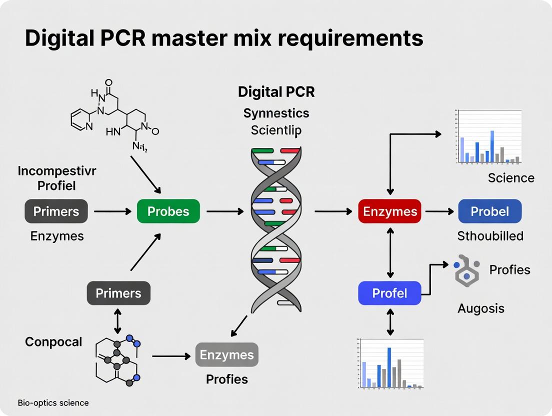
Essential dPCR Master Mix Guide: Key Components, Selection Criteria & Troubleshooting for Precision Genomics
This comprehensive guide details the critical requirements for digital PCR (dPCR) master mixes, tailored for researchers, scientists, and drug development professionals.
Essential dPCR Master Mix Guide: Key Components, Selection Criteria & Troubleshooting for Precision Genomics
Abstract
This comprehensive guide details the critical requirements for digital PCR (dPCR) master mixes, tailored for researchers, scientists, and drug development professionals. It covers foundational principles, from the unique role of master mixes in partitioning and endpoint detection to core component specifications. The article provides practical methodologies for application-specific selection, workflows for gene expression, rare mutation detection, and copy number variation analysis. It addresses common troubleshooting and optimization strategies for sensitivity, precision, and partitioning efficiency. Finally, it explores validation frameworks and comparative analyses against qPCR, equipping readers with the knowledge to select, validate, and optimize dPCR master mixes for robust, reproducible results in biomedical research and clinical diagnostics.
What is a dPCR Master Mix? Core Components, Specifications, and Partitioning Fundamentals
Within the broader thesis on dPCR master mix requirements, this article delineates the critical, non-interchangeable components of digital PCR (dPCR) master mixes that distinguish them from their quantitative PCR (qPCR) counterparts. While qPCR enables quantification via external standards, dPCR achieves absolute quantification through endpoint amplification of partitioned reactions, demanding reagent formulations optimized for partition stability, robust endpoint signal generation, and minimal inhibition. This application note details the specific requirements and provides protocols for evaluating dPCR master mix performance.
Core Composition: A Comparative Analysis
Digital PCR master mixes must address challenges absent in bulk qPCR: partition integrity during thermal cycling, efficient amplification in high-surface-area compartments, and precise endpoint fluorescence measurement. The following table summarizes key differentiating components based on current market and research analyses.
Table 1: Core Component Comparison: qPCR vs. dPCR Master Mix
| Component | Standard qPCR Mix Function | Enhanced Requirement for dPCR Mix | Rationale |
|---|---|---|---|
| Polymerase | Fast, hot-start for specificity & speed. | Ultra-stable, inhibitor-tolerant, with consistent activity across partitions. | Prevents "drop-out" of partitions; ensures uniform amplification efficiency. |
| Passive Reference Dye | Optional for normalization. | Mandatory for partition identification and volume normalization. | Critical for distinguishing partitions from debris and correcting for volume variations. |
| Surfactants/Stabilizers | Minimal or absent. | Optimized type and concentration. | Maintains partition stability (prevents coalescence) throughout thermal cycling. |
| dNTPs | Standard concentration. | Often optimized concentration and purity. | Supports reliable endpoint amplification in nanoliter volumes. |
| MgCl₂ | Standard concentration. | Precisely optimized and often at a higher concentration. | Counteracts chelation by partition matrix materials; crucial for polymerase activity. |
| Enhancers/BSA | Sometimes included. | Almost always included at higher levels. | Mitigates surface adsorption of enzymes/DNA to partition walls; enhances robustness. |
Key Performance Evaluation Protocols
Protocol 1: Assessing Partition Stability and Uniformity
Objective: To evaluate a dPCR master mix's ability to maintain discrete, stable droplets or partitions throughout the thermal cycling process. Materials:
- Test dPCR master mix (with reference dye)
- Droplet generator or chip-based partitioning system
- Thermocycler compatible with dPCR
- Reader (droplet or chip)
- Nuclease-free water (no template control) Procedure:
- Prepare a master mix according to the manufacturer's instructions, using water instead of template.
- Generate partitions using the appropriate system.
- Cycle the partitions using a standard dPCR thermal profile.
- After cycling, before reading: Visually inspect partitions under magnification for coalescence or merging.
- Load partitions into the reader. Analyze the amplitude of the reference dye signal.
- Data Analysis: Calculate the coefficient of variation (CV) of the reference dye signal amplitude across all partitions. A CV < 5% indicates high uniformity. A high rate of partition merger (>2%) indicates poor stability.
Protocol 2: Limit of Detection (LOD) and Poisson Confidence Interval Analysis
Objective: To determine the lowest concentration of target reliably detected and quantify the statistical confidence in copy number measurement. Materials:
- dPCR master mix under test
- Validated assay (primers/probe) for a single-copy gene
- Genomic DNA or synthetic target at precisely known, low concentration (e.g., 0.5, 1, 2, 5 copies/μL in reaction)
- Full dPCR workflow system (partitioner, cycler, reader) Procedure:
- Prepare serial dilutions of the target to the specified low concentrations.
- Set up dPCR reactions for each concentration, with at least 8 replicates per concentration.
- Perform partitioning, thermal cycling, and endpoint reading.
- Data Analysis: Apply Poisson statistics: λ = -ln(1 - p), where p is the fraction of positive partitions. Calculate the 95% confidence intervals for the measured copies/μL. The LOD is the lowest concentration where all replicates return a positive count with 95% confidence intervals excluding zero.
Visualizing dPCR Workflow and Mix Function
Title: Digital PCR Workflow from Mix to Result
Title: Four Pillars of an Optimal dPCR Master Mix
The Scientist's Toolkit: Essential Research Reagent Solutions
Table 2: Key Reagents for dPCR Master Mix Evaluation & Application
| Item | Function in dPCR Research |
|---|---|
| Droplet-Stabilized dPCR Master Mix | Commercial mix optimized for oil-surfactant systems; ensures consistent droplet integrity. |
| Chip-Compatible dPCR Master Mix | Formulated for silicon or polymer chips; often has different wetting properties. |
| UV-Degradable Crosslinker (for droplets) | Used in research to break droplets post-amplification for recovery of amplicons. |
| Inhibition Spike-in Controls | Defined inhibitors (e.g., heparin, humic acid) added to test mix resistance. |
| Reference Dye Calibration Standards | Beads or dyes for calibrating reader fluorescence channels. |
| Partition Number Standard | Reference material with known, low copy number for validating Poisson statistics and partition count. |
| Digital PCR-Specific BSA | High-purity, PCR-inert bovine serum albumin to prevent surface adsorption. |
| Nuclease-Free Water (Graded) | Water certified for absence of contaminants that can destabilize partitions. |
The digital PCR master mix is a specialized reagent system engineered for the physics and statistics of partition-based absolute quantification. It transcends the requirements of qPCR mixes by prioritizing partition stability, signal homogeneity, and amplification robustness in confined volumes. The protocols and analyses outlined here provide a framework for empirically validating these properties, contributing directly to the thesis that dPCR master mix formulation is a critical, standalone variable governing the accuracy and precision of absolute nucleic acid quantification.
Within the broader thesis on Digital PCR (dPCR) master mix optimization, this application note dissects the three core components whose precise interplay dictates the efficiency, specificity, and accuracy of amplification. The shift from quantitative PCR (qPCR) to dPCR places heightened demands on the master mix, requiring exceptional robustness to support endpoint, partition-based quantification without real-time monitoring. This document details the functional requirements, quantitative benchmarks, and experimental protocols for evaluating the polymerase, deoxynucleotide triphosphates (dNTPs), and buffer system.
Polymerase: The Catalytic Engine
The DNA polymerase must exhibit high processivity, fidelity, and resistance to inhibitors commonly found in complex biological samples. For dPCR, where the reaction runs to terminal plateau, robust hot-start capability is non-negotiable to prevent primer-dimer and non-specific amplification during setup.
Key Performance Metrics:
- Processivity: >50 nucleotides/second.
- Fidelity: Error rate < 1 x 10⁻⁶ errors/base.
- Inhibitor Tolerance: Must maintain >90% efficiency in the presence of 2% (v/v) whole blood or 1 mM heparin.
- Optimal Temperature: Stable activity between 60°C and 68°C.
Table 1: Comparison of Common Polymerases for dPCR Applications
| Polymerase | Processivity (nt/sec) | Fidelity (Error Rate) | Hot-Start Mechanism | Recommended dPCR Use Case |
|---|---|---|---|---|
| High-Fidelity (e.g., Pfu) | 20-30 | ~1 x 10⁻⁶ | Antibody or chemical | Absolute quantification (high precision) |
| Fast Taq (Engineered) | 80-100 | ~2 x 10⁻⁵ | Chemical modification | High-throughput screening |
| Bst (for RT-dPCR) | Moderate | ~1 x 10⁻⁴ | N/A (isothermal) | Reverse transcription dPCR (RNA targets) |
Deoxynucleotide Triphosphates (dNTPs): The Building Blocks
dNTP quality and concentration are critical. Imbalances or degradation can lead to misincorporation, truncated products, and reduced amplification efficiency, directly impacting Poisson distribution accuracy in dPCR.
Optimal Concentration Range: 200-400 µM of each dNTP (total 800-1600 µM). Higher concentrations may inhibit some polymerases. Purity Requirement: HPLC-purified, ≥99% purity, free of nuclease contamination. Stability: Use of stabilized, ready-to-use mixes (e.g., with pH indicator) is recommended for reproducible master mix formulation.
Buffer System: The Reaction Environment
The buffer maintains pH, provides essential cofactors (Mg²⁺), and can include additives to enhance specificity and yield. For dPCR, buffer optimization focuses on maximizing the fraction of positive partitions (λ) for low-abundance targets while minimizing false positives.
Core Components:
- Tris-HCl: 10-50 mM, pH 8.0-8.5 at 25°C.
- MgCl₂: 3-6 mM (must be titrated for each primer/template set).
- KCl: 50-100 mM.
- Additives: Betaine (0.5-1.5 M), DMSO (1-5%), BSA (0.1-0.5 mg/mL), Trehalose (0.3-0.6 M).
Table 2: Effect of Common Buffer Additives on dPCR Performance
| Additive | Typical Concentration | Primary Function | Impact on dPCR |
|---|---|---|---|
| Betaine | 1.0 M | Reduces secondary structure, evens dNTP usage | Increases partition positivity for GC-rich targets |
| DMSO | 3% (v/v) | Lowers DNA melting temperature | Improves amplification efficiency of complex templates |
| BSA | 0.2 mg/mL | Binds inhibitors, stabilizes polymerase | Increases robustness in clinical samples (e.g., plasma) |
| Trehalose | 0.4 M | Thermal stabilizer | Enhances reaction stability during chip/plate loading |
Experimental Protocols
Protocol 1: Titration of MgCl₂ Concentration for Optimal dPCR
Objective: Determine the optimal Mg²⁺ concentration for a specific primer/probe set to maximize fluorescence amplitude separation between positive and negative partitions. Materials: dPCR master mix (lacking Mg²⁺), 50 mM MgCl₂ stock, target DNA, primer/probe set, dPCR instrument and consumables. Procedure:
- Prepare a 2X master mix base containing polymerase, dNTPs, buffer (without Mg²⁺), primers, probe, and water.
- Prepare six 0.2 mL PCR tubes. To each, add 15 µL of 2X master mix base and a variable volume of 50 mM MgCl₂ stock to achieve final concentrations of 2.0, 3.0, 4.0, 5.0, 6.0, and 7.0 mM in a 30 µL final reaction. Adjust volume with nuclease-free water.
- Add 15 µL of template DNA (containing ~1000 copies/µL) to each tube. Include a no-template control (NTC) for the 4.0 mM condition.
- Partition and amplify on the dPCR system using the manufacturer's recommended cycling protocol.
- Analysis: Plot the fluorescence amplitude (ΔRn or equivalent) of positive partitions and the calculated copies/µL versus Mg²⁺ concentration. The optimum is the concentration yielding the highest amplitude and copy number without increasing NTC signals.
Protocol 2: Assessing Polymerase Inhibitor Tolerance
Objective: Quantify the resilience of a master mix formulation to common inhibitors. Materials: Optimized master mix, target DNA, inhibitors (e.g., heparin, EDTA, humic acid), dPCR system. Procedure:
- Prepare a serial dilution of the inhibitor in nuclease-free water.
- Formulate master mix reactions containing a constant amount of target DNA (~1000 copies/reaction) and a final concentration range of the inhibitor (e.g., heparin: 0, 0.01, 0.1, 0.5, 1.0 IU/µL).
- Run dPCR amplification.
- Analysis: Calculate the percentage of recovered copies/µL relative to the inhibitor-free control. Plot recovery % vs. inhibitor concentration. The formulation is considered robust if it maintains >90% recovery at clinically relevant inhibitor levels.
Visualizations
Polymerase Function in dPCR Cycle
Buffer System Components and Goals
Digital PCR Experimental Workflow
The Scientist's Toolkit: Research Reagent Solutions
Table 3: Essential Materials for dPCR Master Mix Research
| Item | Function in dPCR Optimization | Example/Note |
|---|---|---|
| Hot-Start High-Fidelity Polymerase | Catalyzes DNA synthesis with minimal errors; hot-start prevents pre-cycling artifacts. | Chemically modified or antibody-bound enzymes (e.g., ThermoFisher's Platinum SuperFi II, NEB's Q5). |
| HPLC-Purified dNTP Mix | Provides balanced, high-purity nucleotide substrates for accurate replication. | 100 mM solutions, pH 7.0, supplied as separate nucleotides or pre-mixed sets. |
| 10X Optimized Reaction Buffer (Mg²⁺ free) | Provides stable pH and ionic environment; allows for flexible Mg²⁺ titration. | Often supplied with the polymerase. |
| MgCl₂ Solution (50 mM) | Essential polymerase cofactor; concentration is critically optimized. | Supplied nuclease-free, certified for molecular biology. |
| PCR Additive Kit (Betaine, DMSO, BSA) | Used empirically to improve amplification of difficult templates or in inhibitory samples. | Commercial kits or individual molecular biology-grade reagents. |
| Digital PCR Chip/Droplet Generator Oil | Creates the partitions essential for absolute quantification. | Instrument-specific consumables (e.g., Bio-Rad's DG8 Cartridges, Thermo Fisher's QuantStudio chips). |
| Nuclease-Free Water | Reaction solvent; must be free of contaminants that degrade enzymes or nucleic acids. | Certified PCR-grade, DEPC-treated or 0.1 µm filtered. |
| Fluorogenic Hydrolysis Probes (e.g., TaqMan) | Provide sequence-specific detection within each partition. | Dual-labeled probes (FAM, HEX/VIC) with appropriate quenchers (e.g., BHQ1). |
The Critical Role of Passive Reference Dyes and Evaporation Inhibitors
This application note, a component of a broader thesis on Digital PCR (dPCR) master mix optimization, addresses two often-overlooked yet critical components: passive reference dyes and evaporation inhibitors. In dPCR, where absolute quantification hinges on the precise partitioning and endpoint fluorescence measurement of thousands of individual reactions, these additives are not merely optional but fundamental to data integrity. This document details their function, provides protocols for evaluation, and presents current data on their impact on assay performance.
Table 1: Impact of Passive Reference Dyes on dPCR Data Normalization and CV Reduction Data synthesized from recent commercial master mix specifications and peer-reviewed evaluations (2023-2024).
| Passive Dye Type | Excitation/Emission (nm) | Primary Function | Reported Reduction in Well-to-Well CV | Compatible Detection Channels |
|---|---|---|---|---|
| ROX (Reference Dye) | ~575/~602 | Fluorescence normalization for pipetting and partition volume variation. | Up to 50% reduction (from 10% to <5%) | ROX, CY5 (depending on filter set) |
| Mustang Purple | ~545/~570 | Normalization in multiplex assays where ROX channel is occupied. | Up to 45% reduction | VIC/HEX, CY3 |
| Internal Fluorescence Standard (IFS) | Varies by formulation | Normalization and direct monitoring of partition integrity. | Enables absolute fluorescence thresholding | Specific to formulation |
Table 2: Efficacy of Evaporation Inhibitors in dPCR Partition Stability Comparative data from studies on partition loss during thermal cycling.
| Inhibitor Class/Example | Concentration Range | Function | Reported Partition Loss Prevention | Key Consideration |
|---|---|---|---|---|
| Low-Molecular-Weight PEG | 0.1-1.0% v/v | Increases viscosity and surface tension at oil-aqueous interface. | Up to 95% reduction in loss (vs. untreated) | Can slightly inhibit polymerase at high conc. |
| Synthetic Polymers | 0.05-0.5% w/v | Forms a protective film at the interface. | 90-98% reduction | Must be non-fluorescent and inert. |
| Combination Formulations (Proprietary) | Proprietary | Multi-modal action (viscosity, surface sealing). | >98% reduction, longest stability (>6 hrs) | Optimized for specific chip/cartridge materials. |
Experimental Protocols
Protocol 1: Evaluating Passive Reference Dye Performance in Multiplex dPCR Objective: To quantify the coefficient of variation (CV) improvement conferred by a passive reference dye in a duplex SARS-CoV-2 assay (targeting ORF1ab and N genes).
Materials: See "The Scientist's Toolkit" below. Workflow:
- Master Mix Preparation: Prepare two identical reaction mixes for the duplex assay (final volume 20 µL). Include in Mix A: 1x dPCR master mix, primers/probes for ORF1ab (FAM) and N (HEX/VIC), template. In Mix B: include all components of Mix A plus a 1x final concentration of ROX passive reference dye.
- Partitioning: Load 20 µL of each mix onto the same dPCR chip/cartridge according to the manufacturer's protocol. Ensure partitions are generated from the same batch.
- Thermal Cycling: Run in the same instrument with standard cycling: 95°C for 10 min, then 45 cycles of 95°C for 15 sec and 60°C for 60 sec.
- Data Analysis: For each well/array, the instrument software will first normalize the FAM and HEX signals to the ROX signal in Mix B. Manually calculate:
- Raw CV: Standard Deviation (FAM positive partitions' amplitude) / Mean (FAM positive partitions' amplitude) for Mix A.
- Normalized CV: Same calculation using the ROX-normalized amplitudes for Mix B.
- Compare CVs and the tightness of the positive/negative clusters between conditions.
Protocol 2: Testing Evaporation Inhibitor Efficacy via Partition Count Monitoring Objective: To measure the rate of partition loss over an extended thermal cycling protocol with and without an evaporation inhibitor.
Materials: See "The Scientist's Toolkit" below. Workflow:
- Master Mix Preparation: Prepare a simple, non-amplifying mix (1x buffer, 50 nM FAM dye, water). Split into two. To the test mix, add a low-MW PEG inhibitor to 0.5% v/v (final). The control mix has no inhibitor.
- Baseline Partitioning: Load each mix onto a dPCR chip (n=4 chips per condition). Immediately image/read to obtain the initial partition count (N_initial). This is the 100% reference.
- Stress Cycling: Subject chips to an extended thermal protocol (e.g., 95°C for 5 min, 50°C for 2 min, repeated 50x) to accelerate evaporation.
- Post-Cycle Imaging: After cycling, re-image the same fields of view on the chip.
- Data Analysis: Calculate partition retention.
- Partition Retention (%) = (Npostcycle / N_initial) * 100.
- Plot retention % over time (or cycle number) for inhibitor vs. control. Statistical significance can be assessed via a t-test comparing the final retention percentages.
Mandatory Visualizations
Diagram 1: Passive Reference Dye Normalization Workflow
Diagram 2: Evaporation Inhibitor Mechanism & Impact
The Scientist's Toolkit: Essential Research Reagent Solutions
Table 3: Key Materials for dPCR Master Mix Optimization Studies
| Reagent/Material | Function in Experiment | Example & Notes |
|---|---|---|
| dPCR Master Mix (Core) | Provides polymerase, dNTPs, buffer, and Mg2+ for amplification. | Commercial mixes (e.g., Bio-Rad ddPCR Supermix, Thermo Fisher QuantStudio) or custom formulations for thesis research. |
| Passive Reference Dye | Fluorescence standard for normalizing target signals across partitions. | ROX, Mustang Purple. Must be spectrally distinct from target probes and stable at PCR temperatures. |
| Evaporation Inhibitor | Prevents loss of aqueous volume from partitions during thermal cycling. | Low-MW PEG 400, specific polymers (e.g., Pluronic F-68). Concentration must be optimized. |
| Fluorogenic Probe(s) | Target-specific detection (e.g., TaqMan). Provides the primary quantitative signal. | FAM, HEX/VIC, CY5 probes. Used in multiplex assays to test passive dye compatibility. |
| Inert Fluorescent Dye | For partition integrity/evaporation assays without amplification. | SYBR Green I, FAM-labeled inert oligonucleotide. Provides a measurable signal in all partitions. |
| Partitioning Device | Creates the nanoscale reaction chambers. | Droplet generator chips (Bio-Rad), microfluidic chips (Stilla), printed arrays (Thermo Fisher). |
| dPCR Instrument | Performs thermal cycling and endpoint fluorescence reading of each partition. | Bio-Rad QX200/QX600, Thermo Fisher QuantStudio, Stilla naica, *Qiagen QIAcuity. |
This application note, framed within a thesis on Digital PCR master mix requirements, details the distinct partitioning chemistries of emulsion-based (droplet) and chip-based digital PCR platforms. The choice of partitioning technology fundamentally dictates the required formulation of the dPCR master mix, impacting assay sensitivity, robustness, and ease of use. We present a comparative analysis of the chemistry, provide validated protocols for assay setup on both systems, and outline key reagent considerations for researchers and drug development professionals.
Digital PCR (dPCR) achieves absolute quantification by partitioning a sample into thousands of individual reactions. The two dominant partitioning methods—water-in-oil emulsion droplets and microfluidic chips—impose unique physical and chemical constraints on the reaction mix. Emulsion-based systems require surfactants and stabilizers to maintain droplet integrity during thermal cycling. In contrast, chip-based systems rely on precise interfacial chemistry to prevent evaporation and ensure uniform filling. This note elucidates these requirements through experimental data and protocols.
Comparative Analysis of Partitioning Chemistry
Table 1: Core Chemical Requirements by Partitioning Method
| Requirement / Component | Emulsion-Based dPCR (e.g., Droplet Digital PCR) | Chip-Based dPCR (e.g., Microfluidic Chip) |
|---|---|---|
| Primary Stabilizer | Surfactant (e.g., PEG-modified fluorosurfactant) at 0.5-2% v/v in carrier oil. | Chip surface treatment (e.g., silane coating); often requires specific additives in the mix (e.g., polymers). |
| Carrier Fluid | Fluorinated oil (e.g., HFE-7500, Fluorinert FC-40). | Air or immiscible, non-volatile filler oil (platform-dependent). |
| Evaporation Prevention | Achieved by the closed emulsion system. | Critical; requires a sealed chamber or a hydration system (e.g., integrated fluid, layered oil). |
| Master Mix Viscosity | Moderate viscosity tolerated. Must not destabilize emulsion. | Often requires lower viscosity for efficient, bubble-free partition loading. |
| Additive Criticality | Surfactant is ABSOLUTELY CRITICAL. Droplets will coalesce without it. | Passivation agents (e.g., BSA, DTT) are HIGHLY CRITICAL to prevent biomolecule adsorption to chip surfaces. |
| Typical Partition Volume | ~0.5 - 1 nL | ~0.5 - 6 nL (generally larger than droplets) |
| Partition Number | 20,000 - 100,000+ | 1,000 - 30,000 |
| Key Chemical Challenge | Maintaining thermostable, uniform droplets; avoiding osmotic imbalance. | Minimizing surface interactions; ensuring consistent thermal contact. |
Table 2: Performance Characteristics & Master Mix Optimization Targets
| Characteristic | Emulsion-Based dPCR | Chip-Based dPCR | Optimal Master Mix Property |
|---|---|---|---|
| Partition Uniformity | High (CV <5% for volume). Dependent on surfactant efficiency. | Very High (defined by chip manufacture). Dependent on loading technique. | Consistent viscosity and surface tension. |
| Dynamic Range | Very High (> 5 logs) due to high partition count. | High (~4-5 logs). Limited by lower partition count. | Enzyme linearity and inhibitor tolerance. |
| Inhibitor Tolerance | Higher. Inhibitors are diluted and compartmentalized. | Lower. Inhibitors are distributed across all partitions. | Enhanced polymerase resilience (e.g., using engineered enzymes). |
| Cross-Contamination Risk | Very Low (partitions are physically isolated). | Low, but requires careful chip cleaning protocols. | N/A (addressed by workflow). |
| Primary Optimization Focus | Emulsion Stability & PCR Efficiency within oil. | Wetting, Surface Passivation & Evaporation Control. | Platform-specific formulation. |
The Scientist's Toolkit: Essential Research Reagent Solutions
Table 3: Key Reagents for dPCR Formulation Research
| Reagent / Material | Function in Formulation | Primary Application |
|---|---|---|
| Fluorosurfactant (e.g., PEG-PFPE) | Stabilizes water-in-oil emulsion; prevents droplet coalescence during thermal cycling. | Emulsion-based dPCR only. Critical component of droplet generation oil. |
| Fluorinated Carrier Oil (HFE-7500) | Inert, non-volatile continuous phase for droplet generation and thermal cycling. | Emulsion-based dPCR. Provides thermal stability and optical clarity. |
| BSA (Molecular Biology Grade) | Passivates surfaces (plastic, silica) to prevent adsorption of polymerase and template DNA. | Critical for Chip-based dPCR. Beneficial additive for emulsion-based to prevent adsorption to tube walls. |
| DTT or Betaine | Reduces secondary structure in DNA/RNA; can improve partition uniformity and amplification efficiency. | Both platforms. Additive for high-GC or complex templates. |
| Polymer Additives (e.g., Ficoll, PEG) | Modifies viscosity and surface tension; aids in uniform partition loading and stability. | Chip-based dPCR. Often included in proprietary master mixes. |
| Passivated DNA Polymerase | Engineered enzyme with reduced surface adsorption and enhanced inhibitor tolerance (e.g., gasket residues). | Both platforms, critical for Chip-based. Maximizes reaction efficiency in constrained environments. |
| Evaporation Sealant (e.g., silicone oil) | Forms a vapor barrier over reactions in open-well chips to prevent volume loss. | Chip-based dPCR with open-chip designs. |
Experimental Protocols
Protocol 1: Formulation Testing for Emulsion Stability (ddPCR)
Objective: To evaluate the performance of a custom or commercial master mix for droplet generation and thermal cycling stability.
Master Mix Preparation:
- Prepare a 1X dPCR master mix (22 µL final volume per reaction) containing:
- 1X PCR Buffer (provided with enzyme)
- 900 nM each forward/reverse primer
- 250 nM TaqMan probe (FAM/HEX)
- 3-5 mM MgCl₂ (optimize)
- 0.1-1 mg/mL BSA (optional, for tube passivation)
- 1 U/µL passivated hot-start DNA polymerase
- Target DNA (e.g., 10-100 copies/µL)
- Nuclease-free water to volume.
- Prepare a 1X dPCR master mix (22 µL final volume per reaction) containing:
Droplet Generation:
- Load 20 µL of master mix into the sample well of a DG8 cartridge.
- Load 70 µL of Droplet Generation Oil (containing surfactant) into the oil well.
- Place the cartridge in the droplet generator. The instrument will produce ~40 µL of droplet emulsion.
Transfer & Sealing:
- Carefully transfer ~40 µL of droplets to a semi-skirted 96-well PCR plate.
- Seal the plate with a foil heat seal using a plate sealer (180°C for 5 seconds).
Thermal Cycling:
- Cycle using standard conditions (e.g., 95°C for 10 min, then 40 cycles of 94°C for 30s and 60°C for 60s, with a 2°C/s ramp rate).
- Hold at 4°C until reading.
Droplet Reading & Analysis:
- Transfer plate to droplet reader.
- Analyze data for: Droplet Count (>15,000 per 20 µL sample is good), Amplitude Separation (clear positive/negative clusters), and Rain (minimal intermediate amplitude droplets).
Protocol 2: Chip Loading Optimization for Microfluidic dPCR
Objective: To ensure uniform, bubble-free loading of partitions on a chip-based system.
Chip Priming & Preparation:
- If required by the platform, prime the microfluidic chip by applying vacuum or pressure to the outlet port to fill channels with the recommended Filler Oil.
- Ensure chip is on a pre-cooled (4°C) thermal block or holder to slow reaction start.
Master Mix Preparation & Loading:
- Prepare master mix as in Protocol 1, but omit BSA if the proprietary mix already contains passivating agents.
- Centrifuge the master mix briefly to remove bubbles.
- Pipette the recommended volume (e.g., 15-35 µL) into the chip's sample inlet port. Avoid introducing air bubbles.
Partitioning & Sealing:
- Engage the instrument's partitioning mechanism. This may involve applying pressure to drive the mix into the nanowell array or closing valves to define chambers.
- For open-array systems, immediately apply the recommended Sealing Oil or sealing gasket to prevent evaporation.
Thermal Cycling & Imaging:
- Place the sealed chip in the thermocycler/imaging instrument.
- Run the optimized cycling protocol. Ramp rates are often slower for chip-based systems to ensure thermal uniformity.
- End-point fluorescence imaging is performed on each partition.
Analysis & Quality Control:
- Analyze images for: Total Active Partitions (should match chip specification), Uniform Fluorescence of negative partitions (low CV indicates good passivation), and Well-to-Well Contamination.
Visualizing dPCR Workflow & Chemistry
(Diagram 1: Comparative dPCR Partitioning Workflows)
(Diagram 2: Chemical Interactions in dPCR Partitions)
Within the broader research thesis on digital PCR (dPCR) master mix requirements, three technical specifications are paramount for robust assay design: inhibitor tolerance, dynamic range, and limit of detection (LoD). This application note details protocols and comparative analyses to evaluate commercial dPCR master mixes against these criteria, providing a framework for researchers and drug development professionals to select optimal reagents for challenging sample matrices and low-abundance target quantification.
Inhibitor Tolerance: Protocols and Comparative Data
Inhibitors co-purified with nucleic acids can severely impede polymerase activity, leading to underestimation of target concentration. This experiment evaluates master mix resilience against common inhibitors.
Protocol 1.1: Inhibitor Spike-in dPCR Assay
Objective: To quantify the reduction in apparent target concentration in the presence of serial dilutions of defined inhibitors. Materials:
- Test dPCR master mixes (A, B, C).
- Standardized gDNA or plasmid target (e.g., 1000 copies/µL).
- Inhibitor stocks: Humic Acid (10 mg/mL), Heparin (1 mg/mL), IgG (20 mg/mL), EDTA (100 mM).
- Droplet or chip-based dPCR system. Method:
- Prepare a master solution containing target nucleic acid (final 500 copies/µL).
- Spike inhibitor stocks into aliquots of the master solution to create a dilution series (e.g., Humic Acid: 0, 10, 50, 100, 200 µg/mL).
- Mix 1:1 with the test dPCR master mixes according to manufacturer protocols.
- Partition and amplify using standardized thermal cycling conditions.
- Analyze using system software. Calculate the percentage recovery: (Concentration with Inhibitor / Concentration without Inhibitor) * 100%.
Table 1: Inhibitor Tolerance of Commercial dPCR Master Mixes
| Master Mix | Humic Acid (100 µg/mL) % Recovery | Heparin (0.5 U/mL) % Recovery | IgG (1 mg/mL) % Recovery | EDTA (1 mM) % Recovery |
|---|---|---|---|---|
| Mix A (Standard) | 45% | 30% | 78% | 15% |
| Mix B (Inhibitor Resistant) | 92% | 85% | 95% | 90% |
| Mix C (High-Fidelity) | 60% | 70% | 90% | 40% |
Dynamic Range: Protocols and Comparative Data
Dynamic range defines the interval over which the measured copy number concentration is linearly related to the expected concentration. A wide dynamic range is critical for quantifying targets with unknown or vastly different abundances.
Protocol 2.1: Log-Linear Dilution Series for Dynamic Range Determination
Objective: To establish the upper and lower bounds of quantitative linearity for a dPCR assay. Materials:
- Test dPCR master mixes.
- Target plasmid DNA.
- Reference dye (if required by master mix). Method:
- Prepare a 10-fold serial dilution of target DNA across 7-8 orders of magnitude (e.g., from 10^6 to 10^0 copies/µL).
- For each dilution, set up dPCR reactions in quintuplicate with each test master mix.
- Perform partitioning and amplification. Record the measured concentration (copies/µL) for each reaction.
- Plot measured concentration (log10) vs. expected concentration (log10). Perform linear regression.
- Define the lower bound as the concentration where R² ≥ 0.99 and accuracy is within ±25%. The upper bound is determined by partition saturation.
Table 2: Dynamic Range of Tested dPCR Master Mixes
| Master Mix | Lower Limit of Quantification (LLOQ) | Upper Limit of Quantification (ULOQ) | Effective Linear Range (Log10) | Regression R² (across range) |
|---|---|---|---|---|
| Mix A | 2 copies/µL | 50,000 copies/µL | 4.4 logs | 0.998 |
| Mix B | 1 copy/µL | 100,000 copies/µL | 5.0 logs | 0.999 |
| Mix C | 5 copies/µL | 20,000 copies/µL | 3.6 logs | 0.997 |
Limit of Detection (LoD): Protocols and Comparative Data
LoD is the lowest concentration of target that can be reliably distinguished from zero. It is a function of partition number, master mix sensitivity, and background.
Protocol 3.1: Probabilistic LoD Determination using Negative Binomial Model
Objective: To statistically determine the 95% detection probability concentration. Materials:
- Test dPCR master mixes.
- Low-concentration target (e.g., 0.5, 1, 2, 5 copies/µL).
- Negative template control (NTC, e.g., nuclease-free water). Method:
- Prepare 24 replicate reactions for each low target concentration and for the NTC using each master mix.
- Run dPCR and record the number of positive and negative partitions for each replicate.
- For each concentration, calculate the proportion of replicates with ≥1 positive partition.
- Fit a negative binomial or Poisson-binomial model to the positive reaction rate vs. concentration data.
- The LoD is the concentration at which detection probability reaches 95%.
Table 3: Experimentally Determined Limit of Detection (95% Probability)
| Master Mix | Partitions per Reaction | False Positive Rate (NTC) | Calculated LoD (copies/µL) | 95% CI for LoD |
|---|---|---|---|---|
| Mix A | 20,000 | 0/24 | 1.8 | (1.3 - 2.5) |
| Mix B | 25,000 | 0/24 | 0.9 | (0.6 - 1.3) |
| Mix C | 15,000 | 0/24 | 3.5 | (2.5 - 4.9) |
The Scientist's Toolkit: Key Reagent Solutions
| Item | Function in dPCR Master Mix Evaluation |
|---|---|
| Inhibitor-Resistant Polymerase | Engineered DNA polymerase that maintains activity in the presence of common PCR inhibitors (e.g., humic acid, heparin). Critical for analyzing crude or complex samples. |
| Passive Reference Dye | A dye that fluoresces independently of amplicon formation. Used to normalize fluorescence signals and identify failed or empty partitions, improving data reliability. |
| Emulsion Stabilizer / Surfactant | A chemical crucial for stable droplet formation and prevention of coalescence in droplet-based dPCR systems. Affects partition uniformity and number. |
| dUTP / Uracil-DNA Glycosylase (UDG) | System to prevent carryover contamination. dUTP is incorporated into amplicons, which can be enzymatically cleaved by UDG prior to amplification of new samples. |
| Competitor DNA (e.g., Salmon Sperm DNA) | Non-specific DNA added to the mix to adsorb non-specific inhibitors and reduce polymerase adsorption to tube walls, potentially improving low-copy detection. |
Visualizations
Title: How Sample Inhibitors Reduce dPCR Accuracy
Title: Workflow to Determine dPCR Dynamic Range
Title: Statistical Determination of dPCR Limit of Detection
Selecting and Applying dPCR Master Mixes: Workflows for Gene Expression, Rare Mutations, and CNV Analysis
Within the broader thesis on Digital PCR (dPCR) master mix requirements, the precise selection of master mix chemistry is paramount for assay accuracy and sensitivity. This application note details the criteria for matching master mix formulation to the target of interest—whether DNA or RNA, wild-type or mutant—in absolute quantification and rare allele detection applications central to modern drug development.
Master Mix Chemistry: Core Components and Selection Criteria
dPCR master mixes are optimized for endpoint, partitioned amplification. The choice hinges on target type and application goal.
Table 1: Master Mix Selection Guide Based on Target and Application
| Target Type | Primary Application | Recommended Master Mix Type | Key Required Enzyme(s) | Critical Additives/Features | Typical LoD (Limit of Detection) |
|---|---|---|---|---|---|
| Genomic DNA | Copy Number Variation (CNV), Rare Mutation Detection | DNA-specific, high-fidelity, inhibitor-tolerant | Hot-start DNA polymerase, dNTPs | PCR inhibitors neutralizers, UNG (optional) | ≤0.1% mutant allele frequency |
| cDNA (from RNA) | Gene Expression, Viral RNA Quantification | Reverse Transcriptase (RT) + dPCR combo mix or separate steps | Reverse Transcriptase, DNA polymerase | RNase inhibitor, sequence-specific or random primers | <5 copies per reaction |
| Wild-type DNA | Reference Gene Quantification, Pathogen Load | Standard DNA dPCR master mix | Standard DNA polymerase, dNTPs | Standard buffer, EvaGreen or probe-compatible | <10 copies per reaction |
| Mutant DNA (SNV, Indel) | Oncology Biomarkers, MRD (Minimal Residual Disease) | Probe-based (e.g., TaqMan), allele-specific | High-specificity DNA polymerase | Allele-specific probes, possibly asymmetric primer ratios | ≤0.01% mutant allele frequency (for optimized assays) |
| miRNA/small RNA | Biomarker Discovery, Regulatory RNA Analysis | Polyadenylation + RT-specific or stem-loop RT | Poly(A) polymerase, specific RT, DNA polymerase | Tailored RT primers, enhanced sensitivity buffers | High sensitivity required; instrument-dependent |
Detailed Experimental Protocols
Protocol 1: Rare Mutant Allele Detection in High Wild-type Background
Objective: Quantify a single nucleotide variant (SNV) at an allele frequency as low as 0.01%. Principle: Use a sequence-specific hydrolysis (TaqMan) probe with a wild-type blocker to suppress amplification of the non-target allele.
Workflow:
- Sample Preparation: Extract genomic DNA from FFPE or plasma samples. Quantify using fluorometry.
- Assay Design: Design a mutant-specific TaqMan probe (e.g., FAM-labeled). Design a wild-type-specific blocking oligonucleotide (C3-spacer at 3' end) that binds with higher affinity to the wild-type sequence.
- Reaction Assembly (20 µL):
- Template DNA: 10-100 ng
- Master Mix: Probe-based dPCR Supermix (e.g., ddPCR Supermix for Probes, Bio-Rad): 10 µL
- Mutant-specific Forward Primer (900 nM final)
- Reverse Primer (900 nM final)
- Mutant-specific FAM-labeled Probe (250 nM final)
- Wild-type Blocker Oligo (optional, 500 nM final)
- Nuclease-free water to volume.
- Partitioning & Amplification: Generate droplets or load into chip. Thermal cycle: 95°C for 10 min (enzyme activation), then 40 cycles of 94°C for 30s and a combined annealing/extension at 58-60°C for 60s (ramp rate 2°C/s).
- Analysis: Read partitions. Set amplitude threshold to distinguish positive (FAM+) from negative partitions. Calculate mutant allele concentration and frequency using Poisson statistics.
Protocol 2: Absolute Quantification of Viral RNA (e.g., SARS-CoV-2)
Objective: Accurately quantify viral RNA copy number without a standard curve. Principle: Use a one-step RT-dPCR assay to minimize handling and maximize accuracy.
Workflow:
- Sample & Control Prep: Isolate viral RNA. Include no-template control (NTC) and positive RNA control.
- Reaction Assembly (20 µL):
- RNA template: 5 µL
- Master Mix: One-step RT-dPCR mix for probes (e.g., ddPCR One-Step RT-ddPCR Advanced Kit for Probes): 10 µL
- Forward/Reverse Primers (900 nM final each)
- Target-specific Probe (250 nM final, HEX or FAM)
- Additives: RNase inhibitor (additional 0.5 U/µL)
- Nuclease-free water to volume.
- Reverse Transcription & Partitioning: Thermal cycle: Reverse Transcription: 50°C for 60 min. Enzyme activation: 95°C for 5-10 min. Partitioning: Perform immediately after RT step if using droplet-based systems.
- Amplification: 40 cycles of 94°C for 30s and 55-60°C for 60s.
- Analysis: Read fluorescence. Set thresholds. Report copies/µL of input RNA extract, factoring in partition volume and sample input.
The Scientist's Toolkit: Key Reagent Solutions
Table 2: Essential Research Reagents for dPCR Applications
| Reagent Category | Specific Example | Critical Function in Application |
|---|---|---|
| Probe-based dPCR Supermix | ddPCR Supermix for Probes (Bio-Rad) | Optimized buffer chemistry for hydrolysis probe assays, essential for high-specificity mutant detection. |
| One-step RT-dPCR Mix | One-Step RT-ddPCR Advanced Kit (Bio-Rad) | Integrates reverse transcription and DNA amplification in a single, partitioned reaction for direct RNA quantification. |
| Evagreen dPCR Supermix | QIAcuity Digital PCR Master Mix (Qiagen) | Intercalating dye chemistry for high-resolution melt analysis or multiplexing where probe channels are limited. |
| Allele-Specific PCR Additive | PerfectMatch PCR Enhancer (Agilent) or competitor oligonucleotides | Increases polymerase fidelity and specificity, critical for distinguishing wild-type from mutant sequences. |
| Inhibitor-Resistant Polymerase | OmniTaq Polymerase (DNA Polymerase Technology) | Tolerant to common inhibitors in crude samples (e.g., blood, soil), improving accuracy of direct quantification. |
| Digital PCR Plates/Chips | QIAcuity Nanoplate (Qiagen), QuantStudio Absolute Q Digital PCR Chip (Thermo Fisher) | Microfluidic devices that create physical partitions for target amplification and endpoint fluorescence reading. |
| Nuclease-free Water & Tubes | Molecular Biology Grade Water (Ambion), Low-binding tubes | Prevents degradation of sensitive RNA/DNA templates and oligonucleotides, ensuring reaction integrity. |
Diagrams of Experimental Workflows and Decision Logic
Diagram 1: dPCR Master Mix Selection Logic
Diagram 2: Protocol for Rare Mutant Detection Workflow
Optimized Protocol for Rare Allele Detection and Liquid Biopsy Applications
Within the broader thesis on Digital PCR (dPCR) master mix requirements, achieving optimal sensitivity and specificity for rare allele detection in liquid biopsies is paramount. This application note details an optimized dPCR protocol designed for the robust detection of low-frequency somatic mutations (e.g., <0.1% variant allele frequency, VAF) from circulating tumor DNA (ctDNA), focusing on critical master mix components and validation data.
Table 1: Comparison of dPCR Master Mix Performance for Rare Allele Detection
| Master Mix Characteristic | Standard EvaGreen Mix | Optimized Probe-Based Mix | Functional Impact |
|---|---|---|---|
| Limit of Detection (LOD) | 0.5% VAF | 0.05% VAF | Enables ultra-rare variant detection |
| False Positive Rate | 0.01 events/μL | <0.001 events/μL | Reduces background in wild-type partitions |
| Partition Number | ~20,000 | ~28,000 | Increases statistical power & precision |
| Inhibition Resistance | Moderate | High | Tolerates common ctDNA contaminants |
| Digital Resolution | 2-plex | 4-plex (multicolor) | Allows for internal controls & multiple targets |
Table 2: Validation Data for KRAS G12D Detection in Simulated Plasma
| Input ctDNA (GE*) | Target VAF | Measured VAF (Optimized Mix) | Measured VAF (Standard Mix) | CV (%) |
|---|---|---|---|---|
| 1000 | 0.1% | 0.098% | Not Detected | 12.5 |
| 1000 | 0.5% | 0.51% | 0.47% | 8.2 |
| 1000 | 1.0% | 0.99% | 0.95% | 6.1 |
*GE = Genome Equivalents
Detailed Experimental Protocols
Protocol 1: ctDNA Extraction and Qualification for dPCR
Objective: Isolate high-integrity, inhibitor-free ctDNA from blood plasma.
- Plasma Collection: Collect blood in cell-stabilization tubes. Perform double centrifugation (1600 x g, 10 min; 16,000 x g, 10 min) to obtain platelet-poor plasma.
- ctDNA Extraction: Use a silica-membrane column-based kit designed for <100 bp fragments. Elute in 20-40 μL of low-EDTA TE buffer or nuclease-free water.
- Quality Control: Quantify using a fluorometer specific for dsDNA. Assess fragment size profile via Bioanalyzer/TapeStation (expected peak ~170 bp).
Protocol 2: Optimized Rare Allele dPCR Assay
Objective: Detect and precisely quantify a target somatic mutation at low VAF. Reagents: Optimized probe-based dPCR master mix, mutant-specific FAM probe/assay, wild-type HEX probe/assay, restriction enzyme (optional), nuclease-free water, target DNA.
- Reaction Setup (20 μL Total Volume):
- 8.0 μL Optimized 4X dPCR Master Mix
- 1.0 μL 20X Mutant Assay (FAM-labeled)
- 1.0 μL 20X Reference Assay (HEX-labeled, wild-type locus)
- 2.0 μL Template DNA (up to 100 ng ctDNA)
- 8.0 μL Nuclease-Free Water
- Partitioning: Load reaction mix into a microfluidic chip or droplet generator according to manufacturer's instructions to create ~28,000 partitions.
- Thermal Cycling:
- 95°C for 10 min (enzyme activation)
- 45 cycles of: 94°C for 30 sec, 60°C for 60 sec (anneal/extend)
- 98°C for 10 min (enzyme deactivation)
- 4°C hold.
- Imaging/Analysis: Read partitions on a compatible fluorescence reader. Apply amplitude thresholding and analyze using Poisson statistics for absolute quantification. VAF = (Mutant copies/mL) / (Total (mutant + wild-type) copies/mL).
Protocol 3: Determination of Limit of Blank (LoB) and LOD
Objective: Empirically define assay sensitivity and specificity.
- LoB: Run no-template control (NTC) and wild-type genomic DNA (≥20 replicates). LoB = Mean false positive mutant count in wild-type samples + 1.645(SD).
- LOD: Create dilution series of synthetic mutant DNA in wild-type background (e.g., 1%, 0.5%, 0.1%, 0.05%). Run 24 replicates per level. LOD is the lowest VAF where ≥95% of replicates have mutant count > LoB.
Visualization: Experimental Workflow
Title: Liquid Biopsy dPCR Workflow for Rare Alleles
The Scientist's Toolkit: Research Reagent Solutions
Table 3: Essential Materials for Optimized Rare Allele dPCR
| Item | Function & Rationale |
|---|---|
| Optimized Probe-Based dPCR Master Mix | Contains high-performance polymerase, balanced dNTPs, and optimized buffer for maximum partition uniformity and low background, critical for low VAF precision. |
| Mutation-Specific TaqMan Assays | FAM-labeled probe/primers for specific mutant allele. MGB or LNA probes enhance discrimination. |
| Reference Assay (Wild-Type) | HEX/VIC-labeled assay for the homologous wild-type locus. Serves as internal control for total DNA input and normalization. |
| Digital PCR Chip/Cartridge System | Microfluidic device generating 20,000+ partitions. High partition count is non-negotiable for rare allele statistics. |
| Inhibition-Resistant Polymerase | Engineered polymerase tolerant to common plasma inhibitors (hemoglobin, heparin, EDTA), reducing false negatives. |
| Fragment-Specific ctDNA Extraction Kit | Optimized for recovery of short (~170 bp) DNA fragments, maximizing yield of tumor-derived ctDNA. |
| Droplet Stabilization Reagent | For droplet-based dPCR, ensures droplet integrity during thermal cycling, preventing coalescence. |
| Nuclease-Free Water (PCR Grade) | Ultrapure water to prevent enzymatic degradation of reagents and template. |
| Synthetic Mutation Standards | Pre-quantified DNA fragments with known mutations for absolute calibration, LOD determination, and run-to-run QC. |
Best Practices for Copy Number Variation (CNV) and Gene Expression Analysis
Within the framework of research on Digital PCR (dPCR) master mix requirements, the analysis of Copy Number Variation (CNV) and gene expression presents unique challenges and opportunities. dPCR's unparalleled precision in absolute quantification makes it the gold standard for these applications. This document outlines best practices, detailed protocols, and critical considerations for robust CNV and gene expression analysis, emphasizing the pivotal role of optimized dPCR master mix composition.
Core Principles and dPCR Master Mix Implications
Accurate CNV and gene expression analysis by dPCR depends on master mix properties that ensure efficient amplification, precise partitioning, and minimal bias.
Key Master Mix Requirements:
- Inhibition Resistance: Must withstand inhibitors common in genomic DNA and cDNA samples.
- Partitioning Efficiency: Formulation should promote consistent droplet or chip well formation without cross-talk.
- High Efficiency & Linear Dynamic Range: Enable accurate quantification across varying target concentrations.
- Reference Assay Compatibility: Support reliable multiplexing for reference/target ratios (CNV) or housekeeping/target genes (expression).
Quantitative Impact of Master Mix Components: Table 1: Impact of dPCR Master Mix Components on CNV/Expression Analysis
| Component | Primary Function | Optimal Characteristic for CNV/Expression | Risk of Sub-Optimal Performance |
|---|---|---|---|
| Polymerase | Catalyzes DNA synthesis | High processivity, inhibitor-resistant (e.g., Glycerol-free). | Reduced amplification efficiency, false-negative partitions. |
| Nucleotide Purity | Building blocks for amplification | Ultra-pure dNTPs, free of contaminants. | Increased background, nonlinear quantification. |
| Stabilizers | Maintain enzyme activity & partition integrity | Biocompatible polymers (e.g., PEG). | Droplet coalescence, well-to-well contamination. |
| Mg2+ Concentration | Cofactor for polymerase | Optimized and precisely defined concentration. | Altered amplification efficiency and assay specificity. |
| Passive Reference Dye | Distinguish partitions | High fluorescence, inert, does not inhibit PCR. | Incorrect partition identification, quantification errors. |
Detailed Experimental Protocols
Protocol 2.1: CNV Analysis Using a Reference Assay (Duplex dPCR)
Objective: Absolute quantification of a target genomic locus relative to a reference (diploid) locus to determine copy number.
Materials (Research Reagent Solutions):
- Optimized dPCR Master Mix: For duplex probe-based assays (e.g., containing dUTP and UDG for carryover prevention).
- Target Locus Assay: FAM-labeled hydrolysis probes/primers.
- Reference Locus Assay: HEX/VIC-labeled hydrolysis probes/primers (e.g., RNase P, TERT).
- DNA Sample: 10-100 ng/µL, high molecular weight genomic DNA.
- Droplet or Chip Generator & Reader: Platform-specific equipment.
- Nuclease-Free Water.
Procedure:
- Sample & Reaction Setup:
- Thaw all reagents and mix gently. Keep on ice.
- Prepare duplex dPCR reaction mix on ice in this order:
- dPCR Master Mix: 11 µL
- Target Locus Assay (20X): 1.1 µL
- Reference Locus Assay (20X): 1.1 µL
- DNA Template (50 ng): 2.2 µL
- Nuclease-Free Water: to a final volume of 22 µL.
- Mix thoroughly by pipetting. Do not vortex after adding master mix.
- Partitioning:
- Load reaction mix into the droplet generator or chip according to the manufacturer's protocol. For droplet systems, generate droplets.
- Transfer droplets or sealed chip to a thermal cycler.
- Amplification:
- Use the following thermal cycling protocol:
- UDG Incubation (if using): 37°C for 10 min.
- Polymerase Activation: 95°C for 10 min.
- 40-45 Cycles of:
- Denaturation: 94°C for 30 sec.
- Annealing/Extension: 60°C for 60 sec (optimize per assay).
- Signal Stabilization: 98°C for 10 min.
- Hold: 4°C or 12°C.
- Use the following thermal cycling protocol:
- Reading & Analysis:
- Read the plate/chip on the dPCR reader.
- Set amplitude thresholds to clearly separate positive and negative partitions for both FAM and HEX/VIC channels.
- Export copy number data (copies/µL) for target and reference.
- Copy Number Calculation:
- Calculate using the formula: CN = 2 × (Target Copies/µL) / (Reference Copies/µL).
- Interpret: CN ~2 (diploid), CN >2 (amplification/gain), CN <2 (deletion/loss). Use confidence intervals from Poisson statistics.
Protocol 2.2: Absolute Gene Expression Analysis (Reverse Transcription-dPCR)
Objective: Absolute quantification of mRNA transcript copy number in a cDNA sample.
Materials (Research Reagent Solutions):
- dPCR Master Mix for cDNA: Optimized for amplification from cDNA templates.
- Gene-Specific Assay: Hydrolysis probes or EVAGreen dye.
- Reverse Transcription Kit: Using random hexamers and/or oligo-dT primers.
- RNA Sample: High-quality, DNase I-treated total RNA.
- No-RT Control: Reaction without reverse transcriptase.
- Droplet or Chip System.
Procedure:
- cDNA Synthesis:
- Perform reverse transcription on 100 ng – 1 µg total RNA using a high-fidelity RT kit.
- Include a No-RT Control (-RT) to monitor genomic DNA contamination.
- Dilute synthesized cDNA 1:5 to 1:10 in nuclease-free water.
- dPCR Reaction Setup:
- Prepare reaction mix on ice:
- dPCR Master Mix: 11 µL
- Gene-Specific Assay (20X): 1.1 µL
- Diluted cDNA Template: 2.2 µL
- Nuclease-Free Water: to 22 µL.
- Set up a separate reaction for the -RT control.
- Mix and partition as in Protocol 2.1.
- Prepare reaction mix on ice:
- Amplification & Detection:
- Use a thermal cycling protocol optimized for the assay (similar to Protocol 2.1, without UDG step unless specified).
- Read the plate and set thresholds. The -RT control should show minimal to no positive partitions.
- Quantification:
- The reader software provides absolute concentration in copies/µL of the dPCR reaction.
- To report as copies/ng input RNA, use:
(copies/µL dPCR) × (Total dPCR rxn vol.) × (cDNA dilution factor) / (ng input RNA).
Visualization of Workflows and Relationships
Title: dPCR Workflow for Copy Number Variation Analysis
Title: Master Mix Role in dPCR Accuracy
The Scientist's Toolkit: Essential Research Reagent Solutions
Table 2: Key Reagents for dPCR-based CNV and Gene Expression Analysis
| Reagent/Material | Function & Importance | Selection Criteria |
|---|---|---|
| Inhibitor-Resistant dPCR Master Mix | Ensures robust amplification from challenging samples (e.g., FFPE DNA, cDNA). Essential for data accuracy. | Choose mixes formulated for high GC content, with inhibitors in mind, and validated for your platform. |
| Hydrolysis Probe Assays (TaqMan) | Provide high specificity for target and reference sequences in duplex reactions. | Verify primer/probe sequences for unique genomic loci. Check for lack of common SNPs in binding sites. |
| EVAGreen or SYBR Green Master Mix | Cost-effective for single-plex gene expression screening. Intercalates into dsDNA. | Requires extensive optimization and melt curve analysis post-dPCR to confirm amplicon specificity. |
| Digital PCR Plates or Cartridges | The consumable for partition formation. Critical for partition integrity and data quality. | Must be compatible with your dPCR system. Lot-to-lot consistency is paramount. |
| Nuclease-Free Water & Tubes | Prevents degradation of primers, probes, and templates. Maintains reaction integrity. | Use certified nuclease-free, molecular biology grade. |
| Quantitative DNA/RNA Standards | For validating assay linearity, dynamic range, and absolute quantification calibration. | Use serially diluted standards of known concentration (e.g., gBlocks, cloned plasmids). |
Within the broader research on Digital PCR (dPCR) master mix requirements, a critical subtopic is the optimization of multiplex assays. Unlike qPCR, dPCR's endpoint, partitioning-based nature allows for higher levels of multiplexing without kinetic bias. However, successful multiplexing hinges on two interdependent pillars: the spectral compatibility of fluorophores and the precise balancing of primer and probe concentrations. This application note details protocols and strategies to achieve robust multiplex dPCR, enabling precise copy number variation analysis, mutation detection, and pathogen identification in complex samples.
Fluorophore Compatibility: Spectral Overlap and Channel Configuration
Effective multiplexing requires fluorophores with minimal spectral cross-talk. dPCR instruments typically have 4-6 optical channels. The selection must account for the instrument's excitation sources and emission filters.
Table 1: Common Fluorophore Combinations for 4-Color dPCR Systems
| Channel (Ex/Em) | Primary Fluorophore | Common Quencher | Compatible Co-Plex Fluorophores | Key Consideration |
|---|---|---|---|---|
| FAM (470/520) | FAM, SYBR Green I | BHQ-1, TAMRA | HEX, VIC, TET, CAL Fluor Gold 540 | Avoid using with high HEX concentration due to spillover. |
| HEX/VIC (535/555) | HEX, VIC, TET | BHQ-1 | FAM, TAMRA, Cy3 | Can often be distinguished from FAM via filter optimization. |
| ROX/Texas Red (580/610) | ROX, Cy3.5, CAL Fluor Red 610 | BHQ-2 | Cy5, Quasar 670 | Good separation from FAM/HEX and far-red channels. |
| Cy5 (635/665) | Cy5, Quasar 670 | BHQ-3 | ROX, Texas Red | Minimal spillover into other common channels. |
Protocol 2.1: Initial Spectral Cross-Talk Assessment
Objective: To quantify and correct for fluorescence bleed-through between channels for a selected fluorophore set.
Materials:
- Single-plex dPCR reactions for each fluorophore.
- dPCR Master Mix (optimized for probe-based assays).
- Target template for each assay.
- dPCR instrument with compatible optical setup.
Procedure:
- Prepare individual single-plex reactions for each target/fluorophore combination. Include a no-template control (NTC) for each.
- Run all reactions on the dPCR instrument, ensuring data is collected in all optical channels.
- Analyze the data. For the reaction containing only Fluorophore A, plot the fluorescence amplitude in its primary channel (Y-axis) against the amplitude in a non-primary channel, e.g., Channel B (X-axis).
- Calculate the percentage of positive partitions for Fluorophore A that are also positive in Channel B. This defines the bleed-through percentage.
- Repeat for all fluorophore/channel combinations. A bleed-through >1% typically requires compensation or reassessment of fluorophore choice.
Diagram 1: Fluorophore Selection & Spectral Overlap Workflow
Concentration Balancing of Assay Components
In a multiplex reaction, all primer pairs and probes compete for master mix components (dNTPs, polymerase, Mg²⁺). Imbalanced concentrations lead to "assay drop-out," where the least efficient assay fails or shows reduced sensitivity.
Table 2: Typical Starting Concentration Ranges for Multiplex dPCR Optimization
| Component | Single-Plex Typical Range | Multiplex Optimization Starting Point | Notes |
|---|---|---|---|
| Primer, Forward/Reverse | 200-900 nM each | 100-400 nM each | Lower concentrations reduce dimer formation and competition. |
| Hydrolysis Probe (FAM) | 100-250 nM | 50-150 nM | The brightest fluorophore (e.g., FAM) can often be used at lower concentrations. |
| Hydrolysis Probe (Darker Dye) | 100-250 nM | 150-300 nM | Dyes like Cy5 may require higher concentrations for clear cluster separation. |
| dPCR Master Mix | 1X | 1X | Ensure master mix is validated for multiplexing (high enzyme processivity, robust buffer). |
Protocol 3.1: Primer and Probe Concentration Titration Matrix
Objective: To empirically determine the optimal concentration for each primer and probe in the multiplex context.
Materials:
- dPCR Master Mix.
- Stock solutions of each primer and probe.
- Template containing all targets at known, moderate copy numbers (~50 copies/µL).
- Nuclease-free water.
Procedure:
- Design a titration matrix. For a duplex, test 3 concentrations of Assay A primer/probe against 3 concentrations of Assay B primer/probe (a 3x3 matrix). For higher plex, use a fractional factorial design.
- Prepare dPCR reactions according to the matrix. Keep master mix volume and template concentration constant.
- Run on a dPCR instrument.
- Analyze the data. For each condition, record: a) Total positive partitions for each target, b) Mean fluorescence amplitude (MFI) of the positive cluster, c) Separation (ΔMFI) between positive and negative partitions.
- The optimal condition is the one that yields the most similar copy number results to the single-plex reference and provides maximal, well-separated positive clusters for all targets.
Diagram 2: Concentration Balancing Optimization Cycle
The Scientist's Toolkit: Key Research Reagent Solutions
Table 3: Essential Materials for Multiplex dPCR Development
| Item | Function in Multiplex dPCR | Key Consideration for Master Mix Research |
|---|---|---|
| Multiplex-Optimized dPCR Master Mix | Provides the core enzymes, buffer, and dNTPs. Must support efficient co-amplification of multiple targets. | Look for mixes specifically advertised for multiplexing. They often have enhanced polymerase processivity and optimized Mg²⁺/salt buffers. |
| Dual-Quenched Hydrolysis Probes (e.g., with internal ZEN/TAO quencher) | Lower background fluorescence, improving signal-to-noise and enabling more fluorophores per channel. | Reduces baseline noise, critical for distinguishing multiple positive clusters in a single channel. |
| UDG/dUTP System | Prevents carryover contamination; UDG is compatible with standard probes. | Essential for clinical diagnostic assay development. Must be inactive at dPCR cycling temperatures. |
| Passive Reference Dye (ROX) | Normalizes for well-to-well fluorescence fluctuations. Not used as a reporter in quantification. | Required for instruments using a reference for droplet/partition identification. Use at manufacturer-specified concentration. |
| Synthetic gBlock Gene Fragments | Defined, multi-target templates for assay development and optimization without genomic DNA variability. | Crucial for creating controlled multiplex validation samples with precise copy number ratios. |
| Nuclease-Free Water (PCR Grade) | Reaction solvent. Must be free of contaminants that degrade enzymes or nucleic acids. | Inconsistent water quality is a major source of failed multiplex reactions and reduced partition yield. |
| Optical Seal or Sealing Foil | Prevents evaporation and cross-contamination during thermal cycling. | Must be compatible with the dPCR instrument's optical system (clear, low autofluorescence). |
Integrating fluorophore compatibility checks with systematic concentration balancing is non-negotiable for developing robust multiplex dPCR assays. This process, framed within the larger thesis of master mix requirements, highlights that the master mix must not only be efficient but also provide a stable, competitive environment for simultaneous amplifications. The protocols outlined here provide a foundational workflow for researchers to expand the multiplexing capability of their dPCR systems, thereby increasing data density and cost-effectiveness for advanced genomic applications.
Sample Preparation and Input Recommendations for Optimal Performance
1. Introduction Within the context of a broader thesis on Digital PCR (dPCR) master mix requirements, sample and input quality are established as the primary determinants of assay precision and accuracy. This document outlines critical sample preparation protocols and nucleic acid input recommendations, substantiated by current experimental data, to ensure optimal dPCR performance.
2. The Scientist's Toolkit: Essential Research Reagent Solutions
| Item | Function in dPCR Sample Preparation |
|---|---|
| Solid-Phase Reversible Immobilization (SPRI) Beads | Selective binding of nucleic acids for size-selective purification and concentration, crucial for removing inhibitors. |
| RNase Inhibitors (e.g., Recombinant Proteins) | Essential for RT-dPCR, protects RNA templates from degradation during reverse transcription and sample handling. |
| Inhibitor-Resistant DNA Polymerases | Engineered enzymes within master mixes that maintain activity in the presence of common sample inhibitors (e.g., heparin, hematin). |
| Fragmentation & Library Prep Kits | For complex samples (e.g., FFPE), standardizes fragment size and adds adapters for targeted sequencing or dPCR analysis. |
| Digital PCR-Specific Master Mix | Contains optimized polymerase, dNTPs, buffers, and often a passive reference dye. Formulated for precise partitioning and endpoint detection. |
| Nucleic Acid Integrity Assessment Kits (e.g., RIN/Qubit) | Quantifies and qualifies input material (RNA Integrity Number, concentration) to guide input normalization. |
3. Quantitative Input Guidelines and Performance Metrics Optimal input amounts balance Poisson statistics for confident low-copy detection with avoidance of droplet saturation. The following table summarizes key parameters.
Table 1: Recommended Input Ranges and Their Impact on dPCR Performance
| Target Application | Recommended DNA Input (Mass) | Recommended DNA Input (Copies) | Optimal Accepted Droplets | Key Performance Metric Impact |
|---|---|---|---|---|
| Rare Variant Detection | 10-100 ng | 3,000 - 30,000* | 15,000 - 20,000 | Maximizes sensitivity for variants at <0.1% allele frequency. |
| Copy Number Variation | 5-50 ng | 1,500 - 15,000* | >10,000 | Ensures precise ratio measurement between target and reference. |
| Absolute Quantification (High Titer) | 1-10 ng | 300 - 3,000* | >8,000 | Provides high precision for viral load or gene expression standards. |
| Microbial Detection (Low Titer) | Up to 100 ng | Up to 30,000* | >15,000 | Increases probability of capturing single, low-abundance targets. |
| RT-dPCR (RNA Input) | 1-100 ng total RNA | Varies by transcript abundance | >10,000 | Requires optimization of reverse transcription step efficiency. |
*Based on human genomic DNA (~3.3 pg/diploid cell).
4. Detailed Experimental Protocols
4.1. Protocol: Validation of Input Linear Range and Inhibition Testing Objective: To establish the optimal input range for a specific assay and test for sample-derived inhibition. Materials: Purified nucleic acid sample, dPCR master mix, assay-specific primers/probes, droplet generator, reader. Method: 1. Prepare a 5-point serial dilution (e.g., 0.1, 1, 10, 50, 100 ng/µL) of the target DNA in nuclease-free water. 2. For inhibition testing, spike a constant amount of a synthetic target or control DNA into each sample dilution. 3. Assemble 20 µL reactions per manufacturer's protocol: 10 µL 2X dPCR master mix, 1 µL 20X primer/probe assay, variable volume of sample dilution, and water to volume. 4. Generate droplets per instrument specifications. 5. Perform PCR amplification with standard cycling conditions. 6. Read droplets and analyze data. Plot measured concentration (copies/µL) vs. expected concentration for the dilution series and the spiked control. Interpretation: The linear range is where the measured target concentration scales proportionally with input. Inhibition is indicated by a significant drop in the measured concentration of the spiked control in undiluted samples.
4.2. Protocol: Best-Practice Nucleic Acid Purification for Inhibitor-Rich Samples Objective: To obtain inhibitor-free nucleic acids from complex matrices (e.g., plasma, soil, FFPE). Materials: SPRI bead solution, 80% ethanol, elution buffer, magnetic stand, sample. Method: 1. Lyse sample using a chaotropic salt-based lysis buffer appropriate for the matrix. 2. Bind nucleic acids to SPRI beads at a defined sample-to-bead ratio (e.g., 1:1.8) to select for desired fragment size. Incubate 5-10 minutes. 3. Place on a magnetic stand until supernatant clears. Discard supernatant. 4. Wash beads twice with 80% ethanol while on the magnet. Air-dry beads for 5 minutes. 5. Elute DNA/RNA in a low-salt elution buffer (e.g., 10 mM Tris-HCl, pH 8.0). Incubate at 55°C for 2 minutes, then place on magnet. Transfer purified eluate to a clean tube. Note: For RT-dPCR, include an RNase inhibitor in the elution buffer or immediately proceed to reverse transcription.
5. Visualization of Workflows and Relationships
dPCR Sample Preparation and Analysis Workflow
Impact of DNA Input Amount on dPCR Results
Troubleshooting dPCR Assays: Solving Common Master Mix Issues for Improved Sensitivity & Precision
Diagnosing and Fixing Poor Partitioning Efficiency (Rain, Low Positive Counts)
This application note addresses the critical challenge of poor partitioning efficiency in digital PCR (dPCR), manifesting as inter-droplet "rain" or low positive counts. Within the broader thesis on dPCR master mix optimization, these phenomena directly indicate suboptimal reagent formulations or reaction conditions. Efficient partitioning is non-negotiable for absolute quantification, especially in low-abundance target applications critical to drug development, such as monitoring minimal residual disease or quantifying viral loads.
Table 1: Common Causes and Quantitative Impacts on Partitioning Efficiency
| Factor | Typical Optimal Range | Deviation Leading to Rain/Low Counts | Observed Impact on Coefficient of Variation (CV) |
|---|---|---|---|
| Input DNA Integrity | DIN ≥ 7.0, RIN ≥ 8.0 | Fragmented DNA (DIN < 5) | CV increase of 15-40% |
| Master Mix Surfactant Concentration | System-specific optimal (e.g., 0.5-2%) | Deviation > ±0.3% from optimal | Partition failure rate increase of 10-60% |
| PCR Inhibitor Carryover | [EDTA] < 0.5 mM, [Hemoglobin] < 0.1 mg/mL | [EDTA] > 1.0 mM, [Hb] > 0.5 mg/mL | False negative increase of 20-80% |
| Partition Generation Pressure/Oil Temp | Manufacturer specified (e.g., 2.5 psi ±0.2) | Deviation > ±0.5 psi or > ±2°C | Irregular partition formation in 5-30% of samples |
| Target Amplicon Length | 60-120 bp (ddPCR) | Length > 150 bp | Positive count reduction of 10-25% |
| Template Concentration | Ideal for Poisson: ~100-1000 copies/20µL | Extremely low (< 10 copies/20µL) | High Poisson error, CV > 20% |
Table 2: Diagnostic Clues from Rain Patterns
| Rain Pattern (2D Amplitude Plot) | Likely Primary Cause | Suggested Master Mix/Protocol Fix |
|---|---|---|
| Vertical streaking (wide cluster in negative dimension) | Inhibitors, poor enzyme kinetics | Optimize Mg²⁺, use inhibitor-resistant polymerase, add BSA |
| Horizontal streaking (wide cluster in positive dimension) | Non-specific amplification, high background | Increase annealing temperature, optimize probe design, use hot-start polymerase |
| Diagonal scattering between clusters | Partition merging or degradation | Adjust surfactant/oil ratio in mix, verify droplet generator seals |
| Overall low amplitude (compressed clusters) | Low PCR efficiency, poor probe cleavage | Re-optimize primer/probe concentrations, check quencher integrity |
Experimental Protocols for Diagnosis and Optimization
Protocol 1: Systematic Diagnosis of Partitioning Failures
Objective: To identify the root cause(s) of rain or low positive counts. Materials: Affected dPCR samples, fresh control master mix, control DNA (wild-type genomic DNA), droplet generator, thermocycler, droplet reader. Procedure:
- Run a Reference Assay: Perform dPCR on a well-characterized control DNA sample using the suspect master mix and protocol. This establishes a baseline.
- Interrogate Template Quality:
- Run sample on a Fragment Analyzer or Bioanalyzer to obtain a DNA Integrity Number (DIN).
- If DIN < 7, perform cleanup using a silica-column or SPRI bead-based kit optimized for short fragments.
- Repeat dPCR with cleaned template.
- Test for Inhibitors via Spike-and-Recovery:
- Dilute the sample template 1:5 in nuclease-free water.
- Spike a known concentration of control target (e.g., 500 copies/µL) into both neat and diluted sample.
- Perform dPCR. Calculate recovery:
(Conc. in spiked neat sample - Conc. in unspiked neat) / Known spike concentration. - Recovery < 80% indicates inhibition. Proceed with inhibitor-removal column or alternative purification.
- Master Mix Stress Test:
- Prepare the same reaction using a fresh batch of master mix or a commercial "rain-resistant" formulation.
- Partition and amplify under identical conditions.
- Compare amplitude plots and CV. Improvement implicates master mix stability or formulation.
Protocol 2: Optimization of Master Mix Surfactant Concentration
Objective: Empirically determine the optimal surfactant concentration for a custom master mix to minimize rain. Materials: Base master mix (without surfactant), concentrated surfactant (e.g., Tween-20, Triton X-100), gradient PCR instrument, droplet generator. Procedure:
- Prepare a 10% (v/v) stock of the surfactant in nuclease-free water.
- Prepare 8 aliquots of the base master mix. Spike surfactant stock to create a concentration series (e.g., 0.1%, 0.25%, 0.5%, 0.75%, 1.0%, 1.5%, 2.0%, 3.0%).
- To each mix, add identical amounts of template and primers/probe for a robust, medium-copy target.
- Generate droplets and perform PCR with a standard thermal profile.
- Analyze results:
- Primary Metric: Plot the number of accepted (monodisperse) partitions vs. surfactant concentration. Identify the plateau peak.
- Secondary Metric: For concentrations at the peak, calculate the separation between positive and negative clusters (ΔRFU). The concentration yielding the highest ΔRFU with maximal partition count is optimal.
Protocol 3: Verification via Limit of Blank (LoB) and Limit of Detection (LoD)
Objective: Quantitatively confirm fix by assessing assay sensitivity and specificity. Procedure:
- Run Limit of Blank (LoB): Perform dPCR (n=12 replicates) using no-template control (NTC) reactions with the optimized master mix and conditions.
- Calculate the 95th percentile of the copies/µL reported for the NTCs. This is the LoB.
- Run Limit of Detection (LoD): Prepare a dilution series of target template at concentrations expected to be near the LoB (e.g., 0.5, 1, 2, 5 copies/µL). Run n=12 replicates per concentration with the optimized setup.
- Determine the concentration where ≥95% of replicates report a concentration > LoB. This is the LoD.
- Compare: A lower LoD and consistent, low counts in NTCs after optimization confirm improved partitioning efficiency and reduced background.
Visualizations
Title: Diagnostic & Optimization Workflow for dPCR Partitioning Issues
Title: Interplay of Factors Affecting dPCR Partitioning Efficiency
The Scientist's Toolkit: Research Reagent Solutions
Table 3: Essential Reagents for Diagnosing and Fixing Partitioning Issues
| Reagent/Material | Primary Function | Key Consideration for Partitioning |
|---|---|---|
| Rain-Resistant dPCR Master Mix (Commercial) | Provides optimized, standardized formulation for robust droplet formation and amplification. | Contains proprietary surfactants and stabilizers designed to widen the "rain-free" amplification window. Use for benchmark comparisons. |
| Inhibitor-Resistant DNA Polymerase | Enzymes engineered to withstand common inhibitors (hemoglobin, EDTA, heparin). | Critical for clinical samples (blood, FFPE). Ensures efficient amplification in all partitions, reducing false negatives. |
| Droplet Generation Oil & Surfactants | Creates stable, monodisperse water-in-oil emulsions. | Oil viscosity and surfactant type/concentration must match master mix. Incompatibility causes droplet coalescence or breakup. |
| Molecular Biology Grade BSA | Stabilizes enzymes, sequesters inhibitors, and can reduce surface adhesion. | Addition (0.1-1.0% w/v) often improves cluster separation and partition stability, especially with custom mixes. |
| SPRI Bead Cleanup Kits | Removes PCR inhibitors and selects for optimal DNA fragment sizes. | Pre-partitioning cleanup is vital. Select beads with a size cutoff that retains short amplicons but removes long fragments that hinder partitioning. |
| Digital PCR Assay Optimization Kits | Pre-formulated mixtures of Mg²⁺, additives, and competitors for assay tuning. | Allows systematic optimization of reaction conditions without reformulating master mix from scratch. |
| Quantitative DNA Integrity Standards | Controls with defined Degradation Scores (e.g., DIN). | Essential for diagnosing template degradation as a source of low efficiency and rain. |
Optimizing Template Input and Master Mix Volume for Maximum Precision
This application note is framed within a broader doctoral thesis investigating the fundamental requirements of Digital PCR (dPCR) master mixes to achieve unparalleled precision in nucleic acid quantification. The core hypothesis is that maximum precision—defined as the minimization of confidence intervals around a target concentration—is not solely a function of the dPCR platform but is critically dependent on the synergistic optimization of template input (copy number per partition) and master mix reaction volume. Suboptimal combinations lead to increased measurement uncertainty, impacting critical applications in liquid biopsy, rare mutation detection, and gene expression analysis in drug development.
Table 1: Impact of Template Input on Assay Precision (Theoretical Poisson Distribution)
| Mean Copies per Partition (λ) | % Partitions Positive | Coefficient of Variation (CV%) | Recommended Use Case |
|---|---|---|---|
| 0.1 - 0.5 | 10% - 40% | >10% | Ultra-rare target detection |
| 0.6 - 1.6 | 45% - 80% | 5% - 10% | Optimum precision range |
| 1.7 - 3.0 | 82% - 95% | 10% - 20% | High-abundance targets |
| >3.0 | >95% | >20% | Not recommended for precise quantitation |
Table 2: Experimental Results: Master Mix Volume vs. Precision Conditions: 20,000 partitions, target λ = 1.0, 5 replicates per condition.
| Master Mix Volume per Partiton (nL) | Observed CV% | Partition Uniformity (CV%) | Remarks |
|---|---|---|---|
| 0.5 | 12.5% | 25% | High volumetric error, low precision |
| 0.8 | 7.2% | 15% | Moderate precision |
| 1.0 | 5.1% | 8% | Platform-optimized, best precision |
| 1.2 | 5.3% | 9% | Slight waste, no precision gain |
Experimental Protocols
Protocol 1: Determining Optimal Template Input Range
Objective: To empirically determine the template input (copies/partition) that yields the lowest CV for a specific assay.
Materials: See "The Scientist's Toolkit" below. Method:
- Prepare Template Dilution Series: Using a quantified DNA standard, prepare a 7-point serial dilution in TE buffer (pH 8.0) to target a range of 0.1 to 3.0 copies per partition.
- Assemble dPCR Reactions: For each dilution, assemble 8 replicate reactions using the optimized master mix volume (e.g., 1.0 nL/partition * total partitions). Keep master mix composition constant.
- Partitioning and Amplification: Load reactions onto the chosen dPCR chip/cartridge. Perform PCR cycling per assay specifications: 95°C for 10 min (enzyme activation), then 45 cycles of [95°C for 15 sec, 60°C for 60 sec].
- Data Analysis: For each dilution, calculate the mean measured concentration and the CV across replicates. Plot CV against the mean copies/partition (λ). The minimum of the curve identifies the optimal input range (typically λ 0.6-1.6).
Protocol 2: Titrating Master Mix Volume for Partition Uniformity
Objective: To identify the master mix volume that provides the most uniform partition filling, minimizing volumetric noise.
Materials: See "The Scientist's Toolkit" below. Method:
- Master Mix Preparation: Prepare a large batch of dPCR master mix containing a fluorescent probe-based assay for a medium-abundance reference gene (targeting λ ~1.0).
- Volume Variation Setup: Aliquot the master mix and adjust the total reaction volume to achieve different per-partition volumes (e.g., 0.5, 0.8, 1.0, 1.2 nL) based on the manufacturer's partition count.
- Partition Imaging (Pre-PCR): For droplet-based systems, use a droplet analyzer to measure droplet diameter and uniformity CV for each volume condition. For chip-based systems, use bright-field imaging to assess chamber filling.
- Amplification and Analysis: Run the dPCR protocol. Measure the fluorescence amplitude (RFU) of positive partitions. The coefficient of variation of the positive cluster's amplitude is inversely related to partition filling uniformity. The condition with the lowest amplitude CV indicates optimal master mix volume.
Visualizations
Title: dPCR Precision Optimization and Failure Pathways
Title: Protocol to Determine Optimal Template Input
The Scientist's Toolkit: Essential Research Reagent Solutions
| Item | Function & Importance for Precision |
|---|---|
| Droplet-Digital PCR (ddPCR) Supermix | Provides optimized reagents for emulsion stability and efficient amplification in oil-water partitions. Critical for low CV. |
| Nuclease-Free Water (PCR Grade) | Solvent for dilutions and master mix. Must be free of contaminants to prevent inhibition or background. |
| Quantified gDNA or cDNA Standard | Essential for creating accurate dilution series to determine the optimal template input range (λ). |
| Target-Specific Assay (Primers/Probes) | Hydrolysis (TaqMan) or EvaGreen assays. High specificity and efficiency minimize false negatives/positives. |
| Droplet Generation Oil / Chip | Platform-specific consumable. Batch consistency is vital for partition uniformity. |
| Droplet Reader Oil / Chip Holder | Ensures accurate droplet positioning for endpoint fluorescence measurement. |
| Digital PCR Plate Sealer | Prevents evaporation and cross-contamination during long cycling protocols, safeguarding reaction volume. |
| Precision Micro-pipettes (2µL, 10µL) | Accurate liquid handling is non-negotiable for master mix assembly and template addition to minimize error. |
Within the broader research on Digital PCR (dPCR) master mix requirements, a paramount challenge is the reliable analysis of target nucleic acids from challenging sample matrices like whole blood and Formalin-Fixed, Paraffin-Embedded (FFPE) tissues. These matrices introduce potent inhibitors that can compromise polymerase activity, leading to underestimation of target concentration, false negatives, and reduced precision. This Application Note details targeted strategies and optimized protocols to overcome inhibition, ensuring accurate, robust dPCR quantification.
Understanding Inhibitors and Their Mechanisms
Inhibitors present in complex samples interfere with the PCR reaction through various mechanisms, which a dPCR-optimized master mix must counteract.
Table 1: Common Inhibitors in Challenging Matrices and Their Modes of Action
| Sample Matrix | Key Inhibitory Compounds | Primary Mechanism of Inhibition | Impact on dPCR |
|---|---|---|---|
| Whole Blood | Hemoglobin, Heparin, Lactoferrin, IgG | Binding to DNA polymerase, degradation of nucleic acids, chelation of Mg²⁺ ions. | Reduced amplification efficiency, increased partition "drop-out," skewed Poisson statistics. |
| FFPE Tissues | Formaldehyde adducts, paraffin, melanin, salts, acidic polysaccharides. | Cross-linking of nucleic acids, fragmentation, polymerase steric hindrance, co-purification. | Lower apparent target copy number, increased variability between replicates, non-amplifying partitions. |
Diagram Title: Inhibitor Sources and dPCR Impact Pathways
Strategic Approaches and Optimized Protocols
Strategy 1: Sample Pre-Treatment and Purification
Effective nucleic acid extraction is the first critical barrier against inhibitors.
Protocol 1.1: Optimized Nucleic Acid Extraction from Whole Blood for dPCR
Objective: Isolate high-purity, inhibitor-free genomic DNA from whole blood. Materials: See "The Scientist's Toolkit" (Table 3). Workflow:
- Lysis: Mix 200 µL of fresh whole blood with 20 µL of Proteinase K and 1 mL of Lysis Buffer BL. Vortex vigorously for 15 seconds. Incubate at 56°C for 10 minutes.
- Precipitation: Add 1 mL of absolute ethanol to the lysate. Mix by inversion until a homogeneous solution forms.
- Binding: Load the mixture onto a HiBind DNA Mini Column (in a 2 mL collection tube). Centrifuge at 10,000 x g for 1 minute. Discard flow-through.
- Wash 1: Add 500 µL of HB Buffer (with ethanol added). Centrifuge at 10,000 x g for 1 minute. Discard flow-through.
- Wash 2: Add 700 µL of DNA Wash Buffer (with ethanol added). Centrifuge at 10,000 x g for 1 minute. Discard flow-through. Repeat this wash step.
- Elution: Transfer the column to a clean 1.5 mL microcentrifuge tube. Apply 50-100 µL of Nuclease-Free Water pre-heated to 70°C directly to the center of the membrane. Incubate at room temperature for 2 minutes. Centrifuge at 10,000 x g for 1 minute to elute DNA.
- dPCR QC: Quantify DNA yield by UV spectrophotometry (A260/A280 ratio of ~1.8). Assess fragmentation and inhibitor presence by running 20 ng on a 1% agarose gel. Proceed to dPCR setup.
Protocol 1.2: DNA Recovery from FFPE Tissue Sections for dPCR
Objective: Recover amplifiable DNA from FFPE tissue, reversing cross-links and removing paraffin. Materials: See "The Scientist's Toolkit" (Table 3). Workflow:
- Dewaxing: Place two 10 µm FFPE tissue sections in a 1.5 mL tube. Add 1 mL of xylene (or xylene substitute). Vortex for 10 seconds. Incubate at 56°C for 3 minutes. Centrifuge at full speed for 2 minutes. Carefully remove and discard supernatant.
- Ethanol Wash: Add 1 mL of 100% ethanol to the pellet. Vortex for 10 seconds. Centrifuge at full speed for 2 minutes. Discard supernatant. Air-dry the pellet for 5-10 minutes.
- Proteinase K Digestion: Add 180 µL of ATL Buffer and 20 µL of Proteinase K to the dried pellet. Vortex. Incubate overnight (~16 hours) at 56°C with agitation (750 rpm).
- Cross-link Reversal/Inhibitor Removal: Add 200 µL of AL Buffer (containing guanidine HCl). Vortex. Incubate at 70°C for 10 minutes.
- Binding & Washing: Add 200 µL of 100% ethanol. Vortex. Follow steps 3-6 from Protocol 1.1, using the same buffers and columns.
- Post-Extraction Repair: Treat 50 µL of eluted DNA with 5 U of Uracil-DNA Glycosylase (UDG) and 1X PreCR Repair Mix for 30 minutes at 37°C to repair deamination and nicks.
- dPCR QC: Assess DNA by gel electrophoresis (expected smear <500 bp). Use a multiplexed dPCR assay with a short (~60 bp) and a long (~150 bp) amplicon to gauge fragmentation impact.
Strategy 2: Master Mix Engineering and Reaction Optimization
The choice and formulation of the dPCR master mix are decisive in tolerating residual inhibitors.
Protocol 2.1: Evaluating and Optimizing Inhibitor-Tolerant dPCR Master Mixes
Objective: Compare commercial master mixes for their resilience to spiked inhibitors using a standardized assay. Materials: Candidate inhibitor-tolerant dPCR master mixes (A, B, C), reference DNA target, inhibitor stocks (Hemoglobin, Heparin, Humic Acid), EvaGreen or probe-based assay. Experimental Design:
- Prepare a 5 ng/µL stock of reference gDNA.
- Prepare serial dilutions of each inhibitor in nuclease-free water.
- For each master mix, set up reactions containing: 1X master mix, 1X assay mix, 10,000 copies of reference DNA, and a variable volume of inhibitor stock to reach the final concentrations listed in Table 2.
- Partition and amplify according to manufacturer's recommendations.
- Analyze recovered concentration (copies/µL) and amplitude (if using probe-based chemistry).
Table 2: Example Data from Master Mix Inhibition Tolerance Test
| Master Mix | Added Inhibitor (Conc.) | Theoretical Conc. (copies/µL) | Measured Conc. ± SD (copies/µL) | % Recovery | Notes (Amplitude, etc.) |
|---|---|---|---|---|---|
| Standard Mix | None | 1000 | 995 ± 45 | 99.5% | Normal amplitude. |
| Standard Mix | Hemoglobin (2 µM) | 1000 | 650 ± 120 | 65.0% | Reduced amplitude, increased rain. |
| Inhibitor-Tolerant Mix A | None | 1000 | 1010 ± 30 | 101.0% | Normal amplitude. |
| Inhibitor-Tolerant Mix A | Hemoglobin (2 µM) | 1000 | 980 ± 40 | 98.0% | Slightly reduced amplitude. |
| Inhibitor-Tolerant Mix A | Heparin (0.1 U/mL) | 1000 | 1020 ± 35 | 102.0% | Normal amplitude. |
Diagram Title: Master Mix Strategy for Robust dPCR
Strategy 3: Post-Purification Additives and Dilution
Direct counteraction of inhibitors within the dPCR reaction.
Protocol 3.1: Using Additives to Rescue Inhibited Reactions
Objective: Systematically test reaction additives to improve dPCR performance from inhibited FFPE samples. Method:
- Prepare a standard dPCR master mix and assay targeting a 100 bp region.
- Aliquot the master mix. To separate aliquots, add one of the following additives to the final concentration indicated:
- Bovine Serum Albumin (BSA): 0.1 - 0.5 µg/µL
- T4 Gene 32 Protein: 0.05 - 0.2 µM
- Betaine: 0.5 - 1.0 M
- Polyvinylpyrrolidone (PVP): 0.5 - 1.0%
- Add 20 ng of FFPE-derived DNA (from Protocol 1.2) to each additive-condition and a no-additive control.
- Run dPCR. Compare measured concentration, cluster separation, and signal amplitude.
The Scientist's Toolkit
Table 3: Key Research Reagent Solutions for Addressing Inhibition
| Item | Function/Description | Example Use Case |
|---|---|---|
| Inhibitor-Tolerant DNA Polymerase | Engineered polymerase resistant to binding by hematin, IgG, and other common inhibitors. | Core component of a robust dPCR master mix for blood analysis. |
| Silica-Based Membrane Spin Columns | Selective binding of nucleic acids in high-salt conditions; washes remove inhibitors. | Purification of DNA from blood or lysed FFPE tissue (Protocols 1.1, 1.2). |
| Proteinase K | Broad-spectrum serine protease that digests proteins and nucleases. | Essential for lysis of blood cells and de-crosslinking of FFPE tissues. |
| Carrier RNA | Co-precipitates with low-concentration nucleic acids, improving yield and consistency. | Added during FFPE DNA extraction when target material is scant. |
| UDG (Uracil-DNA Glycosylase) | Removes uracil bases incorporated from cytosine deamination, common in FFPE DNA. | Post-extraction repair step to reduce C>T artifacts in sequencing or bias in detection. |
| PreCR Repair Mix | Enzyme cocktail (e.g., ligase, polymerase, glycosylase) to repair nicks and damage. | Restores amplifiability of fragmented FFPE DNA prior to dPCR. |
| BSA (Bovine Serum Albumin) | Binds to and neutralizes a wide range of inhibitors, including phenolics and humic acids. | Additive in the dPCR reaction to mitigate residual FFPE-derived inhibitors (Protocol 3.1). |
| T4 Gene 32 Protein | Single-stranded DNA binding protein, stabilizes DNA and displaces weakly bound inhibitors. | Additive to improve amplification efficiency from highly fragmented or damaged DNA. |
Correcting Amplification Bias and Non-Specific Signal in Multiplex Assays
Within the broader research thesis on Digital PCR (dPCR) master mix requirements, a critical challenge is the optimization of multiplex assays. Amplification bias, where different targets amplify with varying efficiencies, and non-specific signal from primer-dimers or off-target binding, compromise quantification accuracy and multiplexing capacity. This application note details protocols and solutions for correcting these issues, enabling robust, high-order multiplex dPCR.
| Source | Primary Impact | Typical Effect on Quantification | Common Correction Strategy |
|---|---|---|---|
| Primer/Probe Concentration Imbalance | Amplification Efficiency Bias | Under/over-estimation of specific targets | Asymmetric Primer Tuning |
| Sequence-Dependent Tm Variation | Differential Amplification | Bias >20% between targets | Competitive PCR Enhancers |
| Primer-Dimer Formation | Non-Specific Fluorescence | False positive counts, reduced dynamic range | Hot-Start Polymerase, Probe-based detection |
| Cross-Talk Between Fluorescent Channels | Spectral Overlap | Inaccurate partition classification | Spectral Calibration with Unmixing Algorithms |
| Template GC-Content Variation | Amplification Efficiency Bias | Poor low-abundance target performance | Balanced Betaine or DMSO additives |
Table 2: Performance of Different dPCR Master Mix Additives for Multiplex Correction
| Additive/Condition | Function | Recommended Concentration | % Reduction in Primer-Dimer Signal (Mean ± SD) | Improvement in Target Balance (CV Reduction) |
|---|---|---|---|---|
| Betaine | Homogenizes DNA melting temp | 0.5 - 1.0 M | 45 ± 12% | 15% |
| DMSO | Reduces secondary structure | 3 - 10% (v/v) | 38 ± 10% | 12% |
| Touchdown PCR | Increases initial stringency | --- | 60 ± 15% | 20% |
| Competitor DNA (e.g., Poly dI:dC) | Binds non-specific primers | 0.1 - 0.5 ng/µL | 70 ± 8% | 5% |
| Enhanced Hot-Start Polymerase | Inhibits activity until denaturation | --- | 85 ± 5% | 8% |
Experimental Protocols
Protocol 1: Optimizing Primer/Probe Concentrations to Correct Amplification Bias
Objective: To empirically determine the optimal primer and probe concentrations for balanced amplification in a duplex or triplex assay. Materials: See "The Scientist's Toolkit" below. Procedure:
- Design Primers/Probes: Ensure amplicons are similar in length (80-150 bp). Label probes with non-overlapping fluorophores (e.g., FAM, HEX/VIC, Cy5).
- Prepare Master Mix Matrix: Create a series of reactions where the primer concentration for the suspected "strong" amplicon is held at a standard concentration (e.g., 900 nM), while the "weak" amplicon primer is titrated from 50 nM to 900 nM. Probe concentrations are typically titrated from 50 nM to 250 nM independently.
- Run dPCR: Partition the reactions using a droplet or chip-based dPCR system. Use a template that contains all targets at approximately equal copy numbers (e.g., 1000 copies/µL each).
- Analyze Data: Calculate the copies/µL for each target. The optimal concentration is where the measured ratio between targets is closest to 1:1 (or the expected ratio) with the lowest coefficient of variation (CV) between replicates.
- Validate: Run the optimized assay with a dilution series of each target to confirm linearity and maintained efficiency.
Protocol 2: Assessing and Suppressing Non-Specific Amplification
Objective: To identify and minimize non-specific signals (primer-dimers, off-target binding) using no-template controls (NTCs) and chemical additives. Materials: See "The Scientist's Toolkit" below. Procedure:
- Establish Baseline with NTCs: Prepare dPCR reactions containing the multiplex assay master mix and all primers/probes, but using nuclease-free water instead of template. Run in at least 8 replicates.
- Analyze NTC Partitions: Plot the fluorescence amplitude of all partitions. Partitions clustering distinctly from the negative population indicate systematic non-specific signal. Note the rate (percentage) of these false-positive partitions.
- Iterative Additive Testing: Repeat the NTC experiment, adding one potential suppressor (e.g., 0.5 M Betaine, 5% DMSO, 0.2 ng/µL Poly dI:dC) to the master mix. Compare the false-positive rate to the baseline.
- Evaluate Impact on Specific Signal: Using a positive template with known low copy number (e.g., 10 copies/µL), confirm that the additive does not significantly suppress the true positive signal or worsen amplification bias.
- Implement Thermal Cycling Adjustments: If non-specific signal persists, employ a touchdown protocol (e.g., start annealing temperature 5°C above calculated Tm, decrease by 0.5°C per cycle for 10 cycles, then hold at the final Tm for remaining cycles).
Visualization of Workflows and Relationships
Diagram 1 Title: Multiplex dPCR Optimization Decision Pathway
Diagram 2 Title: Master Mix Optimization Iterative Workflow
The Scientist's Toolkit
Table 3: Essential Research Reagent Solutions for Multiplex dPCR Optimization
| Item | Function & Relevance to Correction | Example Product/Brand (Research-Use Only) |
|---|---|---|
| Hot-Start DNA Polymerase | Critical for suppressing primer-dimer formation during reaction setup by inhibiting polymerase activity at low temperatures. | Thermostable Hot-Start Pol, UltraPass Hot-Start Pol. |
| dNTP Mix (Balanced) | Provides equimolar deoxynucleotides; imbalances can introduce amplification bias, especially in multiplex. | PCR Grade dNTP Set, 100 mM each. |
| PCR Enhancers/Additives | Chemicals like Betaine, DMSO, or commercial enhancer cocktails homogenize melting temps and reduce secondary structure, correcting bias. | GC-Rich Solution, PCR Enhancer Solution. |
| Competitor DNA (e.g., Poly dI:dC) | Non-specific carrier DNA that binds excess primers, reducing off-target priming and primer-dimer artifacts. | Poly(dI:dC), Salmon Sperm DNA. |
| Fluorogenic Probe Sets | Hydrolysis (TaqMan) or hybridization probes increase specificity over dye-based detection, reducing non-specific signal. | Dual-Labeled Probes (FAM, HEX, Cy5). |
| Digital PCR Supermix (Base) | The core master mix formulation; choosing one optimized for multiplexing (with elevated Mg2+, stabilizers) is foundational. | ddPCR Supermix for Probes, QXDx dPCR Master Mix. |
| Spectral Calibration Dyes/Plate | Essential for setting fluorescence gain and spectral compensation to minimize cross-talk between channels in multiplex assays. | Spectral Calibration Kit, Rainbow Calibration Particles. |
| Nuclease-Free Water (PCR Grade) | The diluent; essential for preventing enzymatic degradation of primers/probes and avoiding background contamination. | Molecular Biology Grade Water. |
Storage, Handling, and Stability Guidelines to Maintain Master Mix Integrity
Application Notes
Within the scope of doctoral research on Digital PCR (dPCR) master mix requirements, maintaining reagent integrity is paramount for ensuring precise and reproducible quantification of nucleic acid targets. Master mixes for dPCR are complex formulations containing thermostable DNA polymerase, dNTPs, salts, stabilizers, and often a passive reference dye. Their performance is acutely sensitive to suboptimal storage and handling, which can lead to enzyme inactivation, nucleotide degradation, and variability in droplet or partition formation, directly impacting the accuracy of absolute quantification. These application notes synthesize current research and empirical data to establish rigorous protocols for master mix integrity.
1. Stability Under Various Storage Conditions
Quantitative stability data for a model dPCR master mix, studied over 12 months, is summarized below. Key metrics include change in measured target concentration (ΔConc.) and change in fluorescence amplitude (ΔAmp.), indicators of polymerase fidelity and reaction efficiency.
Table 1: Long-Term Stability of dPCR Master Mix Under Defined Conditions
| Storage Condition | Temperature | Duration | ΔConc. (%) | ΔAmp. (%) | Recommended? |
|---|---|---|---|---|---|
| Long-Term Storage | -80°C | 12 months | +0.5 | -1.2 | Yes, optimal |
| Long-Term Storage | -20°C | 12 months | +1.8 | -2.5 | Yes |
| Working Aliquot | 4°C | 1 month | +2.1 | -3.8 | Yes, short-term |
| Stress Test | 25°C | 1 week | +15.7 | -22.4 | No |
| Stress Test | 37°C | 48 hours | +42.3 | -35.1 | No |
| After Freeze-Thaw (5 cycles) | -20°C to 4°C | N/A | +8.5 | -12.3 | Avoid |
2. Critical Handling Protocols
Protocol 2.1: Experiment for Evaluating Freeze-Thaw Stability
- Objective: To quantify the impact of repeated freeze-thaw cycles on master mix performance.
- Materials: Aliquoted dPCR master mix, target DNA standard, dPCR instrument, partitioning device/chip.
- Methodology:
- Prepare twenty 100 µL aliquots of master mix from a single lot. Store at -20°C.
- Assign four aliquots to each test group (0, 1, 3, 5, 10 freeze-thaw cycles).
- For cycle groups >0, thaw aliquots at 4°C for 60 minutes, then vortex gently for 5 seconds and briefly centrifuge before returning to -20°C for a minimum of 4 hours.
- After completing assigned cycles, perform dPCR on all aliquots using a standardized template (e.g., 1000 copies of a linearized plasmid) and a no-template control.
- Analysis: Compare the mean measured concentration, coefficient of variation (CV%) between replicates, and endpoint fluorescence amplitude across groups.
Protocol 2.2: Protocol for Minimizing Bench-Top Degradation
- Objective: To maintain activity during preparation for a dPCR run.
- Materials: Pre-chilled cooling block, calibrated pipettes, low-retention tips.
- Workflow:
- Prepare a pre-chilled (4°C) metal or polymer cooling block.
- Retrieve only the necessary number of master mix aliquots from the freezer.
- Thaw aliquots completely on the cooling block. Do not thaw at room temperature.
- Once thawed, mix the aliquot by gently flicking the tube. Vortex only if specified by the manufacturer, and then briefly (<3 seconds).
- Centrifuge briefly (5-10 seconds) to collect contents at the bottom.
- Keep the tube on the cooling block at all times prior to dispensing.
- Assemble the reaction mix in the order: water, master mix, primers/probe, template. Pipette gently to avoid introducing air bubbles.
- Proceed immediately to droplet or partition generation.
3. Visualizing Integrity Factors and Workflow
Diagram 1: Master Mix Integrity Management Workflow
4. The Scientist's Toolkit: Essential Research Reagent Solutions
Table 2: Key Materials for dPCR Master Mix Integrity Research
| Item | Function & Rationale |
|---|---|
| Ultra-Low Temperature Freezer (-80°C) | Provides optimal long-term storage to maximize shelf-life of enzymes and nucleotides by halting all degradative processes. |
| Programmable Freezer (Controlled Rate) | Ensures consistent, slow freezing of master mix aliquots to prevent ice crystal formation and associated damage to protein components. |
| Pre-Chilled Metal Cooling Block (4°C) | Maintains master mix at a stable, cold temperature during thawing and reaction setup, minimizing activity loss. |
| Calibrated, Low-Retention Pipettes & Tips | Ensures accurate and precise volumetric dispensing, critical for reaction stoichiometry, and minimizes master mix loss due to surface adhesion. |
| dPCR-Specific Passive Reference Dye | An inert fluorescent dye used to normalize fluorescence signals and assess partition quality, serving as an internal control for master mix performance. |
| Nuclease-Free Water & Buffers | Guarantees the absence of contaminating nucleases or inhibitors that could degrade master mix components or interfere with the dPCR reaction. |
| Stabilized dNTP Mix | dNTPs formulated with Mg²⁺ and stabilizers to prevent hydrolysis and maintain concentration, which is critical for consistent polymerase extension rates. |
| Fluorometer (e.g., Qubit) | Allows precise quantification of template DNA independently of UV absorbance, ensuring accurate spiking for stability testing experiments. |
Validating dPCR Performance: Benchmarking Master Mixes and Comparative Analysis vs. qPCR
Within the broader thesis on Digital PCR (dPCR) master mix requirements, establishing a robust validation framework is paramount. The performance of any master mix—defined by its enzymes, buffers, and additives—directly impacts the precision, accuracy, and reproducibility of dPCR data. This document provides detailed application notes and protocols for quantifying these critical metrics, enabling standardized comparison and selection of master mix formulations for applications in rare variant detection, copy number variation analysis, and absolute quantification in drug development.
Core Metrics: Definitions and Quantitative Benchmarks
Table 1: Core Validation Metrics for dPCR Master Mix Evaluation
| Metric | Definition | Ideal Target (for Validation) | Key Influencing Master Mix Component |
|---|---|---|---|
| Precision (Repeatability) | Closeness of agreement between replicate measurements under identical conditions (within-run). | CV of copies/μL < 5% for high target concentration. | Polymerase fidelity, buffer stability, inhibitor resilience. |
| Accuracy (Trueness) | Closeness of agreement between the measured value and an accepted reference value. | Bias within ±10% of NIST-traceable standard. | Enzyme specificity, reduction of polymerase errors, absence of bias in partition amplification. |
| Reproducibility | Closeness of agreement between measurements under varied conditions (between-run, between-operator, between-lots). | CV of copies/μL < 10% across all variables. | Master mix lot-to-lot consistency, robust formulation against thermal cycler variations. |
| Linear Dynamic Range | Range over which measured concentration is linearly proportional to input. | 5-6 orders of magnitude (e.g., 10^0 to 10^5 copies/μL). | Master mix efficiency, absence of amplification bias at high or low target concentrations. |
| Limit of Blank (LoB) | Highest apparent concentration expected from a negative control. | < 1 positive partition in no-template control. | Nuclease-free purity, absence of contaminating nucleic acids. |
| Limit of Detection (LoD) | Lowest concentration detectable with ≥95% probability. | Determined statistically from precision profile at low concentration. | Master mix sensitivity, polymerase activity, background signal. |
Detailed Experimental Protocols
Protocol 1: Assessing Precision and Reproducibility
- Objective: Quantify within-run (repeatability) and between-run/between-lot (reproducibility) variance.
- Materials: Test master mix, reference genomic DNA (gDNA) or plasmid, dPCR system, appropriate assay.
- Procedure:
- Prepare a single sample at two concentrations (High: e.g., 100 copies/μL; Low: e.g., 10 copies/μL) using a NIST-traceable standard.
- For within-run precision: Partition and amplify 10 replicate reactions of each concentration in a single run. Calculate the mean, standard deviation (SD), and coefficient of variation (CV%) for the reported copies/μL for each concentration set.
- For intermediate precision/reproducibility: Repeat the entire experiment (Step 2) on three different days, with two different operators, and using two different lots of the master mix. Use a nested ANOVA to partition variance components.
- Data Analysis: Summarize CV% targets as in Table 1. High master mix performance yields low variance components for operator and lot factors.
Protocol 2: Assessing Accuracy (Trueness) and Linearity
- Objective: Determine the bias of the measurement from a known value and establish the dynamic range.
- Materials: Serial dilutions (at least 5 logs) of a certified reference material (CRM), test and control master mixes.
- Procedure:
- Prepare a 10-fold serial dilution series of the CRM, covering the expected dynamic range (e.g., from 10^5 to 10^0 copies/μL).
- Perform dPCR analysis in triplicate for each dilution point using the test master mix.
- Plot the measured concentration (log10) against the expected concentration (log10). Perform linear regression.
- Data Analysis:
- Accuracy: Calculate % bias at each point: [(Measured - Expected) / Expected] * 100.
- Linearity: The slope of the regression line indicates efficiency (ideal = 1.00). R² should be >0.99. The range where bias remains within ±10% defines the validated dynamic range.
Protocol 3: Determining Limit of Detection (LoD)
- Objective: Statistically determine the lowest concentration reliably distinguished from the Limit of Blank.
- Materials: Diluted target near expected LoD, negative control (nuclease-free water).
- Procedure:
- Experimentally determine the LoB by analyzing at least 20 negative control replicates. LoB = 95th percentile of the copies/μL measured from negatives.
- Prepare a low-concentration sample at 2-5x the anticipated LoD. Run at least 20 replicates.
- Calculate the mean and SD of the copies/μL at this low concentration.
- Data Analysis: LoD = LoB + 1.645*(SD of low concentration sample). This represents the concentration detected with 95% probability (per CLSI guidelines).
Visualized Workflows and Relationships
Title: dPCR Master Mix Validation Workflow
Title: Master Mix Components Drive Key Validation Metrics
The Scientist's Toolkit: Key Research Reagent Solutions
Table 2: Essential Materials for dPCR Validation Studies
| Item | Function & Importance in Validation |
|---|---|
| NIST-Traceable Standard Reference Materials (SRMs) | Provides an accuracy anchor. Essential for quantifying bias and establishing the calibration curve for linearity studies. |
| Certified Nuclease-Free Water | Critical negative control matrix for determining LoB and ensuring no contamination inflates false positives. |
| Dilution Buffer with Carrier | A consistent, defined buffer (e.g., with tRNA or salmon sperm DNA) for preparing serial dilutions of target to minimize adsorption losses, crucial for linearity and LoD studies. |
| dPCR-Optimized Probe-Based Assay | A validated, highly specific assay with known efficiency. Changes in master mix should not affect assay performance; a stable assay isolates master mix variables. |
| Partitioning/Chip Stabilizer Oil | For droplet-based systems, a consistent oil is vital for reproducible partition generation, directly impacting precision and reproducibility metrics. |
| Digital PCR System-Specific Reagents | Appropriate consumables (chips, cartridges, supermixes for controls) as per instrument manufacturer to eliminate system-introduced variability. |
Within the broader thesis on Digital PCR master mix requirements, this application note provides a detailed comparative analysis of the chemical formulations, performance characteristics, and optimal use cases for digital PCR (dPCR) master mixes versus standard quantitative PCR (qPCR) master mixes. The transition from bulk-phase qPCR to partitioned dPCR necessitates specific master mix optimizations to ensure accurate absolute quantification, particularly for low-abundance targets and complex backgrounds in drug development.
Key Compositional & Performance Differences
The core components of both mixes share similarities but are optimized for fundamentally different physical and chemical environments during amplification.
Table 1: Comparative Composition of Master Mixes
| Component | Standard qPCR Master Mix | Digital PCR Master Mix | Functional Difference Rationale |
|---|---|---|---|
| Polymerase | Fast, hot-start Taq (e.g., Taq DNA Pol) | Highly processive, inhibitor-tolerant enzyme (e.g., Tth or engineered Taq) | dPCR requires sustained activity in stationary phase; must perform in final partition without refresh. |
| Buffer System | Standard KCl/(NH₄)₂SO₄, ~pH 8.3 | Enhanced stability buffers, often with crowding agents (e.g., BSA, trehalose) | Stabilizes enzymes & DNA during partition formation & endpoint detection. Reduces surface adsorption. |
| dNTPs | Standard concentration (~200 µM each) | Often elevated, balanced concentration (~400-500 µM each) | Supports exhaustive amplification to endpoint within a partition without depletion. |
| MgCl₂ | Optimized for kinetics & probe binding (~3-5 mM) | Tightly controlled, often slightly higher (~5.5-6.5 mM) | Counteracts chelation by dNTPs at higher concentrations; critical for endpoint signal intensity. |
| Surfactants/Additives | Low or none (e.g., Tween-20) | Critical. Non-ionic surfactants (e.g., PCR-grade Tweens, Pluronics) | Prevents coalescence of partitions (oil-water emulsions); ensures uniform partition generation. |
| Dye/Probe System | Intercalating dye (SYBR) or hydrolysable probe (TaqMan) | Almost exclusively hydrolysis probes (TaqMan) or BEAMing probes | Intercalating dyes partition unevenly; probe-based systems are partition-size agnostic & specific. |
| Inhibitor Tolerance | Moderate | Very High (explicitly enhanced) | Samples are not diluted by partitioning; mix must withstand concentrated inhibitors in each reaction. |
Table 2: Quantitative Performance Comparison
| Parameter | Standard qPCR Master Mix | Digital PCR Master Mix | Measurement Context |
|---|---|---|---|
| Dynamic Range | 7-8 logs (relative) | 4-5 logs (absolute) but linear from 0 to >100,000 copies | dPCR is linear without calibration curve. |
| Precision (CV) | ~5-25% (inter-run, depends on target) | <10%, often <5% for copy number | dPCR excels at precise, replicate measurements. |
| Absolute Accuracy | Requires standard curve | Direct absolute quantification | dPCR counts discrete events (positive/negative partitions). |
| Tolerance to PCR Inhibitors (ΔCq shift) | Significant (Cq delay of 2-5 cycles) | Minimal (Cq delay <1 cycle in bulk test) | dPCR mix formulations include potent inhibitor blockers. |
| Optimal Input DNA | 1 pg – 100 ng | 1 ng – 100 ng (for optimal partition occupancy) | Too much DNA oversaturates partitions (>Poisson limit). |
| Reaction Volume | 10-25 µL (bulk phase) | 15-40 µL pre-partition, generates 10,000-20,000 partitions | dPCR mix must be compatible with partition generation physics. |
Detailed Experimental Protocols
Protocol 1: Assessing Inhibitor Tolerance Using a qPCR vs. dPCR Master Mix
Objective: To compare the resistance of each master mix formulation to a common PCR inhibitor (humic acid). Materials: Standard qPCR Master Mix (with SYBR Green), Digital PCR Master Mix (with TaqMan probe), identical primer/probe set for a single-copy gene, gDNA template, humic acid stock (10 mg/mL), qPCR instrument, droplet dPCR system. Procedure:
- Prepare a 2X serial dilution of humic acid in nuclease-free water, from 200 µg/mL to 0 µg/mL (no inhibitor control).
- For each inhibitor concentration, prepare two separate master mixes:
- Mix A (qPCR): 1X qPCR Master Mix, 200 nM primers, 1X SYBR Green, 10 ng gDNA. Final volume 20 µL.
- Mix B (dPCR): 1X dPCR Master Mix, 900 nM primers, 250 nM probe, 10 ng gDNA. Final volume 20 µL.
- For Mix A: Aliquot 20 µL into qPCR tubes, run in triplicate on a qPCR instrument using standard cycling: 95°C for 2 min, 40 cycles of (95°C for 5 sec, 60°C for 30 sec, with plate read).
- For Mix B: Use 20 µL to generate droplets per manufacturer's protocol. Perform PCR in a thermal cycler with a similar two-step profile (no plate reads). Transfer to droplet reader for endpoint analysis.
- Analysis: For qPCR, record Cq values. For dPCR, record copies/µL. Plot Cq shift or % recovery vs. inhibitor concentration.
Protocol 2: Determining Linear Dynamic Range and Limit of Detection
Objective: To evaluate the quantitative response of each system to a serially diluted target. Materials: Certified reference DNA (e.g., NIST standard), qPCR/dPCR master mixes as in Protocol 1, droplet generator & reader. Procedure:
- Prepare a 10-fold serial dilution of reference DNA from 10^6 copies/µL to 10^0 copies/µL.
- qPCR Arm: Use each dilution as template in the qPCR master mix (as per Protocol 1, no inhibitor). Run in octuplicate. Generate a standard curve (Cq vs. log10 input copies).
- dPCR Arm: Use each dilution as template in the dPCR master mix. Generate droplets and perform PCR. Read droplets. Run in quadruplicate.
- Analysis: For qPCR, report R^2, amplification efficiency, and LoD (lowest concentration with 95% detection). For dPCR, plot measured concentration vs. expected concentration. Report R^2, linearity, and LoD (based on Poisson confidence intervals for 0 copies).
Visualization of Experimental Workflows
Title: qPCR vs dPCR Experimental Workflow Comparison
Title: Mechanism of Inhibitor Tolerance in Master Mixes
The Scientist's Toolkit: Research Reagent Solutions
Table 3: Essential Materials for dPCR Master Mix Evaluation
| Reagent / Solution | Function in Analysis | Key Consideration for dPCR |
|---|---|---|
| Partition-Stable DNA Polymerase | Catalyzes DNA synthesis. Must remain active in stationary phase. | Choose enzymes with high processivity and explicit certification for dPCR. |
| PCR-Grade Non-Ionic Surfactant (e.g., Pluronic F-68) | Stabilizes emulsion partitions; prevents droplet coalescence. | Concentration is critical; too little causes coalescence, too much inhibits PCR. |
| Molecular Biology Grade BSA or Recombinant Albumin | Binds inhibitors, stabilizes proteins, reduces surface adhesion. | Essential for challenging samples (e.g., FFPE, blood). Must be non-interfering. |
| UDG/UNG Enzyme & dUTP Mix | Prevents carryover contamination from previous PCR products. | Compatible with dPCR partition chemistry and endpoint reading. |
| TaqMan or Hydrolysis Probes | Target-specific fluorescent detection. | Must be optimized for higher concentration in dPCR mix (≈250-900 nM). |
| Droplet Generation Oil & Surfactant Oil | Creates the immiscible phase for water-in-oil emulsion droplets. | Must be matched to the master mix formulation and instrument. |
| Nuclease-Free TE Buffer (pH 8.0) | Diluent for DNA standards and sample preparation. | Ensures template stability and accurate dilution series for LoD experiments. |
| Certified Reference Standard DNA | Provides absolute copy number standard for qPCR curve and dPCR calibration. | Traceable to national standards (e.g., NIST SRM) for method validation. |
The choice between a dPCR master mix and a standard qPCR master mix is not merely procedural but fundamental to assay design. As outlined in this thesis-focused analysis, dPCR mixes are engineered for endpoint amplification in confined partitions, prioritizing inhibitor tolerance, signal stability, and compatibility with partition physics. For applications demanding absolute quantification, rare allele detection, or analysis of highly inhibited samples, the optimized dPCR master mix is indispensable. Standard qPCR mixes, optimized for kinetics and efficiency in a bulk reaction, remain the workhorse for high-throughput relative quantification. Selecting the appropriate master mix is the first critical step in ensuring data fidelity for research and drug development.
1. Introduction Within the broader thesis research on dPCR master mix requirements, a critical need exists for a standardized, empirical comparison of leading commercial offerings. This application note details the methodology and results of a benchmarking study evaluating key performance characteristics, including sensitivity, precision, resistance to inhibitors, and multiplexing capability.
2. Research Reagent Solutions Toolkit
| Product Category | Example Items | Primary Function in dPCR |
|---|---|---|
| dPCR Master Mix | ddPCR Supermix for Probes (Bio-Rad), QuantStudio Absolute Q dPCR Master Mix (Thermo Fisher), QIAcuity dPCR Master Mix (QIAGEN) | Provides optimized polymerase, nucleotides, and buffers for partitioning and amplification. |
| Hydrolysis (TaqMan) Probes | FAM, HEX/VIC, Cy5-labeled probes | Sequence-specific detection with high specificity via 5' nuclease activity. |
| Evagreen Dye | EvaGreen dye | Intercalating dye for dsDNA detection, enabling high-resolution melting analysis. |
| Reference Assay | RNase P or GAPDH Copy Number Assay | Provides a reference target for data normalization and quality control. |
| Inhibitor Stocks | Humic Acid, Hematin, EDTA | Used in spike-in experiments to assess master mix robustness. |
| dPCR Plates/Chips | DG32 Cartridge, QIAcuity Nanoplate, Absolute Q Plate | Microfluidic devices for generating thousands of discrete partitions. |
3. Experimental Protocols
3.1 Protocol: Limit of Detection (LoD) and Precision Analysis Objective: Determine the lowest detectable copy number and inter-assay precision for each master mix.
- Standard Preparation: Serially dilute a quantified gDNA or synthetic target standard across 6 orders of magnitude (e.g., from 10^5 to 10^0 copies/µL).
- Reaction Assembly: For each master mix, assemble 20 µL reactions containing 1X master mix, 1X primer-probe assay (FAM channel), and 5 µL of each standard dilution. Include 8 technical replicates for the low-copy samples (≤10 copies/µL).
- Partitioning & Amplification: Load reactions onto the respective instrument platform (e.g., QX200 Droplet Generator, QIAcuity, or Absolute Q). Use manufacturer-recommended cycling conditions.
- Data Analysis: Calculate the measured copies/µL for each replicate. Determine the LoD as the lowest concentration where ≥95% of replicates are positive. Calculate the Coefficient of Variation (%CV) for the measured concentration across replicates at each dilution.
3.2 Protocol: Inhibitor Resistance Testing Objective: Evaluate the resilience of each master mix to common PCR inhibitors.
- Inhibitor Spike-in: Prepare a constant target concentration (e.g., 1000 copies/µL). Add humic acid (0-200 µg/mL) or hematin (0-100 µM) to the master mix prior to adding template.
- Reaction Assembly: Assemble reactions as in 3.1, with 4 replicates per inhibitor concentration.
- Amplification & Analysis: Perform dPCR. Plot the measured concentration (as % of the uninhibited control) against inhibitor concentration. The mix showing the smallest deviation at high inhibitor levels is the most robust.
3.3 Protocol: Multiplexing Efficiency Objective: Assess the ability to perform duplex (2-plex) detection without signal crosstalk or loss of efficiency.
- Assay Design: Use two validated assays for different targets, one with a FAM probe and one with a HEX/VIC probe.
- Template Combination: Test reactions with: (A) Target 1 only, (B) Target 2 only, (C) Both targets.
- Reaction Assembly: Assemble reactions with 1X master mix and both primer-probe sets at their optimal final concentrations.
- Analysis: Quantify copies/µL in each channel. Calculate the ratio of measured concentration in the duplex (C) to the single-plex (A or B). Efficient multiplexing yields ratios close to 1.0.
4. Benchmarking Data Summary
Table 1: Performance Characteristics of Commercial dPCR Master Mixes
| Master Mix (Manufacturer) | LoD (copies/µL) | Precision (%CV at 10 cp/µL) | Inhibitor Resistance (Humic Acid IC₅₀) | Duplex Efficiency (FAM Recovery %) | Duplex Efficiency (HEX Recovery %) |
|---|---|---|---|---|---|
| ddPCR Supermix for Probes (Bio-Rad) | 0.5 | 12% | 85 µg/mL | 98% | 95% |
| Absolute Q dPCR MM (Thermo Fisher) | 0.8 | 15% | 120 µg/mL | 92% | 90% |
| QIAcuity dPCR Probe MM (QIAGEN) | 0.6 | 10% | 70 µg/mL | 101% | 99% |
| Twin.tec dPCR Probe MM (Eppendorf) | 1.0 | 18% | 95 µg/mL | 88% | 85% |
IC₅₀: Concentration of inhibitor that reduces measured copies by 50%. Data is representative; actual results may vary based on assay and instrument.
5. Visualized Workflows & Relationships
Title: dPCR Benchmarking Experimental Workflow
Title: Master Mix Selection Guide Based on Application
This application note, framed within a thesis on Digital PCR (dPCR) master mix requirements research, investigates the critical impact of master mix formulation on the validation parameters of a clinical assay for In Vitro Diagnostic (IVD) development. Using a model KRAS G12C mutation detection assay, we compare a standard master mix against a next-generation, inhibitor-resistant formulation. Data demonstrates that master mix selection directly influences key validation metrics including precision, accuracy, sensitivity, and robustness, ultimately determining the success of an IVD product in meeting regulatory standards.
In dPCR-based IVD development, the master mix is not merely a reagent but a fundamental component defining assay performance. This study quantifies how master mix chemistry affects validation outcomes, providing a data-driven framework for selection within the rigorous context of clinical diagnostic development.
Materials & The Scientist's Toolkit
Research Reagent Solutions
| Item | Function in dPCR IVD Assay Validation |
|---|---|
| dPCR Supermix A (Standard) | Contains standard DNA polymerase, buffers, and dNTPs. Baseline for performance comparison. |
| dPCR Supermix B (Inhibitor-Resistant) | Contains engineered polymerase and enhancers for robust amplification in complex matrices (e.g., FFPE). |
| Hydrolysis (TaqMan) Probe Assay | Target-specific primers and FAM-labeled probe for KRAS G12C mutation detection. |
| Reference Assay (HEX) | VIC/HEX-labeled assay for a reference gene (e.g., RNase P) for normalization and quality control. |
| Synthetic gDNA Controls | Precisely quantified wild-type and KRAS G12C mutant templates for accuracy and LOD studies. |
| Clinical Sample Matrix | Formalin-Fixed, Paraffin-Embedded (FFPE) derived DNA, representing a challenging real-world matrix. |
| Droplet Generation Oil | For creating stable, monodisperse water-in-oil droplets for partitioning. |
| Droplet Reader Oil | Clear oil for stable droplet reading in the fluorescence detector. |
Experimental Protocols
Protocol 1: Precision (Repeatability & Reproducibility) Testing
Objective: Assess intra-run and inter-run precision using both master mixes.
- Prepare three concentration levels of KRAS G12C synthetic template: High (10%), Low (1%), and Limit of Blank (LOB, 0.1%).
- For each master mix (A & B), prepare a single reaction plate with 24 replicates per concentration level.
- Perform droplet generation and PCR amplification using a standard thermal profile: 95°C for 10 min, then 40 cycles of 94°C for 30 sec and 60°C for 60 sec.
- Read plate on droplet reader. Analyze copies/μL for each well.
- Repeat the entire experiment on three separate days by two independent operators (Reproducibility).
- Data Analysis: Calculate mean, standard deviation (SD), and % Coefficient of Variation (%CV) for copies/μL for each condition.
Protocol 2: Accuracy and Limit of Detection (LOD) Determination
Objective: Determine bias from expected value and the lowest concentration detected with ≥95% probability.
- Prepare a dilution series of mutant template in wild-type background: 10%, 5%, 1%, 0.5%, 0.1%, 0.05%.
- For each master mix, run 20 replicates of each dilution, plus 20 no-template controls (NTCs).
- Run dPCR as per Protocol 1.
- Accuracy Analysis: For 10% and 1% levels, calculate % recovery (Observed/Expected * 100).
- LOD Analysis: Using a Probit or similar regression model, determine the concentration at which 95% of replicates are positive (positive = measured concentration > LOB).
Protocol 3: Robustness Testing with Challenging Matrices
Objective: Evaluate resistance to PCR inhibitors present in FFPE-derived DNA.
- Extract DNA from 10 different FFPE tissue blocks using two different extraction kits.
- Spike-in a constant 1% KRAS G12C mutant template into each extracted sample.
- Run dPCR with both master mixes (8 replicates per sample/master mix combination).
- Data Analysis: Calculate mean mutant copies/μL and %CV for each condition. Compare recovery relative to the same spike-in performed in clean, synthetic DNA.
Results & Data Presentation
Table 1: Precision Analysis (%CV)
| Template Level | Master Mix | Intra-run (Repeatability) %CV (n=24) | Inter-run (Reproducibility) %CV (n=72) |
|---|---|---|---|
| High (10%) | Mix A | 5.2% | 8.7% |
| Mix B | 3.1% | 4.5% | |
| Low (1%) | Mix A | 12.5% | 18.3% |
| Mix B | 6.8% | 9.2% | |
| LOB (0.1%) | Mix A | 35.6% (12/24 pos) | 41.2% (40/72 pos) |
| Mix B | 15.4% (24/24 pos) | 18.9% (72/72 pos) |
Table 2: Accuracy and Sensitivity
| Parameter | Master Mix A | Master Mix B |
|---|---|---|
| Accuracy (% Recovery at 1%) | 85% ± 12% | 98% ± 6% |
| Limit of Detection (LOD) | 0.25% Mutant Allele Fraction | 0.08% Mutant Allele Fraction |
| Inhibition Resistance (Recovery in FFPE) | 55-80% (High variability) | 92-105% (Consistent) |
Discussion & Visualized Workflows
The data unequivocally demonstrates that Master Mix B, formulated with enhanced polymerase and buffer chemistry, delivers superior performance critical for IVD validation. It achieves lower %CV (enhanced precision), higher accuracy, a lower LOD (greater sensitivity), and exceptional robustness in the presence of inhibitors. This directly translates to a higher probability of successful assay validation under regulatory guidelines (e.g., CLSI EP05, EP06, EP17).
Master Mix Drives Key IVD Validation Parameters
Mechanism of Inhibitor Resistance in dPCR
This application note, framed within a broader thesis on Digital PCR (dPCR) master mix requirements, examines the critical trade-off between reagent cost and the resulting data quality and experimental throughput. For researchers and drug development professionals, selecting the optimal dPCR master mix involves balancing budgetary constraints against the need for precise, reproducible, and high-throughput data essential for applications like rare variant detection, absolute quantification, and nucleic acid characterization.
Table 1: Comparative Analysis of Leading dPCR Master Mixes (Q1 2024)
| Master Mix (Supplier) | Cost per Reaction (USD) | Precision (%CV) | Dynamic Range (logs) | Time to Result (min) | Recommended Partition Count | Inhibitor Tolerance |
|---|---|---|---|---|---|---|
| Mix A (Premium) | 4.50 | <5% | 6 | 120 | 20,000+ | High |
| Mix B (Standard) | 2.80 | 5-10% | 5 | 150 | 15,000-20,000 | Medium |
| Mix C (Economy) | 1.75 | 10-15% | 4 | 180 | 8,000-15,000 | Low |
| Mix D (High-Throughput) | 3.25 | <7% | 5.5 | 100 | 10,000-15,000 | Medium-High |
Table 2: Impact on Project Economics (10,000 Reaction Study)
| Cost Factor | Mix A (Premium) | Mix B (Standard) | Mix C (Economy) | Mix D (High-Throughput) |
|---|---|---|---|---|
| Total Reagent Cost | $45,000 | $28,000 | $17,500 | $32,500 |
| Estimated Repeat Rate | 2% | 5% | 15% | 3% |
| Effective Cost w/ Repeats | $45,900 | $29,400 | $20,125 | $33,475 |
| Instrument Occupancy (hrs) | 200 | 250 | 300 | 167 |
Experimental Protocols
Protocol 1: Assessing dPCR Master Mix Performance for Rare Allele Detection
Objective: To evaluate the limit of detection (LOD) and precision of different master mixes at detecting a 0.1% mutant allele in a wild-type background.
Materials:
- Tested dPCR master mixes (A, B, C, D).
- Reference genomic DNA (wild-type).
- Synthetic mutant allele target.
- dPCR instrument (e.g., Bio-Rad QX200, Thermo Fisher QuantStudio).
- Droplet generator or chip loader (as per system).
- FAM/HEX or equivalent probe-based assay.
Procedure:
- Spike-in Sample Preparation: Prepare a serial dilution of the mutant allele in wild-type background DNA to create a 0.1% mutant mixture. Confirm concentration via spectrophotometry.
- Reaction Assembly: For each master mix, assemble 20 replicates of the 0.1% mixture. Each 20 µL reaction should contain: 1X master mix, 900 nM primers, 250 nM probes, and 10 ng template DNA.
- Partitioning: Generate droplets or partitions according to the manufacturer's protocol for each system. Load partitions into the dPCR instrument.
- Amplification: Run the following thermocycling protocol: 10 min at 95°C (enzyme activation), 40 cycles of 30 sec at 94°C and 60 sec at 60°C, followed by a 98°C hold for 10 min. Ramp rate: 2°C/sec.
- Data Analysis: Use the instrument's native software to analyze partitions. Calculate the observed mutant copies per partition (λ), the measured variant frequency, and the coefficient of variation (CV%) across the 20 replicates for each mix.
Protocol 2: Throughput and Workflow Efficiency Benchmarking
Objective: To measure the total hands-on and instrument time required to process a 96-well plate using different master mixes and their compatible workflows.
Materials:
- As in Protocol 1.
- 96-well dPCR plates or droplet generation cartridges.
- Multichannel pipettes and automated liquid handlers (if available).
- Timer.
Procedure:
- Workflow Mapping: Break down the process into stages: reaction mix assembly, partitioning/loading, thermocycling, and data acquisition.
- Timed Trial: For each master mix system, perform a full 96-well plate run using a standardized assay and template. Record the hands-on time for each stage and the total instrument run time.
- Error Tracking: Document any procedural errors (e.g., failed partitioning, clogging) that require rework.
- Calculation: Compute total "time to result" and "effective throughput" (successful wells per unit time) for each system.
Diagrams
Master Mix Selection Decision Tree (100 chars)
Cost, Quality, Throughput Trade-off Relationships (99 chars)
The Scientist's Toolkit: Key Research Reagent Solutions
Table 3: Essential Materials for dPCR Master Mix Evaluation
| Item | Function / Relevance |
|---|---|
| Droplet Generator / Chip Loader | Creates the nanoscale partitions essential for dPCR. Performance can vary by master mix viscosity. |
| Evaporation Seal | Prevents reaction mix evaporation during long thermocycling, critical for reproducibility. |
| Passive Reference Dye | Allows for normalization of fluorescence amplitude across partitions and wells. |
| UDG Enzyme & dUTP | Carryover contamination prevention system; its inclusion varies and affects cost. |
| Hot-Start Polymerase | High-fidelity, thermally-activated enzyme critical for specificity and low CV%. |
| Inhibitor-Resistant Polymerase | Essential for analyzing challenging samples (e.g., FFPE, blood) without extensive purification. |
| Optical Grade Oil | For droplet systems: ensures stable droplet formation and clear fluorescence reading. |
| Quantitative DNA Reference Standard | Traceable standard mandatory for validating absolute quantification performance of a master mix. |
Conclusion
Selecting and optimizing the digital PCR master mix is a fundamental determinant of assay success, impacting sensitivity, precision, and reproducibility. From understanding the foundational chemistry tailored for partitioning to applying methodological best practices for specific genomic targets, each step is crucial. Effective troubleshooting ensures data reliability, while rigorous validation and comparative analysis provide confidence for critical research and diagnostic applications. As dPCR adoption grows in precision medicine, liquid biopsy, and microbiome analysis, future master mix developments will likely focus on enhanced multiplexing, greater inhibitor tolerance, and streamlined workflows for clinical translation. A strategic approach to master mix requirements empowers researchers to fully harness the absolute quantification power of dPCR, advancing discoveries and diagnostics in biomedicine.