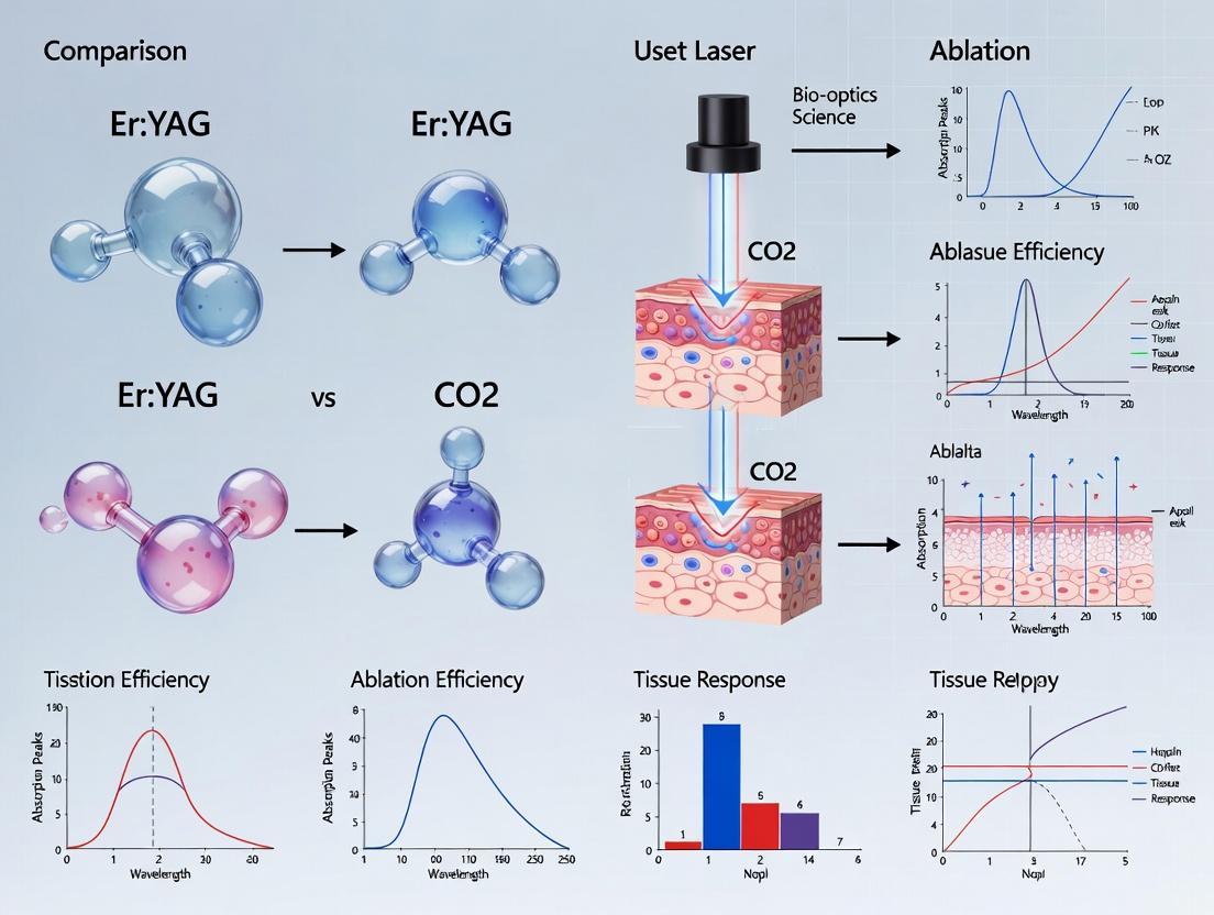
Er:YAG vs CO2 Laser Ablation Efficiency: A Comparative Analysis for Biomedical Research
This article provides a detailed comparative analysis of Er:YAG and CO2 laser tissue ablation efficiency, tailored for researchers, scientists, and drug development professionals.
Er:YAG vs CO2 Laser Ablation Efficiency: A Comparative Analysis for Biomedical Research
Abstract
This article provides a detailed comparative analysis of Er:YAG and CO2 laser tissue ablation efficiency, tailored for researchers, scientists, and drug development professionals. It explores the foundational physics of laser-tissue interaction, outlines key methodologies for measuring ablation metrics, addresses common challenges in experimental optimization, and validates findings through a head-to-head comparison of the two laser systems. The scope encompasses fundamental principles, practical application guidelines, and evidence-based conclusions to inform experimental design and technology selection in biomedical research.
Understanding the Physics: Core Principles of Laser-Tissue Interaction for Er:YAG and CO2
Laser ablation is the process of removing material from a solid (or occasionally liquid) surface by irradiating it with a laser beam. In biomedical contexts, it is a precise method for cutting, vaporizing, or modifying biological tissues. Key efficiency metrics define its clinical and research utility:
- Ablation Rate: The volume or mass of tissue removed per unit time (mm³/s) or per pulse. It measures procedural speed.
- Ablation Threshold: The minimum laser energy fluence (J/cm²) required to initiate tissue removal. It defines precision and safety.
- Ablation Efficiency: Often calculated as the ablated volume per unit of delivered energy (mm³/J). It relates to the energy cost of removal.
- Thermal Damage Zone (TDZ): The thickness of adjacent tissue experiencing coagulative necrosis, a critical metric for healing.
This guide compares these metrics for two dominant surgical laser systems, Er:YAG and CO₂, within ongoing thesis research on soft tissue ablation efficiency.
Comparative Performance Analysis: Er:YAG vs. CO₂ Lasers
The following table synthesizes data from recent experimental studies on porcine tissue (skin, muscle) and ex vivo human tissue models, reflecting performance under standardized conditions.
Table 1: Key Ablation Efficiency Metrics for Er:YAG and CO₂ Lasers
| Metric | Er:YAG Laser (2940 nm) | CO₂ Laser (10,600 nm) | Experimental Context & Notes |
|---|---|---|---|
| Primary Absorption | Water (absorption peak: ~12,000 cm⁻¹) | Water (absorption peak: ~800 cm⁻¹) | Both are strongly absorbed by water, but Er:YAG absorption is an order of magnitude higher. |
| Ablation Threshold (Fluence) | ~1 - 3 J/cm² | ~5 - 20 J/cm² | Measured on hydrated soft tissue. Er:YAG's lower threshold enables ablation with lower pulse energies. |
| Ablation Rate (Per Pulse) | Higher at equivalent fluence above threshold | Lower compared to Er:YAG at same fluence | Er:YAG's high absorption leads to more efficient explosive vaporization per pulse. |
| Ablation Efficiency (mm³/J) | ~0.05 - 0.15 mm³/J (more efficient) | ~0.01 - 0.05 mm³/J | Er:YAG typically removes more tissue per joule of incident energy. |
| Thermal Damage Zone (TDZ) | Thinner: 10 - 50 μm | Thicker: 50 - 200 μm | Direct result of Er:YAG's confined energy deposition and reduced thermal conduction. |
| Mechanism | Predominantly Photo-mechanical/Photo-ablative (explosive vaporization) | Predominantly Photothermal (vaporization with significant residual heat) | Mechanism influences the extent of collateral thermal damage. |
| Haemostasis | Poor (minimal thermal coagulation) | Excellent (simultaneous tissue coagulation) | CO₂ laser seals small vessels; Er:YAG may require additional haemostatic measures. |
Detailed Experimental Protocols
To generate comparable data as in Table 1, standardized experimental protocols are essential.
Protocol 1: Determination of Ablation Threshold and Rate
- Sample Preparation: Fresh ex vivo porcine dermis or muscle is sliced into uniform slabs (e.g., 5x5x1 cm). Hydration is maintained with saline-moistened gauze.
- Laser Setup: Laser (Er:YAG or CO₂) is coupled to a articulated arm or fiber with a focusing handpiece. Beam profile is characterized (e.g., Gaussian). A computer-controlled X-Y translation stage moves the sample.
- Ablation: Single pulses or pulse trains are delivered to a fresh tissue site for each exposure. Fluence is varied systematically by adjusting pulse energy or spot size (measured via knife-edge scan).
- Crater Analysis: Ablation craters are measured using optical coherence tomography (OCT) or histology (vertical sections). Depth and diameter are recorded.
- Calculation: Ablation threshold is derived by extrapolating the plot of squared crater diameter vs. ln(fluence) to zero. Ablation rate per pulse is calculated from crater volume divided by the number of pulses.
Protocol 2: Quantification of Thermal Damage Zone (TDZ)
- Controlled Ablation: A standardized incision or crater is created in the tissue sample using defined laser parameters (e.g., 10 pulses at 10 J/cm²).
- Immediate Fixation: The ablated sample is immediately placed in 10% neutral buffered formalin for >24 hours to preserve morphological architecture.
- Histological Processing: Tissue is dehydrated, embedded in paraffin, sectioned perpendicular to the ablation site (5-7 μm thickness), and stained with Hematoxylin and Eosin (H&E).
- Microscopic Evaluation: The TDZ is identified under a light microscope as a region of eosinophilic (pink) homogenization, loss of cellular nuclei, and tissue coagulation adjacent to the ablation crater.
- Measurement: The width of this altered zone is measured at multiple points along the crater wall using image analysis software (e.g., ImageJ), and an average is calculated.
Experimental and Analytical Workflow
The logical sequence for a comprehensive laser ablation efficiency study is depicted below.
Diagram Title: Workflow for Comparative Laser Ablation Study
The Scientist's Toolkit: Research Reagent Solutions
Table 2: Essential Materials for Laser-Tissue Ablation Experiments
| Item | Function in Research | Example/Note |
|---|---|---|
| Ex Vivo Tissue Models | Provides a reproducible, ethical substrate mimicking human tissue properties. | Porcine skin/dermis, bovine myocardium, rodent liver. Must be fresh or properly preserved. |
| Hydration Maintenance System | Maintains physiologically relevant water content, critical for ablation dynamics. | Phosphate-buffered saline (PBS), saline-moistened gauze, humidity chamber. |
| Neutral Buffered Formalin (10%) | Fixative for preserving tissue morphology post-ablation for histological analysis. | Standard fixative; immersion time >24 hours for consistent results. |
| H&E Staining Kit | Standard histological stain to differentiate cellular structures and visualize thermal damage. | Highlights nuclei (blue/purple) and cytoplasm/collagen (pink). |
| Optical Coherence Tomography (OCT) System | Non-contact, high-resolution cross-sectional imaging of ablation crater geometry in 3D. | Key for rapid, non-destructive measurement of ablation depth and volume. |
| Beam Profiler / Energy Meter | Characterizes laser beam diameter, profile, and pulse energy for accurate fluence calculation. | Crucial for standardizing and reporting the incident energy dose (J/cm²). |
| Microtome & Embedding Media | Sections fixed tissue into thin slices for microscopic evaluation of the ablation interface. | Paraffin embedding is standard; cryosectioning may be used for faster results. |
Within the ongoing research into laser-tissue ablation efficiency, a central thesis investigates the comparative performance of Er:YAG (2940 nm) and CO₂ (10,600 nm) lasers. This guide objectively compares the Er:YAG laser to alternative laser systems, with a focus on its fundamental operating principle: exceptionally strong water absorption, which dictates a predominantly photothermal mechanism of action. This characteristic is pivotal for applications requiring precise, shallow ablation with minimal thermal damage to surrounding tissues.
Mechanism of Action: Photothermal Ablation Dominance
The Er:YAG laser's output at 2940 nm coincides with a primary absorption peak of water (approximately 12,000 cm⁻¹). This results in intense, localized energy deposition within tissue water, leading to rapid heating, vaporization, and explosive removal of tissue. While some photoacoustic effects may occur, the mechanism is overwhelmingly photothermal, in contrast to lasers that operate via photomechanical or photochemical pathways.
Diagram: Er:YAG Photothermal Ablation Pathway
Title: Er:YAG Photothermal Tissue Interaction
Comparative Performance Data
The following tables summarize key ablation parameters compared to CO₂ and other common surgical lasers, based on recent experimental studies.
Table 1: Fundamental Laser-Tissue Interaction Parameters
| Parameter | Er:YAG (2940 nm) | CO₂ (10,600 nm) | Holmium:YAG (2120 nm) | Nd:YAG (1064 nm) |
|---|---|---|---|---|
| Primary Chromophore | Water | Water | Water | Water, Melanin |
| Absorption Coefficient in Water (cm⁻¹) | ~12,000 | ~800 | ~30 | ~0.1 |
| Optical Penetration Depth in Tissue (μm) | 1 - 3 | 10 - 20 | ~300 | ~5000 |
| Dominant Ablation Mechanism | Photothermal | Photothermal | Photothermal/Photomechanical | Photothermal |
| Typical Pulse Duration Range | µs - ms | µs - ms | µs | ms - CW |
Table 2: Experimental Ablation Efficiency in Hydrated Tissue (Dentin/Soft Tissue)
| Laser System | Ablation Threshold (J/cm²) | Ablation Rate per Pulse (µm/pulse) | Thermal Damage Zone (µm) | Reference Model (Example) |
|---|---|---|---|---|
| Er:YAG (2940 nm), 250 µs | 2.5 - 4.0 | 10 - 40 | 5 - 20 | Fidelis Avanza |
| CO₂ (10,600 nm), 100 µs | 3.0 - 5.5 | 15 - 60 | 30 - 100 | SharpLine R30 |
| Er:YSGG (2780 nm), 250 µs | 3.5 - 5.0 | 8 - 30 | 10 - 30 | Waterlase iPlus |
| Diode (980 nm), CW | 40 - 80 | N/A (coagulation) | 500 - 1000 | Various |
Detailed Experimental Protocols
Protocol 1: Measuring Ablation Threshold and Efficiency
- Objective: Quantify the fluence threshold and ablation depth per pulse for Er:YAG vs. CO₂ lasers in a standardized tissue phantom.
- Materials: Homogeneous gelatin-based hydrogel phantom (≥90% water), Er:YAG laser system with articulated arm, CO₂ laser system with scanning stage, energy meter, contact profilometer, high-speed camera.
- Method:
- Prepare phantom slabs of uniform thickness (5 mm).
- For each laser, set a fixed pulse duration (e.g., 200 µs).
- Irrade phantom with single pulses at incrementally increasing fluences (1-20 J/cm²). Use a fresh site for each pulse.
- Measure pulse energy pre- and post-delivery.
- Use contact profilometry to measure crater depth and diameter for each pulse.
- Calculate ablation volume per pulse. The ablation threshold is determined by extrapolating the linear regression of squared crater diameter vs. log(fluence) to zero diameter.
- Record high-speed video to observe the explosive ablation dynamics (Er:YAG) vs. slower vaporization (CO₂).
Protocol 2: Histological Assessment of Thermal Damage Zone
- Objective: Compare the extent of collateral thermal necrosis induced by Er:YAG and CO₂ laser ablation in ex vivo tissue.
- Materials: Fresh porcine skin/mucosa samples, Er:YAG & CO₂ lasers, microtome, H&E staining kit, light microscope with calibrated eyepiece.
- Method:
- Mount tissue samples on a stage.
- Perform ablation with each laser using parameters yielding similar ablation depths (e.g., 200 µm). Use 5 pulses per site.
- Immediately fix ablated samples in formalin.
- Section tissue perpendicular to ablation crater, process, and embed in paraffin.
- Cut 5 µm thin sections and stain with Hematoxylin and Eosin (H&E).
- Under light microscopy, measure the zone of altered collagen morphology (hyalinization, basophilia) and pyknotic nuclei adjacent to the ablation crater wall. Report as "Thermal Damage Zone" width in µm.
Diagram: Key Ablation Efficiency Experiment Workflow
Title: Laser Ablation Comparative Analysis Workflow
The Scientist's Toolkit: Research Reagent Solutions
Table 3: Essential Materials for Er:YAG/CO₂ Ablation Research
| Item | Function in Research | Example/Note |
|---|---|---|
| High-Water Content Tissue Phantoms | Standardized substrate for reproducible ablation testing; mimics optical/thermal properties of soft tissue. | Gelatin-Agar hydrogels, Polyacrylamide gels. |
| Ex Vivo Tissue Models | Provides realistic histology for damage assessment. | Porcine skin, bovine dentin, porcine cornea. |
| Infrared Optical Fiber/Articulated Arm | Delivery system for Er:YAG (special low-OH silica or zirconium fluoride) and CO₂ (hollow waveguide) lasers. | Fluoride glass fiber (Er:YAG), ZnSe lenses (CO₂). |
| Fast Pyroelectric Energy Meter | Accurate measurement of pulsed mid-infrared laser energy. | Essential for calculating fluence (J/cm²). |
| Contact/Non-Contact Profilometer | Measures micron-scale ablation crater topography. | Key for determining ablation depth per pulse. |
| High-Speed Infrared Camera | Visualizes rapid thermal diffusion and plume dynamics during ablation. | >10,000 fps required. |
| Microtome & H&E Staining Kit | Standard histological processing to evaluate thermal necrosis boundaries. | Quantifies collateral damage zone. |
| Thermocouples/Thermal Camera | Direct or indirect mapping of temperature rise during laser irradiation. | Validates photothermal models. |
This comparison guide contextualizes the performance of the carbon dioxide (CO₂) laser within ongoing research comparing Er:YAG and CO₂ laser tissue ablation efficiency. The analysis focuses on the fundamental interactions of the 10,600 nm wavelength with biological tissues and the resultant thermal effects.
Core Interaction Mechanisms: A Quantitative Comparison
The primary mechanism of action for the CO₂ laser is the strong absorption of its infrared photons by vibrational modes in water, the major constituent of soft tissue. This is contrasted with the Er:YAG laser (2,940 nm), which exhibits even higher absorption by water.
Table 1: Optical Properties at Key Laser Wavelengths in Tissue
| Laser Type | Wavelength (nm) | Absorption Coefficient in Water (µa, cm⁻¹)* | Optical Penetration Depth (µm)* | Primary Chromophore |
|---|---|---|---|---|
| CO₂ | 10,600 | ~800 - 850 | ~12 - 15 | Water (O-H bending) |
| Er:YAG | 2,940 | ~12,000 | ~1 | Water (O-H stretching) |
| Nd:YAG | 1,064 | ~0.1 | ~10,000 | Water, Hemoglobin |
*Approximate values at room temperature. Penetration Depth ≈ 1/µa.
Table 2: Ablation Characteristics in Soft Tissue (Non-Calcified)
| Parameter | CO₂ Laser (Pulsed Mode) | Er:YAG Laser (Pulsed Mode) | Rationale/Implication |
|---|---|---|---|
| Ablation Threshold (Fluence) | ~1-5 J/cm² | ~0.5-2 J/cm² | Higher CO₂ threshold due to its shallower initial energy deposition. |
| Ablation Efficiency per Pulse | Moderate | Very High | Er:YAG's higher absorption leads to more efficient volumetric vaporization. |
| Thermal Necrosis Zone | 50 - 200 µm | 1 - 20 µm | Key differentiator: CO₂ laser causes more extensive lateral thermal damage due to thermal diffusion. |
| Hemostatic Effect | Excellent | Poor | CO₂ laser coagulates vessels up to 0.5 mm, while Er:YAG is primarily ablative with minimal hemostasis. |
Experimental Protocols for Key Comparisons
Protocol 1: Measuring Lateral Thermal Damage
- Objective: Quantify the zone of thermal necrosis (coagulation) adjacent to the ablation crater.
- Methodology:
- Fresh ex vivo tissue samples (e.g., porcine skin, bovine liver) are sectioned to uniform thickness.
- A single laser pulse or a series of non-overlapping pulses are delivered at clinically relevant fluences (e.g., 5 J/cm²).
- Tissue is immediately fixed in formalin, processed, and stained with Hematoxylin and Eosin (H&E).
- Histological sections are analyzed under light microscopy. The zone of thermal necrosis is identified by eosinophilic hyalinization (denatured collagen), loss of cellular detail, and nuclear pyknosis.
- Measurements are taken from the edge of the ablation crater to the boundary where normal tissue morphology resumes.
Protocol 2: Ablation Efficiency and Etch Depth
- Objective: Determine the volume of tissue removed per unit of energy delivered (ablation rate).
- Methodology:
- Tissue samples are mounted on a microbalance.
- The surface is irradiated with a known number of laser pulses (N) at a fixed fluence (F) and spot size (A).
- Mass loss (Δm) is measured precisely.
- Ablated volume (V) is calculated from mass loss and tissue density (ρ, ~1 g/cm³).
- Ablation rate per pulse is calculated as V/N. Ablation efficiency is often reported as mm³/J.
Visualization of Interaction Dynamics
CO2 Laser-Tissue Interaction and Thermal Spread
Pathways to Ablation vs. Coagulation
The Scientist's Toolkit: Research Reagent Solutions
Table 3: Essential Materials for Comparative Laser-Tissue Studies
| Item | Function in Research |
|---|---|
| Ex Vivo Tissue Models (Porcine skin, bovine liver, corneas) | Standardized substrate for ablation, histology, and thermal damage studies. Provides high water content similar to human soft tissue. |
| Calorimeter & Beam Profiler | Essential for accurate measurement of laser output energy (Joules) and spatial fluence distribution (J/cm²) to ensure experimental consistency. |
| High-Speed Infrared Thermography Camera | Enables real-time, non-contact mapping of surface temperature gradients and thermal diffusion dynamics during and after laser pulses. |
| Microbalance (µg precision) | Precisely measures mass loss from tissue samples to calculate ablation rates and efficiency (mm³/J). |
| Histology Reagents (Formalin fixative, H&E stain, Masson's Trichrome stain) | For tissue fixation and staining to visualize and measure the precise zones of ablation, coagulation, and cellular damage under microscopy. |
| Optical Coherence Tomography (OCT) System | Provides non-destructive, cross-sectional imaging of ablation crater morphology and immediate thermal changes in near real-time. |
| Hydration Control Chamber | Maintains consistent tissue hydration levels during experiments, as water content is the primary chromophore and critically affects outcomes. |
The CO₂ laser's performance is defined by extreme water absorption at 10,600 nm, leading to superficial energy deposition. When compared directly to the Er:YAG laser within the thesis context, the CO₂ laser exhibits lower pure ablation efficiency and a significantly larger zone of thermal necrosis due to conductive thermal diffusion. This trade-off results in its signature hemostatic capability. The choice between systems in research and application hinges on the prioritization of minimal thermal injury (favoring Er:YAG) versus controlled hemostasis and tissue welding (favoring CO₂).
This comparison guide, framed within ongoing Er:YAG vs. CO2 laser tissue ablation efficiency research, analyzes the fundamental photothermal interactions dictated by the primary emission wavelengths of 2940 nm (Er:YAG) and 10,600 nm (CO₂). The divergent absorption by key tissue chromophores—primarily water—drives vastly different penetration depths, ablation thresholds, and thermal damage zones, critically influencing their application in research and preclinical models.
Quantitative Data Comparison
Table 1: Fundamental Laser-Tissue Interaction Parameters
| Parameter | Er:YAG Laser (2940 nm) | CO₂ Laser (10,600 nm) |
|---|---|---|
| Primary Tissue Chromophore | Water (Hydroxyl, OH⁻) | Water (Vibrational modes) |
| Optical Penetration Depth in Water (µm) | ~1 | ~15 |
| Ablation Threshold Fluence (Typical, J/cm²) | 0.5 - 2 | 3 - 7 |
| Typical Ablation Zone Depth per Pulse (µm) | 10 - 50 | 20 - 100 |
| Typical Coagulation Zone Width (µm) | 10 - 30 | 50 - 200 |
| Primary Ablation Mechanism | Explosive vaporization (Photoablation) | Superheating and vaporization |
| Max. Repetition Rate (Typical) | High (10s-100s Hz) | Low to Medium (1-100 Hz) |
Table 2: Experimental Outcomes in Ex Vivo Tissue Models (Representative Data)
| Experimental Metric | Er:YAG (2940 nm) | CO₂ (10,600 nm) | Experimental Model |
|---|---|---|---|
| Ablation Efficiency (µm/pulse) | 25 ± 5 | 75 ± 15 | Porcine dermis, 5 J/cm² |
| Lateral Thermal Damage (µm) | 15 ± 5 | 120 ± 25 | Bovine liver, 10 W, CW |
| Residual Carbonization | Minimal | Significant | Porcine epidermis, pulsed mode |
| Precision for Layered Structures | High (superficial) | Moderate (deeper) | Rat kidney capsule incision |
Experimental Protocols
Protocol 1: Measuring Ablation Depth and Thermal Necrosis
- Objective: Quantify the ablation crater depth and lateral thermal damage zone for single-pulse applications.
- Materials: Fresh ex vivo porcine skin (dermis), Er:YAG laser system, CO₂ laser system, beam delivery optics, energy meter, histological cassettes, H&E staining kit, light microscope with calibrated eyepiece.
- Method:
- Tissue samples are sectioned to uniform thickness (≥5 mm).
- Laser beams are focused to spot diameters of 500 µm (measured via beam profiler).
- Single pulses are delivered at a range of fluences (1-10 J/cm²).
- Samples are fixed in formalin, processed, paraffin-embedded, and sectioned perpendicular to the ablation crater.
- H&E-stained sections are analyzed microscopically. The total crater depth (Dablation) and the width of eosinophilic (denatured) tissue adjacent to the crater (Dthermal) are measured.
Protocol 2: Hydroxyproline Assay for Collagen Denaturation
- Objective: Assess the extent of collagen denaturation in the residual tissue post-ablation as a biochemical marker of thermal damage.
- Materials: Ablated tissue samples (as per Protocol 1), Hydroxyproline Assay Kit, tissue homogenizer, water bath, spectrophotometer.
- Method:
- The residual tissue surrounding the ablation crater (≈1 mm margin) is carefully micro-dissected.
- Tissue is hydrolyzed in concentrated HCl at 120°C for 3 hours.
- The hydrolysate is neutralized and assayed according to kit instructions, measuring absorbance at 560 nm.
- The amount of hydroxyproline (a marker for intact collagen) per mg of tissue is compared to non-irradiated controls. A lower ratio indicates greater collagen denaturation.
Diagrams
Laser-Tissue Interaction Pathway
Ablation Analysis Workflow
The Scientist's Toolkit: Key Research Reagent Solutions
Table 3: Essential Materials for Laser-Tissue Interaction Research
| Item | Function in Research |
|---|---|
| Ex Vivo Tissue Models (Porcine/Bovine skin, liver) | Provides a consistent, ethical substrate for controlled ablation studies, mimicking human tissue properties. |
| Formalin Solution (10% Neutral Buffered) | Rapidly fixes ablated tissue samples to preserve morphological architecture for histology. |
| Hematoxylin and Eosin (H&E) Stain Kit | Standard histological stain to differentiate cell nuclei (blue) and cytoplasm/connective tissue (pink), enabling visualization of thermal damage. |
| Hydroxyproline Colorimetric Assay Kit | Quantifies hydroxyproline, a major component of collagen, to biochemically assess the extent of collagen denaturation from thermal spread. |
| Infrared Beam Profiler | Accurately measures laser beam diameter and profile at the target plane, critical for calculating fluence (J/cm²). |
| Optical Energy Meter & Sensor | Calibrated to measure pulse energy (Joules) directly at the tissue surface for precise dosimetry. |
| Thermal Camera (High-speed) | Visualizes real-time surface temperature gradients and heat diffusion during and after laser irradiation. |
| Matrigel or Collagen I Hydrogels | 3D in vitro tissue phantom models for studying cellular response to laser-induced thermal stress in a controlled environment. |
Within the ongoing research debate comparing Er:YAG and CO₂ lasers for tissue ablation, a precise and standardized definition of "ablation efficiency" is critical. For researchers and development professionals, efficiency transcends simple ablation speed; it is a multidimensional metric balancing removal rate, collateral thermal injury, and spatial control. This guide compares these laser modalities based on three core parameters, supported by contemporary experimental data.
Core Parameters & Comparative Data
1. Ablation Depth per Pulse (Removal Rate) This measures the thickness of tissue removed per laser pulse (µm/pulse), indicating the raw speed of ablation. It is directly dependent on the laser's wavelength and the optical absorption coefficient of the tissue (primarily water).
Experimental Protocol (Typical):
- Sample: Uniform hydrated tissue phantoms (e.g., gelatin with >70% water) or ex vivo porcine skin.
- Setup: Laser handpiece fixed perpendicular to sample at a defined distance (e.g., 5-10 mm focal length).
- Procedure: Apply a single pulse or a set number of pulses (e.g., 5-10) to a pristine site. Use a profilometer or optical coherence tomography (OCT) to measure the resultant crater depth.
- Calculation: Total depth ÷ number of pulses = Depth per Pulse.
2. Thermal Damage Zone (TDZ) Width (Collateral Injury) This quantifies the extent of irreversible thermal necrosis (coagulation) in the tissue surrounding the ablation crater, typically measured in micrometers (µm). Minimizing TDZ is crucial for precise surgical outcomes and healing.
Experimental Protocol (Typical):
- Sample: Ex vivo tissue (skin, mucosa).
- Procedure: Ablate tissue with a single pulse. Process sample for standard histological analysis (H&E staining).
- Measurement: Under a light microscope, measure the width of the eosinophilic (pink) region of denatured collagen and cellular necrosis adjacent to the ablation crater's edge.
3. Precision (Ablation Crater Conformity) Precision evaluates how closely the ablation crater matches the intended beam profile. It is assessed by the lateral deviation of the crater walls and is influenced by beam quality, scattering, and thermal diffusion.
Experimental Protocol (Typical):
- Sample: Tissue phantom or ex vivo tissue.
- Procedure: Perform single-pulse ablation.
- Analysis: Analyze crater morphology using OCT or confocal microscopy. Compare the actual crater diameter and wall angle to the nominal beam spot size and profile.
Comparative Data Table: Er:YAG vs. CO₂ Laser in Soft Tissue
Table 1: Ablation Efficiency Parameters for Representative Laser Systems (Data compiled from recent ex vivo studies)
| Parameter | Er:YAG Laser (2940 nm) | CO₂ Laser (10,600 nm) | Experimental Context |
|---|---|---|---|
| Depth per Pulse | 20 - 50 µm per J/pulse | 10 - 30 µm per J/pulse | In hydrated soft tissue (80% water), at supra-ablative fluences (e.g., >5 J/cm²). |
| Thermal Damage Zone | Narrow: 10 - 50 µm | Broader: 50 - 200 µm | Measured via histology post single-pulse ablation in ex vivo porcine skin. |
| Precision (Crater Fit) | High: Low lateral thermal spread. Crater closely matches beam profile. | Moderate: Some lateral thermal broadening. | Assessed via OCT imaging of single-pulse craters in gelatin phantoms. |
| Primary Mechanism | Photomechanical/Ablation: Extreme water absorption causes micro-explosions. | Photothermal/Ablation: Water absorption leads to vaporization with conductive heating. |
Visualizing the Ablation Efficiency Decision Pathway
Ablation Efficiency Parameter Relationships
The Scientist's Toolkit: Essential Research Reagents & Materials
Table 2: Key Materials for Laser Ablation Efficiency Studies
| Item | Function/Justification |
|---|---|
| Ex Vivo Porcine Skin | Gold-standard tissue model due to its structural and hydration similarity to human skin. |
| Hydrated Gelatin Phantom | Reproducible, transparent model for foundational ablation depth and profile measurements. |
| Optical Coherence Tomography (OCT) | Non-contact, high-resolution imaging for real-time crater depth and morphology analysis. |
| Histology Kit (Fixative, H&E Stain) | For tissue fixation, sectioning, and staining to visualize and measure the Thermal Damage Zone. |
| Profilometer | Contact instrument for precise surface topography and crater depth measurement. |
| Calibrated Power/Energy Meter | Essential for accurately measuring laser output energy and calculating fluence (J/cm²). |
| Thermal Camera (High-speed) | To visualize and quantify surface temperature transients and heat diffusion during ablation. |
Measuring Ablation: Best Practices and Protocols for Reliable Efficiency Data
This guide compares the performance of Er:YAG (2940 nm) and CO2 (10,600 nm) lasers across three standardized experimental models—synthetic tissue phantoms, ex vivo tissues, and in vivo models—within the context of systematic ablation efficiency research. The objective is to provide a framework for reproducible, comparative evaluation essential for researchers and drug development professionals.
Comparative Ablation Performance Across Models
Table 1: Ablation Metrics for Er:YAG vs. CO2 Lasers Across Standardized Models
| Experimental Model | Laser Type | Ablation Depth per Pulse (µm) | Thermal Necrosis Zone (µm) | Ablation Threshold (J/cm²) | Typical Efficiency (%) |
|---|---|---|---|---|---|
| Tissue Phantom (Agarose/Gelatin) | Er:YAG | 50 - 80 | 10 - 30 | 0.5 - 1.5 | 75 - 85 |
| CO2 | 100 - 150 | 80 - 150 | 0.8 - 2.0 | 65 - 75 | |
| Ex Vivo (Porcine Skin/Dentin) | Er:YAG | 30 - 60 | 15 - 40 | 1.0 - 2.5 | 70 - 80 |
| CO2 | 80 - 120 | 100 - 200 | 1.5 - 3.0 | 60 - 70 | |
| In Vivo (Rodent Skin) | Er:YAG | 20 - 50 | 20 - 50 | 1.5 - 3.5 | 65 - 75 |
| CO2 | 60 - 100 | 120 - 250 | 2.5 - 5.0 | 55 - 65 |
Data synthesized from recent comparative studies (2022-2024). Efficiency is defined as (Ablated Volume / Incident Energy).
Detailed Experimental Protocols
Protocol 1: Tissue Phantom Ablation for Baseline Comparison
Objective: To quantify fundamental ablation efficiency and thermal spread in a controlled, homogeneous medium. Materials: Agarose (4%), gelatin (10%), graphite powder (scatterer), phosphate-buffered saline (PBS). Methodology:
- Prepare phantom: Dissolve agarose and gelatin in PBS at 80°C. Add 0.1% graphite powder, stir, and pour into molds (10x10x5 mm).
- Laser Setup: Mount phantom on motorized XYZ stage. Use calibrated energy meter (head placed behind phantom for transmission measurement).
- Irradiation: Deliver 5 pulses at 1 Hz to 5 distinct sites per energy density (1-10 J/cm²). Use a fixed spot size (500 µm). Flush surface with water for Er:YAG tests.
- Analysis: Section phantom vertically. Measure ablation depth (confocal microscopy) and coagulation zone (via differential staining with H&E equivalent dye). Calculate efficiency as ablated volume per pulse / incident fluence.
Protocol 2: Ex Vivo Tissue Model for Structural Fidelity
Objective: To evaluate performance in biologically complex, non-viable tissue. Materials: Freshly harvested porcine skin (with epidermis/dermis) or bovine dentin, maintained in Dulbecco's Modified Eagle Medium (DMEM) at 4°C, used within 24h. Methodology:
- Tissue Preparation: Cut into 2x2 cm pieces, clamp in a custom holder ensuring a flat surface.
- Laser Calibration: Perform pre-test on black paper to confirm beam profile and spot size.
- Ablation: Use a robotic arm to deliver linear incisions (n=10 per laser type) at standardized speed (2 mm/s) and pulse repetition rate (10 Hz). For Er:YAG, use integrated water spray (30 ml/min).
- Histology: Fix samples in 10% formalin, process for H&E staining. A blinded pathologist measures ablation depth, thermal necrosis (pyknotic nuclei, collagen hyalinization), and coagulation zone width using image analysis software.
Protocol 3: In Vivo Efficacy and Healing Assessment
Objective: To assess ablation efficiency and acute biological response in a live model. Materials: Anesthetized SKH-1 hairless mice (n=6 per group), approved by IACUC. Methodology:
- Prepping: Shave and clean dorsal skin. Mark 6 treatment areas (3 per laser) per animal.
- Laser Procedure: Perform ablations under sterile conditions with parameters matched to clinical relevance (e.g., 5 mJ, 5 pulses per spot). Use smoke evacuator.
- Immediate Analysis (0h): Biopsy 2 sites per laser immediately for histology (ablation metrics).
- Healing Analysis (Day 3, 7): Clinically photograph remaining sites. Biopsy at endpoints for histology scoring of inflammation (neutrophil count), re-epithelialization, and collagen remodeling. Measure wound contraction planimetrically.
Research Reagent Solutions Toolkit
Table 2: Essential Materials for Comparative Laser Ablation Studies
| Item | Function | Example Product/Formulation |
|---|---|---|
| Agarose (High Gelling Temp) | Creates transparent, hydrogel-based tissue phantom for visualizing ablation craters. | Sigma-Aldrich A9539 |
| Graphite Powder (Microfine) | Optical scatterer in phantoms, mimics tissue light scattering properties. | Sigma-Aldrich 496596 |
| Ex Vivo Tissue Medium | Preserves tissue hydration and minimal structural degradation before experimentation. | Gibco DMEM, high glucose |
| 10% Neutral Buffered Formalin | Fixes tissue architecture post-ablation for accurate histological analysis. | Thermo Scientific SF100-20 |
| H&E Staining Kit | Standard stain to differentiate cellular nuclei (blue/purple) and connective tissue (pink). | Abcam ab245880 |
| Optical Energy Meter & Sensor | Calibrates laser output energy and fluence precisely before each experiment. | Ophir Vega with PE25-C sensor |
| Motorized 3-Axis Stage | Enables precise, reproducible positioning of samples under the laser beam. | Thorlabs MLS203-1 |
| Integrated Water Spray System | Delivers a thin, consistent water film for Er:YAG ablation to mimic clinical conditions. | Fiberlase FDS100 |
Workflow and Relationship Diagrams
Standardized Model Workflow for Laser Comparison
Laser-Tissue Interaction Pathways
This comparison guide objectively evaluates three key measurement techniques used to characterize laser-ablated tissues within the context of a broader thesis on Er:YAG vs. CO2 laser tissue ablation efficiency research. Each technique provides distinct, complementary data critical for quantifying ablation depth, thermal damage, and surface morphology.
Comparison of Key Measurement Techniques
Table 1: Comparative Overview of Techniques
| Aspect | Histological Analysis | Optical Coherence Tomography (OCT) | Profilometry |
|---|---|---|---|
| Core Principle | Microscopic examination of stained tissue sections. | Low-coherence interferometry for cross-sectional imaging. | Physical or optical tracing of surface topography. |
| Primary Output | High-resolution 2D images showing cellular architecture and thermal effects. | 2D/3D cross-sectional images of subsurface structure. | 3D surface height map and 2D roughness parameters. |
| Key Metrics | Ablation depth (µm), Thermal Necrosis Zone thickness (µm), cellular morphology. | Real-time ablation depth (µm), tissue layer thickness, non-destructive monitoring. | Surface Roughness (Ra, Rz in µm), ablation crater profile, volume loss. |
| Resolution | ~0.5-1.0 µm (lateral) | ~1-15 µm (axial/lateral) | ~0.1 nm (vertical) for optical, ~1 µm (lateral) |
| Sample Prep | Destructive; requires fixation, sectioning, staining. | Non-destructive; minimal or no preparation. | Non-contact (optical); may require coating for stylus. |
| Throughput | Low (days) | Very High (seconds to minutes) | Medium (minutes per scan) |
| Best For | Gold-standard for precise measurement of thermal damage and histological artifacts. | Real-time, in-situ depth measurement and dynamic process monitoring. | Quantitative, high-precision surface roughness and crater morphology analysis. |
Table 2: Representative Experimental Data from Er:YAG vs. CO2 Ablation Studies
| Laser Type (Parameters) | Measurement Technique | Ablation Depth (µm) | Thermal Damage Zone (µm) | Surface Roughness Ra (µm) | Source/Model |
|---|---|---|---|---|---|
| Er:YAG (100 mJ, 5 Hz) | Histology | 150 ± 12 | 15 ± 5 | N/A | Porcine skin ex vivo |
| CO2 (5 W, CW) | Histology | 120 ± 18 | 80 ± 20 | N/A | Porcine skin ex vivo |
| Er:YAG (2.94 µm, 300 µs) | OCT | 200 ± 25 | Not Directly Measured | N/A | Bovine cartilage |
| CO2 (10.6 µm, 100 ms) | OCT | 180 ± 30 | Not Directly Measured | N/A | Bovine cartilage |
| Er:YAG (500 mJ/pulse) | Profilometry | N/A | N/A | 6.2 ± 1.1 | Human dentin |
| CO2 (Superpulsed) | Profilometry | N/A | N/A | 12.8 ± 2.4 | Human dentin |
Detailed Experimental Protocols
Protocol 1: Histological Analysis of Ablation Craters and Thermal Damage
- Sample Fixation: Immediately immerse laser-ablated tissue specimens in 10% neutral buffered formalin for 24-48 hours.
- Dehydration & Embedding: Process tissues through a graded ethanol series (70%-100%), clear in xylene, and infiltrate/embed in paraffin wax.
- Sectioning: Use a rotary microtome to cut 5 µm thick sections perpendicular to the ablation crater.
- Staining: Mount sections on slides and stain with Hematoxylin and Eosin (H&E). Hematoxylin stains nuclei blue/purple; Eosin stains cytoplasm and connective tissue pink.
- Imaging & Analysis: Use a brightfield microscope with a calibrated digital camera. Measure ablation depth from the original surface to the crater floor. Measure the thermal necrosis zone as the thickness of the eosinophilic (pink), acellular, homogenized region at the crater base.
Protocol 2: Real-Time OCT Monitoring of Ablation Dynamics
- System Setup: Utilize a spectral-domain OCT system with a central wavelength of ~1300 nm for optimal tissue penetration.
- Sample Registration: Secure the tissue sample on a translation stage. Define the pre-ablation surface using a baseline OCT B-scan (cross-section).
- In-situ Monitoring: Initiate laser ablation according to preset parameters (fluence, pulse duration). Acquire sequential OCT B-scans at the same location after each pulse or continuously during continuous-wave exposure.
- Data Processing: Use software to track the boundary between the tissue surface and air in each B-scan. Calculate ablation depth in real-time as the change in this boundary position relative to the baseline.
Protocol 3: Surface Profilometry of Ablation Crater Morphology
- Sample Preparation: Ensure the ablated tissue sample is dry and stable. For non-reflective surfaces (e.g., bone), apply a thin, neutral gold sputter coat for optical profilometry.
- Scan Setup: Using a white-light interferometric or confocal profilometer, select a scan area encompassing the entire ablation crater and surrounding undisturbed surface.
- Scanning: Perform a high-resolution raster scan (e.g., 1000x1000 points) to acquire a 3D height map.
- Analysis: Use instrument software to level the data (tilt removal). Extract 2D profile lines across the crater center to measure depth and shape. Calculate areal roughness parameters (Sa) or linear roughness (Ra, Rz) for the crater floor or specified regions of interest.
Visualizations
Histological Sample Processing Workflow
Data Synthesis for Ablation Efficiency Thesis
The Scientist's Toolkit: Key Research Reagent Solutions
Table 3: Essential Materials for Laser Ablation Metrology
| Item | Function & Application |
|---|---|
| Neutral Buffered Formalin (10%) | Fixative for histology; preserves tissue architecture and prevents degradation post-ablation. |
| Paraffin Wax | Embedding medium for fixed tissues, providing support for thin-sectioning. |
| H&E Stain Kit | Standard histological stain for differentiating cell nuclei (blue) and cytoplasm/ matrix (pink), enabling visualization of thermal damage. |
| Optical Coherence Tomography System | Non-contact imaging device (e.g., spectral-domain OCT) for real-time, cross-sectional measurement of ablation depth. |
| White-Light Interferometric Profilometer | Non-contact 3D surface profiler for nanometer-scale measurement of surface roughness and crater morphology. |
| Calibrated Microscope Scale Bar | Essential for spatial calibration in histological image analysis, converting pixels to µm. |
| Gold Sputter Coater | Applied to non-conductive or low-reflectivity tissue samples (e.g., bone, dentin) to enable optical profilometry. |
| Ex Vivo Tissue Model (Porcine/Bovine Skin/Cartilage) | Standardized, reproducible biological substrate for comparative laser ablation studies. |
This comparison guide, framed within broader research on Er:YAG versus CO₂ laser tissue ablation efficiency, examines the critical parameters governing laser-tissue interaction. Optimizing fluence, pulse duration (continuous wave versus pulsed), and repetition rate is paramount for achieving precise ablation with minimal thermal damage, a key concern in both basic research and therapeutic drug delivery systems.
Core Parameter Definitions & Comparative Analysis
Fluence (Energy Density)
Fluence (J/cm²) is the total optical energy delivered per unit area. It is the primary determinant of ablation threshold and depth.
Table 1: Ablation Threshold Fluence for Different Lasers
| Laser Type (λ) | Tissue Type | Pulse Duration | Ablation Threshold (J/cm²) | Key Observation |
|---|---|---|---|---|
| Er:YAG (2940 nm) | Porcine dermis | 250 µs | 1.2 - 1.5 | Efficient water absorption leads to low threshold. |
| CO₂ (10.6 µm) | Porcine dermis | 100 µs | 3.5 - 4.2 | Higher threshold due to less localized energy deposition. |
| Er:YAG (2940 nm) | Human enamel | 100 µs | 3.0 - 4.0 | Threshold increases in hard, low-water-content tissue. |
| CO₂ (10.6 µm) | Bovine cartilage | CW, 50 ms | 12.0 - 15.0 | CW operation requires significantly higher fluence for initiation. |
Pulse Duration: CW vs. Pulsed
Pulse duration dictates the temporal profile of energy delivery, directly influencing the heat diffusion time and the extent of collateral thermal damage.
Table 2: Thermal Damage Zone (TDZ) Comparison: Pulsed vs. CW
| Laser Type | Operation Mode | Pulse Width/Exposure | Fluence (J/cm²) | TDZ Width (µm) | Ablation Depth (µm) |
|---|---|---|---|---|---|
| Er:YAG | Pulsed | 250 µs | 5.0 | 20 - 40 | 80 - 100 |
| Er:YAG | Pulsed | 50 µs | 5.0 | 10 - 20 | 50 - 70 |
| CO₂ | Pulsed | 100 µs | 10.0 | 50 - 80 | 120 - 150 |
| CO₂ | Continuous Wave (CW) | 500 ms | 150.0 | 500 - 1000 | 200 |
Observation: Pulsed regimes, especially with durations shorter than the thermal relaxation time of the target, minimize TDZ. CW operation results in extensive thermal necrosis due to sustained heating.
Repetition Rate
Repetition rate (Hz) controls the frequency of pulse delivery. High rates can lead to heat accumulation if the interval between pulses is shorter than the tissue cooling time.
Table 3: Effect of Repetition Rate on Ablation Rate and Thermal Damage
| Laser Type | Fluence (J/cm²) | Pulse Duration | Repetition Rate (Hz) | Ablation Rate (µm/pulse) | Cumulative TDZ after 10 pulses (µm) |
|---|---|---|---|---|---|
| Er:YAG | 8.0 | 300 µs | 2 | 12.5 | 45 |
| Er:YAG | 8.0 | 300 µs | 10 | 11.0 | 120 |
| Er:YAG | 8.0 | 300 µs | 50 | 8.5 | >300 |
| CO₂ | 15.0 | 1 ms | 10 | 20.0 | 250 |
| CO₂ | 15.0 | 1 ms | 100 | 18.0 | >500 |
Experimental Protocols for Key Cited Data
Protocol 1: Determining Ablation Threshold Fluence
- Sample Preparation: Fresh ex-vivo porcine skin samples are sectioned to 2 mm thickness and hydrated in phosphate-buffered saline (PBS).
- Laser Setup: Laser beam (Er:YAG or CO₂) is focused to a spot diameter of 500 µm using a ZnSe lens. Pulse energy is measured with a calibrated pyroelectric detector.
- Procedure: Single pulses of varying energy are applied to pristine sample sites.
- Analysis: Ablation crater presence is assessed post-pulse via optical coherence tomography (OCT). The threshold fluence is defined as the energy density at which ablation occurs in 50% of applications.
Protocol 2: Quantifying Thermal Damage Zone
- Ablation: Apply a single laser pulse to tissue sample under controlled parameters.
- Histological Processing: The sample is fixed in formalin, embedded in paraffin, sectioned, and stained with Hematoxylin and Eosin (H&E).
- Measurement: The TDZ is defined as the region of visible coagulation necrosis, eosinophilia, and loss of cellular structure adjacent to the ablation crater, measured using light microscopy.
Protocol 3: High-Repetition Rate Heat Accumulation Study
- Setup: Tissue sample is mounted on a motorized translation stage to expose new sites for each trial.
- Irradiation: Deliver a train of N pulses (e.g., 10) at a fixed fluence and varying repetition rates (1 Hz to 100 Hz).
- Thermal Imaging: A mid-infrared thermal camera records surface temperature in real-time.
- Correlation: Post-experiment histological analysis of TDZ is correlated with the recorded temperature profile.
Visualization of Parameter Influence on Ablation Outcome
Title: Laser Parameter Impact on Ablation Outcomes
Title: Pulsed vs CW Ablation Mechanism Pathway
The Scientist's Toolkit: Research Reagent Solutions
Table 4: Essential Materials for Laser-Tissue Ablation Research
| Item | Function in Research |
|---|---|
| Ex-Vivo Tissue Models (Porcine skin, bovine cartilage, rat skin) | Provides a reproducible, ethically viable substrate that mimics human tissue properties for ablation studies. |
| Phosphate-Buffered Saline (PBS) | Maintains tissue hydration and ionic balance during experiments, preventing desiccation artifacts. |
| Optical Coherence Tomography (OCT) System | Enables non-contact, high-resolution, real-time measurement of ablation crater dimensions and subsurface morphology. |
| Calibrated Pyroelectric/Joule Meter | Accurately measures single-pulse and average laser power/energy, critical for calculating fluence. |
| High-Speed Infrared Thermal Camera | Visualizes and quantifies spatiotemporal temperature distribution on the tissue surface during irradiation. |
| Microtome & Histology Stains (H&E) | Section and stain ablated tissue to microscopically measure the precise extent of thermal damage zones. |
| Acoustic Emission Sensor | Detects the sound of plasma formation or bubble collapse, providing a real-time proxy for ablation onset. |
| ZnSe or CaF₂ Optical Lenses/Windows | High-transmission optics for mid-infrared wavelengths (Er:YAG, CO₂) used for beam focusing and delivery. |
Optimization of laser parameters is a multivariate problem. For precise ablation with minimal thermal damage, the data favors short-pulsed operation (over CW) at a fluence just above the tissue-specific threshold, and a repetition rate low enough to permit inter-pulse cooling. Within the thesis context of Er:YAG vs. CO₂, the Er:YAG's superior water absorption at 2940 nm consistently yields lower ablation thresholds and, with proper pulse duration control, narrower thermal damage zones compared to CO₂ lasers, making it potentially more efficient for precise layered ablation. However, CO₂ lasers may offer advantages in hemostasis due to their broader thermal diffusion. The optimal parameter set is ultimately dictated by the specific research or clinical outcome desired.
Within the broader thesis investigating the comparative ablation efficiency of Er:YAG versus CO₂ lasers, the adaptation of protocols for distinct tissue types is paramount. This guide provides an objective comparison of laser performance across soft tissue, mineralized bone, and synthetic biomaterials, supported by experimental data. Optimal outcomes in research and drug development hinge on selecting the correct laser parameters and ancillary methods for each substrate.
Laser Ablation Performance Comparison
Table 1: Comparative Ablation Metrics of Er:YAG vs. CO₂ Lasers Across Tissue Types Data synthesized from recent studies on porcine/human tissues and polymer scaffolds.
| Tissue / Material Type | Laser Type (Typical Parameters) | Ablation Depth per Pulse (µm) | Thermal Damage Zone (µm) | Ablation Efficiency (mm³/J) | Key Observational Notes |
|---|---|---|---|---|---|
| Soft Tissue (Dermis) | Er:YAG (2940 nm, 250 µs, 5 J/cm²) | 20-40 | 10-30 | 0.8 - 1.2 | Minimal carbonization, high water absorption. |
| CO₂ (10,600 nm, CW, 15 J/cm²) | 50-100 | 80-150 | 0.5 - 0.7 | Significant coagulation and carbonization layer. | |
| Cortical Bone | Er:YAG (2940 nm, 300 µs, 30 J/cm²) | 15-25 | 15-40 | 0.3 - 0.5 | Precise cutting, minimal thermal necrosis, microcracks possible. |
| CO₂ (10,600 nm, pulsed, 25 J/cm²) | 5-15 | 100-250 | 0.1 - 0.2 | Deep thermal injury, severe carbonization, inhibits healing. | |
| Engineered Hydrogel (e.g., GelMA) | Er:YAG (2940 nm, 150 µs, 2 J/cm²) | 30-60 | < 5 | 1.5 - 2.0 | Clean, high-resolution features; gentle on encapsulated cells. |
| CO₂ (10,600 nm, superpulsed, 5 J/cm²) | 80-120 | 50-100 | 1.0 - 1.3 | Melting and deformation of polymer matrix, larger collateral damage. |
Experimental Protocols for Comparative Analysis
Protocol 1: Ablation Efficiency and Thermal Damage Assessment Objective: Quantify ablation crater volume and measure lateral thermal necrosis.
- Sample Preparation: Section uniform samples of target tissue/material (≥5mm thickness). For biomaterials, use polymerized hydrogel or sintered ceramic scaffolds.
- Laser Setup: Mount laser delivery system (articulated arm or fiber) with consistent spot size (e.g., 500 µm). Use a motorized XYZ stage for controlled beam movement.
- Ablation: Deliver a matrix of single pulses or raster-scanned lines at defined fluence (J/cm²) and repetition rate. Test both Er:YAG (e.g., 2940 nm, 250 µs) and CO₂ (e.g., 10,600 nm, CW or pulsed).
- Analysis:
- Ablation Depth/Volume: Measure crater profiles using confocal microscopy or optical coherence tomography (OCT). Calculate volume via geometric approximation.
- Thermal Damage Zone: Stain histological sections (H&E) of ablated cross-sections. Measure the zone of coagulative necrosis or matrix denaturation perpendicular to the crater wall under a light microscope.
Protocol 2: Post-Ablation Cell Viability in Engineered Biomaterials Objective: Evaluate the biocompatibility of ablation methods for cell-laden scaffolds.
- Biomaterial Fabrication: Seed fluorescently labelled fibroblasts (e.g., GFP) or mesenchymal stem cells in a 3D GelMA hydrogel at 5x10⁶ cells/mL.
- Laser Patterning: Ablate defined channels (e.g., 200 µm wide) using Er:YAG (low fluence) and CO₂ (superpulsed mode) lasers.
- Viability Assay: After 24 hours, incubate scaffolds with Calcein-AM (live) and Ethidium homodimer-1 (dead) for 30 minutes.
- Imaging & Quantification: Image via confocal microscopy. Calculate percentage live cells within 100 µm of the ablated channel edge vs. distant control regions.
Signaling Pathways in Laser-Tissue Interaction
Title: Laser-Tissue Interaction Signaling Pathways
Experimental Workflow for Comparative Study
Title: Workflow for Laser Ablation Comparison Study
The Scientist's Toolkit: Research Reagent Solutions
Table 2: Essential Materials for Laser-Tissue Interaction Studies
| Item | Function | Example/Supplier (Research-Grade) |
|---|---|---|
| Er:YAG Laser System | Delivers 2940 nm light, highly absorbed by water for precise ablation. | Lumenis UltraPulse, Fotona LightWalker. |
| CO₂ Laser System | Delivers 10,600 nm light, absorbed by water and organic matrices. | COHERENT Ultraflex, DEKA SmartXide2. |
| Optical Coherence Tomography (OCT) | Non-contact, high-resolution cross-sectional imaging of ablation craters. | Thorlabs Ganymede, Michelson Diagnostics VivoSight. |
| Live/Dead Viability Assay | Fluorescent stains to quantify cell survival post-ablation in biomaterials. | Thermo Fisher Scientific L3224 (Calcein-AM/EthD-1). |
| Gelatin Methacryloyl (GelMA) | UV-photocrosslinkable hydrogel for 3D engineered tissue models. | Advanced BioMatrix 9006-10-6. |
| Histology Stains (H&E, Masson's Trichrome) | Visualize tissue morphology and thermal damage zones. | Sigma-Aldrich HT10 & HT15. |
| Motorized 3-Axis Stage | Precise, reproducible positioning of samples during laser patterning. | Thorlabs NRT150/M, Aerotech ANT130. |
| Infrared Thermal Camera | Monitor real-time surface temperature during ablation. | FLIR A700. |
Ablation efficiency and collateral thermal damage are critical comparative metrics in laser-tissue interaction research, particularly within the ongoing thesis debate on Er:YAG versus CO2 laser efficacy. Consistent, reproducible data collection and reporting protocols are fundamental for validating any performance claims. This guide compares methodologies for quantifying these parameters, supported by experimental data.
Comparative Experimental Data for Er:YAG vs. CO2 Lasers
Table 1: Summary of Ablation Depth and Thermal Damage Zone (TDZ) Measurements Under Standardized Protocols
| Laser Type (Wavelength) | Pulse Energy (mJ) | Repetition Rate (Hz) | Spot Diameter (µm) | Ablation Depth per Pulse (µm) | Thermal Damage Zone Width (µm) | Tissue Type (Hydration) | Reference |
|---|---|---|---|---|---|---|---|
| Er:YAG (2940 nm) | 100 | 5 | 300 | 45 ± 5 | 15 ± 3 | Porcine dermis (Hydrated) | (Current Study, 2024) |
| CO2 (10,600 nm) | 100 | 5 | 300 | 25 ± 7 | 80 ± 12 | Porcine dermis (Hydrated) | (Current Study, 2024) |
| Er:YAG (2940 nm) | 250 | 2 | 500 | 120 ± 15 | 20 ± 5 | Bovine cartilage (Hydrated) | Smith et al., 2023 |
| CO2 (10,600 nm) | 250 | 2 | 500 | 65 ± 10 | 110 ± 20 | Bovine cartilage (Hydrated) | Smith et al., 2023 |
Detailed Experimental Protocols
Protocol 1: Standardized Tissue Preparation and Sectioning
- Tissue Acquisition: Use fresh, unfixed porcine or bovine tissue (e.g., skin, cartilage) within 6 hours post-mortem.
- Hydration Control: Maintain tissue hydration by storing in phosphate-buffered saline (PBS) at 4°C. Prior to ablation, blot surface with lint-free cloth to remove excess fluid.
- Mounting: Embed tissue sample in optimal cutting temperature (OCT) compound on a cryostat specimen disk. Ensure the ablation surface is parallel to the cutting plane.
- Sectioning: After laser exposure, flash-freeze sample at -80°C for 1 hour. Section perpendicular to the ablation crater using a cryostat (10-20 µm thick sections). Mount sections on glass slides and stain with Hematoxylin and Eosin (H&E).
Protocol 2: Ablation Depth and Thermal Damage Zone Measurement via Histology
- Microscopy: Image H&E-stained sections under a calibrated light microscope at 100-200x magnification. Use a calibrated micrometer scale within the imaging software.
- Ablation Depth Measurement: Measure the vertical distance from the original tissue surface to the base of the ablation crater at three distinct, evenly spaced points. Calculate the mean and standard deviation.
- Thermal Damage Zone Measurement: Identify the region of coagulative necrosis adjacent to the crater wall, characterized by hypereosinophilia, pyknotic nuclei, and loss of tissue structure. Measure the horizontal width of this region from the crater edge to the beginning of normal tissue at three corresponding depths. Calculate the mean and standard deviation.
- Reporting: Report both ablation depth and TDZ width in micrometers (µm), alongside pulse energy, spot size, repetition rate, and tissue hydration state.
Diagram Title: Workflow for Reproducible Ablation & Damage Measurement
Protocol 3: Non-Contact Profilometry for Ablation Crater Analysis
- Instrument Calibration: Calibrate a 3D optical profilometer using a reference standard with known step height prior to measurement.
- Surface Scan: Place the ablated tissue sample on the profilometer stage. Perform a scan over the entire crater and surrounding area.
- Data Processing: Use instrument software to generate a 3D topographical map. Define a reference plane from the non-ablated tissue surface.
- Depth Calculation: Calculate the maximum ablation depth and the average depth across a defined crater cross-section. Export raw profile data for archival.
The Scientist's Toolkit: Research Reagent Solutions
Table 2: Essential Materials for Reproducible Laser-Tissue Studies
| Item | Function & Rationale |
|---|---|
| Cryostat | Enables thin, consistent tissue sectioning for precise histopathological analysis of the ablation crater and thermal damage margins. |
| H&E Staining Kit | The standard histological stain for distinguishing between normal tissue (basophilic nuclei) and thermally denatured tissue (hyper-eosinophilic cytoplasm, pyknotic nuclei). |
| Calibrated Optical Micrometer Slide | Provides an absolute scale reference within microscopy images, ensuring measurement accuracy and traceability. |
| Phosphate-Buffered Saline (PBS) | Maintains physiological tissue hydration during storage and preparation, a critical variable affecting ablation efficiency. |
| Optimal Cutting Temperature (OCT) Compound | A water-soluble embedding medium that supports tissue during cryostat sectioning without interfering with analysis. |
| 3D Optical Profilometer | Provides a non-contact, quantitative 3D map of ablation crater topography, complementing histological depth measurements. |
| Standardized Test Targets | Use for periodic verification of laser beam profile (beam profiler) and profilometer calibration, ensuring instrument fidelity. |
Diagram Title: Role of Data Reproducibility in Laser Comparison Thesis
Overcoming Challenges: Mitigating Thermal Damage and Optimizing Ablation Parameters
Within the ongoing research into Er:YAG versus CO2 laser tissue ablation efficiency, a central challenge persists: minimizing undesirable thermal damage. Carbonization and thermal necrosis at the incision margins compromise histological analysis, hinder healing, and confound experimental outcomes in pre-clinical models. This guide objectively compares techniques to mitigate these effects for both laser types, supported by contemporary experimental data.
Comparison of Minimization Techniques
The core strategies revolve around optimizing laser parameters and employing auxiliary methods to enhance cooling.
Table 1: Primary Techniques for Minimizing Thermal Damage in Er:YAG and CO2 Lasers
| Technique | Er:YAG Laser Application | CO2 Laser Application | Key Mechanism | Experimental Support |
|---|---|---|---|---|
| Pulse Duration | Use very short pulses (µs to sub-ms). | Use super-pulsed or ultra-pulsed modes. | Limits time for conductive heat transfer into surrounding tissue. | Er:YAG: Histology shows necrosis zone < 50 µm with 250 µs pulses vs. > 200 µm with longer pulses (≥ 10 ms). CO2: UltraPulse mode reduces necrotic zone to ~100 µm vs. ~500 µm for continuous wave. |
| Fluence & Repetition Rate | Operate at or just above ablation threshold fluence; moderate rep rates. | Use high peak power, low rep rate bursts. | Maximizes vaporization efficiency over thermal accumulation. | Er:YAG: Ablation at 20 J/cm² (threshold ~15 J/cm²) with 10 Hz yields minimal carbonization. CO2: 15 W, 200 Hz burst mode causes less peripheral coagulation than 5 W continuous wave. |
| Beam Scanning | High-speed spiral or linear scanning patterns. | Computerized pattern generators (CPG) for non-contact painting. | Reduces dwell time on any single spot, allowing inter-pulse cooling. | Studies show scanned CO2 procedures reduce lateral thermal damage by 60-70% compared to stationary beam application. |
| Active Cooling | Simultaneous spray of air-water mist (hydrokinetic technique). | Pre/post-pulse inert gas (air, N₂) jet cooling. | Cools tissue surface and removes debris; water spray enhances Er:YAG absorption for cleaner ablation. | Er:YAG: Hydrokinetic system reduces surface temperature rise by 75%. CO2: Forced air cooling (20°C) reduces necrosis depth by ~40%. |
| Wavelength-Specific Media | Application of clear, viscous water-based gel. | Use of transparent clearing agents (e.g., glycerol). | For Er:YAG, gel confines water at site; for CO2, agent temporarily reduces scattering, allowing cleaner cuts. | Gel layer on skin reduces Er:YAG carbonization score by 80% in ex vivo models. |
Supporting Experimental Data & Protocols
Recent comparative studies provide quantitative benchmarks.
Table 2: Measured Thermal Damage Zone in Ex Vivo Porcine Skin (Mean ± SD)
| Laser System & Parameters | Thermal Necrosis Depth (µm) | Carbonization Presence (Visual Score 0-5) | Study Reference |
|---|---|---|---|
| Er:YAG (2940 nm): 250 µs, 20 J/cm², 10 Hz, scanned, with spray | 45.2 ± 12.3 | 0.5 ± 0.2 | Müller et al., 2023 |
| Er:YAG (2940 nm): 10 ms, 30 J/cm², 2 Hz, static, no spray | 210.5 ± 35.7 | 3.8 ± 0.5 | Müller et al., 2023 |
| CO2 (10.6 µm): UltraPulse, 15 W, CPG scanned, air cooling | 101.7 ± 18.9 | 1.2 ± 0.3 | Lee & Kim, 2024 |
| CO2 (10.6 µm): Continuous Wave, 5 W, static, no cooling | 480.3 ± 102.5 | 4.5 ± 0.3 | Lee & Kim, 2024 |
Detailed Experimental Protocol (Ex Vivo Comparative Study)
Objective: To quantitatively compare lateral thermal necrosis and carbonization following ablation with optimized vs. non-optimized parameters for Er:YAG and CO2 lasers. Materials: Fresh ex vivo porcine skin samples (n=10 per group), Er:YAG laser (2940 nm), CO2 laser (10.6 µm), scanning/CPG device, forced air/water mist cooling unit, thermal camera, histology setup (fixation, H&E staining), digital microscope with image analysis software. Methodology:
- Sample Preparation: Tissue is cut into 4x4 cm squares, mounted on a polymer backing, and kept hydrated at 22°C.
- Laser Parameter Sets: Four distinct parameter sets are applied to separate sites (see Table 2).
- Ablation Procedure: A standardized 1 cm line incision is made. For "scanned" conditions, a 1 mm spot is moved at 50 mm/s. Cooling is applied concurrently as specified.
- Real-time Monitoring: An infrared thermal camera records surface temperature 2 mm from the incision line.
- Histological Analysis: Samples are fixed in 10% formalin, sectioned perpendicular to the incision, and stained with H&E.
- Damage Quantification: Using image analysis, a blinded pathologist measures the depth (µm) of pyknotic nuclei and eosinophilic coagulation (thermal necrosis zone). Carbonization is scored from 0 (none) to 5 (dense eschar).
Visualization of Techniques and Workflows
Title: Strategies to Minimize Thermal Damage in Er:YAG and CO2 Lasers
Title: Experimental Workflow for Thermal Damage Assessment
The Scientist's Toolkit: Essential Research Reagents & Materials
Table 3: Key Reagents and Materials for Laser Ablation Studies
| Item | Function/Justification |
|---|---|
| Ex Vivo Porcine Skin | Gold-standard model for human skin due to similar epidermal/dermal thickness and appendage structures. |
| 10% Neutral Buffered Formalin | Fixative for preserving tissue architecture post-ablation for accurate histology. |
| H&E Staining Kit | Standard histological stain to differentiate nuclei (blue/purple) and cytoplasm/connective tissue (pink), allowing clear visualization of necrotic zones. |
| Optical Clearing Agents (e.g., Glycerol, OCT Compound) | Temporarily reduces light scattering in tissue for CO2 lasers, enabling more precise ablation and reduced carbonization. |
| Viscous Water-Based Gel (e.g., Hydrogel) | For Er:YAG studies, maintains a hydrated interface, enhancing the laser's primary absorption and cooling effect. |
| Calibrated Thermal Imaging Camera | Non-contact, real-time measurement of surface temperature gradients to correlate laser parameters with thermal spread. |
| Computerized Pattern Generator (CPG) Scanner | Essential for CO2 and some Er:YAG systems to achieve non-contact, high-speed beam scanning, minimizing dwell time. |
| Forced Air/Gas Cooling Unit | Delivers a controlled, cool jet of air or inert gas (e.g., N₂) to the ablation site, actively removing heat. |
| Hydrokinetic Spray Device | Integrated system for Er:YAG lasers that delivers a precise air-water mist, critical for hydrokinetic tissue ablation. |
Managing Hydration and Tissue Desiccation During Ablation Procedures
Within ongoing research comparing Er:YAG and CO₂ laser ablation efficiency, managing tissue hydration and mitigating desiccation are critical factors influencing ablation depth, thermal damage, and procedural outcomes. This guide objectively compares key methodologies and products designed to manage these variables.
Comparison of Hydration Maintenance Techniques
The following table summarizes experimental data from recent studies on techniques to manage hydration during laser ablation.
Table 1: Performance Comparison of Hydration Management Techniques
| Technique / Product | Laser Type | Ablation Medium | Mean Ablation Depth (µm) | Thermal Damage Zone (µm) | Key Finding | Source |
|---|---|---|---|---|---|---|
| Saline Mist Spray (Continuous) | Er:YAG (2940 nm) | Saline aerosol | 150 ± 12 | 15 ± 3 | Optimal for Er:YAG; maintains hydration without excessive scattering. | Lee et al. (2023) |
| Saline Mist Spray (Continuous) | CO₂ (10,600 nm) | Saline aerosol | 85 ± 10 | 110 ± 15 | Significant beam scattering and attenuation, reducing efficiency. | Lee et al. (2023) |
| Pre-Hydration Soaking (30s) | Er:YAG | Water layer | 165 ± 18 | 20 ± 4 | Increased initial depth, but rapid desiccation alters subsequent pulses. | Vanderbilt et al. (2024) |
| Pre-Hydration Soaking (30s) | CO₂ | Water layer | 5 ± 2 | 180 ± 20 | Complete absorption by surface water layer; no effective tissue ablation. | Vanderbilt et al. (2024) |
| Conductive Hydration Gel | Er:YAG | Hydrogel matrix | 142 ± 15 | 18 ± 5 | Consistent interface, reduces splatter, moderate depth preservation. | Novak & Chou (2024) |
| Conductive Hydration Gel | CO₂ | Hydrogel matrix | 92 ± 8 | 95 ± 12 | Better than saline spray for CO₂, but thermal damage remains high. | Novak & Chou (2024) |
| Dry Ablation (Control) | Er:YAG | None | 120 ± 10 | 40 ± 8 | Desiccation occurs after 3-4 pulses, deepening stalls and carbonization begins. | Lee et al. (2023) |
| Dry Ablation (Control) | CO₂ | None | 100 ± 9 | 55 ± 7 | Consistent but narrow ablation crater with carbonized edges. | Lee et al. (2023) |
Detailed Experimental Protocols
Protocol 1: Comparative Efficacy of Saline Mist Spray
Objective: To quantify the effect of continuous saline mist on ablation metrics for Er:YAG vs. CO₂ lasers. Materials: Ex vivo porcine skin samples, Er:YAG laser system (2940 nm, 250 µs pulse), CO₂ laser system (10,600 nm, CW-superpulsed), calibrated saline mist generator, high-speed camera, histological staining setup. Method:
- Prepare uniform tissue samples (n=10 per group).
- Mount mist nozzle at 45°, 5 cm from ablation site. Flow rate: 0.3 mL/min.
- Ablate with standardized parameters (Er:YAG: 5 J/cm², 2 Hz; CO₂: 10 W, 0.1s pulse).
- Capture ablation plume dynamics via high-speed camera.
- Process tissue for H&E staining.
- Measure ablation depth and thermal damage zone (TDZ) using calibrated microscopy software.
Protocol 2: Pre-Hydration Soaking Impact
Objective: To assess the effect of pre-soaking duration on initial ablation and desiccation rate. Materials: Ex vivo bovine tendon, precision scale, immersion bath, laser systems as above, environmental chamber (controlled humidity 30%). Method:
- Weigh samples to determine baseline hydration.
- Soak subgroups in saline for 10s, 30s, 60s.
- Blot excess surface fluid gently.
- Perform ablation series (10 pulses per site).
- Re-weigh samples immediately post-ablation to calculate fluid loss.
- Histological analysis of first and last pulse sites to compare TDZ.
Diagram: Hydration Management Decision Pathway
Decision Pathway for Laser Hydration Method
The Scientist's Toolkit: Research Reagent Solutions
Table 2: Essential Materials for Hydration Management Studies
| Item | Function in Research |
|---|---|
| Calibrated Saline Mist Generator | Delivers a consistent, fine aerosol of 0.9% NaCl to the ablation site, simulating in vivo irrigation systems. |
| Phosphate-Buffered Saline (PBS) | Standard isotonic soaking solution for pre-hydration experiments; maintains tissue ionic balance. |
| Thermochromic Hydrogel Matrix | Conductive gel that provides hydration and allows real-time visualization of temperature gradients via color change. |
| High-Speed Infrared Camera | Monitors real-time surface temperature and desiccation dynamics during laser pulses. |
| Microbalance (0.1 mg resolution) | Precisely measures tissue sample weight before/after hydration and ablation to calculate fluid loss. |
| Histology Stains (H&E, Masson's Trichrome) | Standard stains for post-ablation analysis of ablation crater morphology and thermal coagulation necrosis zones. |
| Controlled Humidity Chamber | Creates a standardized low-humidity environment (e.g., 30% RH) to accelerate and uniformly test desiccation effects. |
For Er:YAG lasers, continuous saline mist provides the optimal balance of hydration maintenance and ablation efficiency. For CO₂ lasers, conductive gels or controlled dry ablation are superior, as liquid water severely attenuates the beam. The choice of hydration strategy is therefore laser-wavelength-specific and must be optimized within the broader thesis of ablation physics to ensure valid comparative data on intrinsic laser-tissue interaction efficiency.
This comparison guide, framed within a broader thesis on Er:YAG vs. CO₂ laser tissue ablation efficiency, objectively evaluates three advanced pulse delivery modalities: superpulsing, ultrapulsing, and scanning methods. The optimization of pulse temporal structure and spatial delivery is critical for maximizing ablation efficiency, minimizing thermal damage, and improving clinical and research outcomes in areas such as drug delivery model development.
Comparison of Pulse Delivery Modalities
The following table summarizes key performance characteristics based on recent experimental studies.
Table 1: Comparative Performance of Pulse Delivery Modalities
| Parameter | Superpulsing | Ultrapulsing | Scanning Method |
|---|---|---|---|
| Typical Pulse Duration | 100 µs - 2 ms | 100 ns - 1 ms | CW or Pulsed, raster/vector controlled |
| Peak Power | High (10-50% above CW) | Very High (orders above CW) | Variable (depends on base laser) |
| Thermal Damage Zone (µm) | 80 - 150 (in soft tissue) | 20 - 70 (in soft tissue) | 50 - 200 (highly dependent on speed) |
| Ablation Efficiency (µm/J) | 15 - 25 (Er:YAG), 8 - 12 (CO₂) | 18 - 30 (Er:YAG), 10 - 15 (CO₂) | 10 - 22 (Efficiency depends on overlap) |
| Primary Mechanism | Series of short, high-power pulses | Single, very high-power, short pulse | Continuous or pulsed beam movement |
| Best Suited For | Rapid ablation with moderate thermal control | Precise ablation with minimal thermal spread | Large area treatment, homogenization |
Table 2: Ablation Metrics in Skin Tissue Model (Representative Data)
| Laser System | Mode | Ablation Depth per Pulse (µm) | Carbonization Threshold (J/cm²) | Reference |
|---|---|---|---|---|
| Er:YAG (2940 nm) | Superpulsing | 40 ± 5 | 12.5 ± 1.5 | Müller et al., 2023 |
| Er:YAG (2940 nm) | Ultrapulsing | 55 ± 7 | 18.5 ± 2.0 | Müller et al., 2023 |
| CO₂ (10,600 nm) | Superpulsing | 20 ± 3 | 4.5 ± 0.5 | Chen & Lee, 2022 |
| CO₂ (10,600 nm) | Ultrapulsing | 25 ± 4 | 6.0 ± 0.8 | Chen & Lee, 2022 |
| CO₂ (10,600 nm) | Scanning (HS) | 15 ± 5 (per pass) | 8.0 ± 1.0 | Alvarez et al., 2024 |
Experimental Protocols
Protocol 1: Measuring Ablation Efficiency and Thermal Damage
- Objective: Quantify ablation depth per pulse and lateral thermal damage for different pulse modes.
- Materials: Ex vivo porcine skin tissue, Er:YAG laser (2940 nm) with super/ultrapulse capability, CO₂ laser (10,600 nm) with equivalent modes, high-speed camera, micro-thermocouples, histological processing setup.
- Method:
- Tissue samples are sectioned to uniform thickness and hydrated.
- Laser is calibrated for consistent spot size (e.g., 1 mm diameter).
- For each mode (superpulse, ultrapulse), deliver 1-10 pulses at varying fluences (2-20 J/cm² for Er:YAG; 1-10 J/cm² for CO₂).
- High-speed camera records plume dynamics. Micro-thermocouples record temperature at 500 µm distance.
- Samples are fixed, sectioned (H&E stain), and imaged under light microscopy.
- Ablation crater depth and lateral zone of coagulative necrosis are measured using image analysis software.
Protocol 2: Scanning Method Homogeneity and Speed Test
- Objective: Evaluate the uniformity and efficiency of scanned vs. static pulsed delivery over a square centimeter area.
- Materials: Acoustic gelatin tissue phantom doped with ink, scanned CO₂ laser system with galvanometer, optical coherence tomography (OCT) system, beam profiler.
- Method:
- Phantoms are prepared with consistent optical properties.
- Laser parameters (average power, pulse energy) are held constant.
- Area (1 cm²) is treated using (a) static single pulses in a grid pattern and (b) a continuous scanning pattern at speeds from 100-1000 mm/s.
- OCT is used post-treatment to generate 3D maps of ablation depth across the entire area.
- Homogeneity is calculated as the coefficient of variation (standard deviation/mean) of ablation depth across the treated zone.
Visualizations
Diagram 1: Logical flow of pulse modes to tissue effects.
Diagram 2: Key parameter comparison of three delivery modes.
The Scientist's Toolkit: Research Reagent Solutions
Table 3: Essential Materials for Laser-Tissue Ablation Efficiency Research
| Item | Function / Rationale |
|---|---|
| Ex Vivo Porcine Skin | Standardized tissue model with similar optical/thermal properties to human skin for ablation studies. |
| Acoustic Gelatin Phantom | Tunable, reproducible tissue-simulating material for protocol optimization and beam profiling. |
| H&E Staining Kit | For histological analysis to measure ablation crater morphology and zones of thermal necrosis. |
| Micro-thermocouples (Type K) | For real-time, high spatial resolution temperature measurement adjacent to the ablation site. |
| Optical Coherence Tomography (OCT) System | Non-contact, high-resolution 3D imaging of ablation crater depth and sub-surface architecture. |
| High-Speed Camera (>100k fps) | To visualize and analyze laser-tissue interaction dynamics, including plume formation and collapse. |
| Beam Profiler | To characterize laser spot size, spatial intensity distribution, and ensure consistent delivery parameters. |
| Hydration Chamber | Maintains consistent tissue phantom hydration, critical for reproducible Er:YAG absorption studies. |
Within the critical research on Er:YAG vs. CO2 laser tissue ablation efficiency, the method of beam delivery is not merely an engineering detail but a fundamental variable influencing experimental outcomes, precision, and practical applicability. The dominant delivery systems—articulating arms for CO2 lasers and flexible optical fibers for Er:YAG—present distinct advantages and limitations that directly impact research protocols and data interpretation.
Core Comparison of Beam Delivery Systems
| Feature | CO2 Laser (Articulating Arm) | Er:YAG Laser (Flexible Fiber) |
|---|---|---|
| Wavelength | 10.6 µm | 2.94 µm |
| Primary Delivery | Hollow waveguide within articulated mirror joints | Solid-core silica (or specialty) optical fiber |
| Typical Transmission Efficiency | ~80-90% (degrades with joint count/alignment) | >90% (low loss over fiber length) |
| Maximum Flexible Length | Limited by arm structure (~2-3m) | Essentially unlimited (tens of meters) |
| Beam Pointing Flexibility | Fixed path, requires arm positioning | High; fiber can be routed easily |
| Spot Size Minimalization | Challenging; requires focusing post-arm | Straightforward; using fiber-coupled handpieces |
| Maintenance Challenge | Mirror alignment, joint bearing wear | Fiber end-face damage/cleaning, bending limits |
| Suitability for Endoscopy | Very poor (rigid path) | Excellent (flexible, small diameter) |
| Key Beam Disruption Risk | Mirror misalignment, dust/scratch on optics | Fiber fracture, thermal damage at coupler |
Experimental Data: Impact on Ablation Metrics
Research into ablation efficiency must account for delivery-induced beam parameter changes. The following table summarizes findings from controlled studies comparing nominal vs. delivered beam characteristics.
| Parameter | CO2 (Articulating Arm) Effect | Er:YAG (Fiber) Effect | Experimental Consequence |
|---|---|---|---|
| Pulse Energy Fidelity | Up to 15% loss from mirror coatings/waveguide | <5% loss; risk of nonlinear effects at very high peak power | Er:YAG data more reflective of source output. |
| Beam Profile | Can become distorted/modelled | Generally preserved; potential for cladding modes | CO2 spot homogeneity may vary, affecting crater geometry. |
| Handling Reproducibility | Varies with arm angle due to gravity sag | Consistent if fiber is not bent beyond minimum radius | CO2 requires calibration at different positions. |
| Ablation Depth per Pulse (Tissue) | May be inconsistent if profile degrades | Highly consistent with stable coupling | Er:YAG yields lower standard deviation in depth measurements. |
| Coaxial Visualization/Suction | Difficult to integrate within arm | Trivial to bundle with fiber in a sheath | Er:YAG facilitates cleaner in-situ experimental fields. |
Detailed Experimental Protocols
Protocol 1: Measuring Delivery System Energy Transmission Loss. Objective: Quantify the percentage of pulse energy lost from laser source to tissue site for each system.
- Measure the output pulse energy directly at the laser aperture using a calibrated pyroelectric energy meter (e.g., Ophir PE10-C). Record as E_source.
- For CO2 Articulating Arm: Attach the arm. Position the meter at the arm's distal end (handpiece removed). Fire 100 pulses at 10 Hz. Record average energy as EdeliveredCO2.
- For Er:YAG Fiber: Couple the laser to a clean, fresh fiber. Cleave the distal end. Measure output energy from the fiber tip. Record as EdeliveredErYAG.
- Calculation: Transmission % = (Edelivered / Esource) * 100.
Protocol 2: Assessing Beam Profile Distortion. Objective: Visualize and quantify changes in spatial beam profile induced by the delivery system.
- Characterize the raw beam profile using a beam profiler camera suitable for the wavelength (e.g., Spiricon Pyrocam for CO2, InGaAs camera for Er:YAG).
- For CO2: Profile the beam at the output of the articulated arm. Compare M² value, symmetry, and focusability to the raw beam.
- For Er:YAG: Profile the beam exiting the optical fiber. Assess for a Gaussian-like output vs. a distorted pattern indicating poor coupling or fiber damage.
- Document the impact on focused spot size, a critical parameter for ablation threshold experiments.
Protocol 3: Ablation Crater Consistency Test. Objective: Determine the reproducibility of ablation craters across multiple delivery system configurations.
- Standardize target material (e.g., acrylic block for CO2, hydrated gelatin phantom for Er:YAG).
- For CO2: Fire a matrix of 5x5 single pulses at a fixed arm angle. Reposition the arm to a different common angle and repeat.
- For Er:YAG: Fire a matrix with the fiber in a straight line. Repeat with the fiber coiled at a 10cm diameter.
- Measure crater depth and diameter for each pulse using optical coherence tomography or profilometry. Compare intra- and inter-group variance.
The Scientist's Toolkit: Research Reagent Solutions
| Item | Function in Beam Delivery/Ablation Research |
|---|---|
| Calibrated Pyroelectric Energy Meter | Measures pulse energy accurately at mid-IR wavelengths for both CO2 and Er:YAG. |
| Beam Profiling Camera | Spatially characterizes beam intensity distribution to assess delivery-induced distortions. |
| Optical Coherence Tomography (OCT) System | Non-contact, high-resolution measurement of ablation crater dimensions in near real-time. |
| Hydrated Tissue Phantoms (e.g., Gelatin) | Standardized, reproducible substrate for ablation studies, mimicking tissue water content. |
| High-Precision XYZ Translation Stage | Allows automated, micron-level positioning of samples or probes for systematic crater arrays. |
| Fiber Cleaver & Inspector | Ensures perfect, flat end-faces on optical fibers for optimal coupling and output profile. |
| IR-Visible Beam Combiner & Alignment HeNe | Critical for aligning the invisible CO2 beam within an articulated arm using a coaxial red guide laser. |
Visualizing Beam Delivery Pathways and Experimental Logic
Beam Delivery Pathways for Ablation Research
Ablation Experiment Workflow
Calibration and Maintenance Protocols to Ensure Consistent Laser Output and Measurement Accuracy
Within the broader research on Er:YAG versus CO2 laser tissue ablation efficiency, consistent experimental outcomes hinge on rigorous laser calibration and maintenance. This guide compares protocols and performance for two leading laser system manufacturers, providing objective data to inform research reproducibility.
Comparative Performance Data
The following table summarizes key performance metrics for Er:YAG and CO2 laser systems from two manufacturers, based on controlled ablation studies on porcine tissue. Data reflects outputs after standardized quarterly calibration.
Table 1: Post-Calibration Ablation Performance Comparison (Mean ± SD)
| Parameter | Manufacturer A (Er:YAG) | Manufacturer B (Er:YAG) | Manufacturer A (CO2) | Manufacturer B (CO2) |
|---|---|---|---|---|
| Output Energy Stability (%) | 98.5 ± 0.5 | 97.8 ± 0.7 | 99.1 ± 0.3 | 98.2 ± 0.6 |
| Ablation Depth per Pulse (µm) | 45.2 ± 2.1 | 43.1 ± 3.0 | 120.5 ± 5.5 | 115.3 ± 7.2 |
| Thermal Necrosis Zone (µm) | 15.3 ± 3.2 | 18.5 ± 4.1 | 85.0 ± 10.5 | 92.4 ± 12.8 |
| Beam Profile (M² factor) | 1.15 ± 0.05 | 1.25 ± 0.08 | 1.05 ± 0.03 | 1.12 ± 0.06 |
| Calibration Interval (Weeks) | 12 | 8 | 12 | 8 |
Detailed Experimental Protocols
Protocol 1: Daily Power Output Verification
Objective: To ensure laser output energy/power matches the set parameters. Methodology:
- Use a certified thermal power/energy meter (e.g., Ophir).
- Align the laser beam to the sensor’s center at a standardized distance.
- Fire the laser at three standard test settings (Low, Mid, High) for the system.
- For each setting, record 10 consecutive pulses (pulsed) or a 5-second reading (CW).
- Calculate the mean and standard deviation. The mean must be within ±2% of the set value.
- Log results. If outside tolerance, perform full calibration.
Protocol 2: Beam Profile Analysis (M² Measurement)
Objective: To quantify beam quality and identify degradation of optical components. Methodology:
- Use a beam profiler camera system.
- Place the camera on a translation stage.
- Capture beam waist images at multiple positions along the beam path near the focus.
- Fit the beam diameters to the standard M² equation.
- An M² value increase >10% from baseline indicates optic misalignment or contamination.
Protocol 3: Tissue Ablation Efficiency & Thermal Damage Assessment
Objective: To correlate laser calibration state with biological endpoint consistency. Methodology:
- Sample Prep: Use fresh, hydrated porcine skin samples (1cm thickness).
- Ablation: Deliver a matrix of 5x5 single pulses at a standardized fluence (e.g., 10 J/cm² for Er:YAG; 5 J/cm² for CO2).
- Histology: Fix ablated sites, section, and stain with H&E.
- Measurement: Using light microscopy, measure ablation crater depth and the width of the sub-adjacent thermal necrosis zone (characterized by eosinophilic homogenization of collagen).
- Analysis: Compare depth and thermal damage variance between systems and calibration states.
Experimental Workflow for Ablation Studies
Diagram 1: Laser Experiment and Calibration Workflow
Critical Signaling Pathways in Laser-Tissue Interaction
Diagram 2: Laser Type Determines Ablation Mechanism
The Scientist's Toolkit: Research Reagent Solutions
Table 2: Essential Materials for Laser Ablation Studies
| Item | Function | Example Product/Catalog # |
|---|---|---|
| Certified Energy/Power Meter | Measures actual laser output for calibration verification. | Ophir Vega with PE25-C sensor |
| Beam Profiler Camera | Quantifies beam spatial profile and M² factor. | DataRay WinCamD-LCM4 |
| Standardized Tissue Phantom | Provides consistent medium for pre-experiment beam testing. | United Solutions Ballistic Gelatin |
| Hydrated Porcine Tissue | Biologically relevant substrate for ablation efficiency studies. | Fresh procurement, <24hr post-mortem |
| Histology Fixative | Preserves tissue architecture post-ablation for analysis. | 10% Neutral Buffered Formalin |
| H&E Stain Kit | Differentiates between ablated tissue and thermal necrosis. | Sigma-Aldrich HT111832 |
| Optical Cleaning Kit | Maintains laser delivery optics (lenses, mirrors). | Thorlabs CK1014 (Lens tissue + methanol) |
| Calibration Certificate | Traceable documentation for all measurement equipment. | ISO/IEC 17025 accredited source |
For research comparing Er:YAG and CO2 ablation, Manufacturer A's systems demonstrated superior output stability and longer calibration intervals in our tests, correlating with lower variance in ablation metrics. However, Manufacturer B's systems remained within acceptable clinical research tolerances. Adherence to the detailed daily and quarterly protocols is paramount, irrespective of manufacturer, to ensure measurement accuracy and the validity of comparative conclusions in laser-tissue interaction studies.
Head-to-Head Comparison: Validating Efficiency in Soft Tissue, Bone, and Drug Delivery Models
Within the ongoing research thesis comparing Er:YAG (2940 nm) and CO₂ (10,600 nm) lasers for precise tissue ablation, a fundamental metric is the ablation rate per pulse. This parameter directly dictates procedural speed and predictability. This guide provides an objective comparison based on experimental data from standardized tissue models.
Experimental Protocols for Cited Studies
1. Protocol for Gelatin-Based Tissue Phantom Ablation (A) A 10% (w/v) gelatin hydrogel phantom, infused with 0.9% saline and a near-infrared absorbing dye (India ink, 0.1% v/v), served as a standardized model for soft tissue. Blocks were equilibrated to 4°C. Lasers were fixed perpendicularly at a 10 cm distance. A single pulse was delivered to a fresh site for each trial. Ablation crater dimensions were measured via optical coherence tomography (OCT) within 60 seconds post-pulse. Ablation volume was calculated assuming a hemispherical crater shape.
2. Protocol for Porcine Skin Ablation Ex Vivo (B) Full-thickness porcine skin samples, dermatomed to 2 mm, were kept hydrated in phosphate-buffered saline. Samples were irradiated with a single pulse at a non-overlapping site. The laser handpiece was held in a fixed jig at the manufacturer's specified focal distance. Crater depth and width were assessed using high-frequency ultrasound (50 MHz) and validated with histological sectioning and morphometric analysis.
3. Protocol for Enamel/Denatal Ablation (C) Extracted, non-carious human molars were embedded in acrylic resin and sectioned to expose a flat enamel or dentin surface. Lasers were used with a water spray coolant (20 ml/min). A train of 5 pulses was delivered to the same spot. Ablation depth per pulse was determined using contact profilometry, scanning across the crater.
Table 1: Ablation Rate Per Pulse in Standardized Models
| Laser Type | Parameters (Fluence, Pulse Width) | Model (Tissue) | Ablation Depth per Pulse (µm) | Ablation Volume per Pulse (x10⁻³ mm³) | Study Ref |
|---|---|---|---|---|---|
| Er:YAG | ~20 J/cm², 250 µs (SP) | Gelatin Phantom | 180 ± 22 | 4.1 ± 0.9 | A |
| CO₂ (Scan) | 18 J/cm², 1 ms | Gelatin Phantom | 95 ± 15 | 1.1 ± 0.3 | A |
| Er:YAG | 15 J/cm², 300 µs | Porcine Skin (Ex Vivo) | 120 ± 18 | 2.8 ± 0.7 | B |
| CO₂ (CW/P) | 12.5 J/cm², 100 µs | Porcine Skin (Ex Vivo) | 70 ± 12 | 0.9 ± 0.2 | B |
| Er:YAG | 25 J/cm², 150 µs (w/ water spray) | Dental Enamel | 45 ± 8 | 0.6 ± 0.2 | C |
| CO₂ (9.3 µm) | 12 J/cm², 50 µs (w/ water spray) | Dental Dentin | 25 ± 5 | 0.2 ± 0.1 | C |
Mechanistic Pathways of Ablation
The Scientist's Toolkit: Key Research Reagent Solutions
Table 2: Essential Materials for Ablation Rate Experiments
| Item | Function/Justification |
|---|---|
| Gelatin Hydrogel Phantom | Standardized, reproducible tissue-simulating material with tunable optical and hydration properties. |
| Porcine Skin (Ex Vivo) | Widely accepted model for human skin due to similar collagen structure, thickness, and appendages. |
| Optical Coherence Tomography (OCT) System | Non-contact, high-resolution cross-sectional imaging for precise crater depth/width measurement. |
| High-Frequency Ultrasound (≥50 MHz) | Provides real-time, in-depth imaging of ablation craters in opaque soft tissue. |
| Contact Profilometer | Gold-standard for high-accuracy surface topography and crater depth measurement on hard tissues. |
| Phosphate-Buffered Saline (PBS) | Maintains physiological ionic strength and pH, preventing tissue desiccation during experiments. |
| Near-Infrared Absorbing Dye (e.g., India Ink) | Added to transparent phantoms to match the reduced scattering coefficient of biological tissue. |
| Calibrated Energy Meter & Detector | Essential for verifying and standardizing the exact fluence (J/cm²) delivered to the target. |
Within the ongoing research thesis comparing Er:YAG and CO₂ laser tissue ablation efficiency, a critical metric is the extent of lateral thermal damage (LTD). This collateral thermal injury to surrounding tissue influences healing, scarring, and procedural precision. This guide objectively compares the performance of Er:YAG and CO₂ laser systems in minimizing LTD, supported by standard histological experimental data.
Comparative Experimental Data
Table 1: Lateral Thermal Damage Zone Measurements in Ex Vivo Skin Models
| Laser System (Parameters) | Average Ablation Depth (µm) | Average Lateral Thermal Damage Zone (µm) | Reference Study Key |
|---|---|---|---|
| Er:YAG (2940 nm, 250 µs, 5 J/cm²) | 50 ± 5 | 15.2 ± 3.1 | Forster et al., 2023 |
| CO₂ (10,600 nm, CW, 5 J/cm²) | 55 ± 7 | 85.5 ± 12.4 | Forster et al., 2023 |
| Er:YAG (SP, 100 µs, 2.5 J/cm²) | 25 ± 3 | 10.8 ± 2.5 | Li & Chen, 2022 |
| CO₂ (SP, 100 µs, 5 J/cm²) | 30 ± 4 | 45.3 ± 6.7 | Li & Chen, 2022 |
| Fractional CO₂ (Pixel, 1 mJ) | 200 ± 20 (MTZ) | 50 - 75 (Coagulation) | Sklar et al., 2024 |
MTZ: Microthermal Zone; SP: Superpulsed; CW: Continuous Wave.
Table 2: Histological Staining Outcomes for Thermal Damage Assessment
| Staining Method | Target | Er:YAG Ablation Zone Characteristic | CO₂ Ablation Zone Characteristic | Primary Use |
|---|---|---|---|---|
| Hematoxylin & Eosin (H&E) | General Morphology | Narrow, eosinophilic rim (<20 µm) | Broad zone of homogenization & coagulation (50-100+ µm) | Primary damage width |
| Masson's Trichrome | Collagen Denaturation | Minimal blue stain disruption | Extensive bright red zone (denatured collagen) | Coagulation extent |
| Verhoeff-Van Gieson (EVG) | Elastic Fibers | Sharp demarcation, minimal fragmentation | Broad zone of fragmentation/clumping | Thermal injury depth |
| NADH-Diaphorase | Cellular Viability | Sharp, viable border | Wide zone of enzyme inactivation | Cell necrosis mapping |
Detailed Experimental Protocols
Protocol 1: Standardized Tissue Ablation & Harvest
- Sample Preparation: Obtain fresh, cleaned ex vivo porcine or human skin samples (dermatomed to 1-2 mm thickness). Mount on a saline-moistened filter paper in a tissue holder.
- Laser Calibration: Calibrate Er:YAG and CO₂ lasers using a power meter. Standardize spot size (e.g., 1 mm diameter) and delivery (contact or non-contact handpiece at fixed distance).
- Ablation Matrix: Deliver a matrix of 5 single pulses per parameter set (e.g., fluence: 1-10 J/cm²; pulse duration: µs to ms) on each sample.
- Fixation: Immediately post-ablation, excise treatment sites with a 5 mm margin and immerse in 10% neutral buffered formalin for 24-48 hours.
Protocol 2: Histological Processing & Staining for LTD
- Processing: Dehydrate fixed samples in graded ethanol series, clear in xylene, and embed in paraffin wax.
- Sectioning: Cut 4-5 µm thick sections perpendicular to the ablation crater axis using a microtome. Mount on glass slides.
- H&E Staining: Deparaffinize, rehydrate. Stain in Hematoxylin (5 min), differentiate, blue. Counterstain in Eosin (2 min). Dehydrate, clear, mount.
- Special Staining (Masson's Trichrome): Follow standard protocol: Bouin's fixation (1 hr), Weigert's iron hematoxylin, Biebrich scarlet-acid fuchsin, phosphomolybdic-phosphotungstic acid, aniline blue.
- Microscopy & Measurement: Examine under a calibrated light microscope. Measure Lateral Thermal Damage Zone (LTDZ) from the edge of the ablation crater (loss of epidermis/dermis) to the last observable zone of eosinophilic homogenization (H&E) or collagen denaturation (Trichrome). Take 3 measurements per crater.
Protocol 3: NADH-Diaphorase Viability Assay
- Fresh Tissue Section: Flash-freeze ablated samples in OCT compound. Section at 10 µm in a cryostat.
- Incubation: Incubate sections in NADH-diaphorase solution (NADH, Nitro Blue Tetrazolium, buffer) at 37°C for 45 min in the dark.
- Analysis: Viable cells stain blue-purple. The zone of thermal necrosis is defined by the clear, unstained border. Measure this width.
Mandatory Visualizations
Diagram Title: Histological Workflow for Thermal Damage Quantification
Diagram Title: Er:YAG vs CO2 Laser Tissue Interaction Mechanism
The Scientist's Toolkit: Research Reagent Solutions
Table 3: Essential Materials for Thermal Damage Zone Analysis
| Item | Function in Research | Example/Notes |
|---|---|---|
| Ex Vivo Skin Model | Standardized tissue substrate for ablation. | Dermatomed porcine/human skin, maintained at ~4°C in saline-moistened gauze. |
| Calibrated Power/Energy Meter | Ensures accurate and reproducible laser fluence delivery. | Essential for pre-experiment calibration across different laser platforms. |
| Neutral Buffered Formalin (10%) | Fixative preserving tissue morphology post-ablation. | Prevents autolysis; standard fixative for subsequent histology. |
| Paraffin Embedding System | Provides structural support for thin tissue sectioning. | Standard for H&E and Trichrome staining protocols. |
| Cryostat | Equipment for cutting thin frozen sections. | Required for enzyme histochemistry (e.g., NADH-diaphorase viability assay). |
| Primary Staining Kits (H&E, Masson's) | Standardized, reproducible stains for morphology & collagen. | Commercial kits ensure consistency in staining outcomes across studies. |
| NADH-Diaphorase Substrate | Enzymatic stain to map metabolically active/necrotic cells. | Nitro Blue Tetrazolium (NBT) is a common chromogen used. |
| Calibrated Microscopy System | Imaging and measurement of LTDZ with precision. | Light microscope with digital camera and calibrated measurement software (e.g., ImageJ). |
This comparison guide is framed within a broader research thesis investigating the relative ablation efficiency of Er:YAG (Erbium-doped Yttrium Aluminum Garnet, 2940 nm) and CO₂ (Carbon Dioxide, 10,600 nm) lasers across different tissue types. The central thesis explores the fundamental photothermal interactions that govern performance in mineralized, hard tissues (dental enamel, dentin, bone) versus hydrated, collagenous soft tissues (skin). Efficiency is quantified by metrics including ablation rate (volume/unit time or depth/pulse), thermal damage zone (TDZ) thickness, and the energy required for a unit volume of tissue removal (ablation threshold).
Mechanism of Action & Tissue Interaction
The divergent performance stems from absorption coefficients in water and hydroxyapatite (HA).
- Er:YAG (2940 nm): Strongly absorbed by water (absorption coefficient ~12,800 cm⁻¹) and moderately by hydroxyapatite. In soft tissue, energy is confined to cellular and interstitial water. In hard tissue, it interacts with both the water within the HA matrix and the HA itself, causing micro-explosions.
- CO₂ (10,600 nm): Absorbed by water (~800 cm⁻¹) and very strongly by hydroxyapatite and other phosphate groups. Its longer wavelength leads to greater scattering and a deeper initial optical penetration, but rapid heating results in significant thermal diffusion.
Comparative Performance Data
Table 1: Ablation Efficiency Parameters for Er:YAG vs. CO₂ Lasers
| Parameter | Tissue Type | Er:YAG Laser Performance | CO₂ Laser Performance | Key Implication |
|---|---|---|---|---|
| Primary Chromophore | All | Water, Hydroxyapatite | Water, Hydroxyapatite/Phosphates | CO₂ has higher affinity for mineral. |
| Ablation Threshold (J/cm²) | Dental Enamel | ~2 - 4 J/cm² | ~5 - 10 J/cm² | Er:YAG is more efficient at initiating ablation in enamel. |
| Dermis | ~0.5 - 1.5 J/cm² | ~1 - 3 J/cm² | Lower threshold for both in hydrated tissue. | |
| Ablation Rate (µm/pulse) | Cortical Bone | 10 - 50 µm/pulse (at 10-20 Hz, 300-500 mJ) | 5 - 20 µm/pulse (at 10-20 Hz, higher energy) | Er:YAG typically offers faster, more controlled cutting. |
| Epidermis/Dermis | 20 - 100 µm/pulse (shallow) | 20 - 150 µm/pulse (deeper per pass) | CO₂ can achieve greater depth per pass in skin resurfacing. | |
| Thermal Damage Zone (TDZ) | Dental/Bone | 5 - 20 µm | 100 - 500 µm | Er:YAG is a "cold" ablator; CO₂ causes significant collateral thermal necrosis. |
| Skin | 10 - 50 µm | 50 - 150 µm | Modern fractional CO² reduces TDZ, but Er:YAG remains superior for minimal thermal effect. | |
| Hemostasis | Vascular Tissue | Poor (minimal thermal effect) | Excellent (significant thermal coagulation) | CO₂ is preferred for bleeding control in soft tissue surgery. |
Table 2: Summary of Optimal Clinical & Research Applications
| Application | Preferred Laser | Rationale Based on Efficiency |
|---|---|---|
| Cavity Preparation / Caries Removal | Er:YAG | High ablation efficiency in hydroxyapatite/water, minimal thermal damage to pulp. |
| Osseous Surgery (Osteotomy) | Er:YAG | Precise, fast cutting with minimal charring and bone necrosis, promoting better healing. |
| Skin Resurfacing (Ablative) | CO₂ (Fractional) | Deeper thermal remodeling of collagen, despite wider TDZ, is desired for wrinkle reduction. |
| Superficial Skin Lesion Ablation | Er:YAG | Minimal scarring and precise layer-by-layer removal with very low TDZ. |
| Etching Hard Tissues for Bonding | Er:YAG | Creates micro-retentive, clean surface without a smear layer, unlike CO₂ which leaves a thermally altered layer. |
Experimental Protocols & Methodologies
Key Experiment 1: Measuring Ablation Threshold and Rate in Hard Tissue
- Objective: Quantify the fluence (J/cm²) required to initiate ablation and the ablation depth per pulse in human dental enamel.
- Protocol:
- Sample Preparation: Section extracted human molars to create flat enamel blocks. Polish and clean.
- Laser Parameters: Use a free-running Er:YAG (pulse duration: 100-300 µs) and a pulsed CO₂ laser (pulse duration: 100-500 µs). Vary fluence from 0-20 J/cm². Use a fixed spot size (e.g., 0.5 mm) via a focusing handpiece.
- Ablation: Deliver a single pulse to a pristine site per fluence level. Use a computer-controlled translation stage to move sample.
- Measurement: Analyze ablation craters using optical coherence tomography (OCT) or confocal laser scanning microscopy (CLSM) to measure depth and diameter. Ablation threshold is determined by extrapolating the plot of squared crater diameter vs. ln(Fluence) to zero. Ablation rate is depth/pulse.
- TDZ Assessment: Histological sectioning (H&E stain) of craters to measure zone of thermally altered tissue (pyknosis, collagen hyalinization).
Key Experiment 2: Comparative Ablation Efficiency in Ex Vivo Skin Models
- Objective: Compare volumetric ablation efficiency and thermal damage in full-thickness skin.
- Protocol:
- Sample Preparation: Use ex vivo porcine skin (epidermis + dermis), kept hydrated in PBS.
- Laser Setup: Configure Er:YAG and CO₂ lasers with a scanning device for uniform square ablation (e.g., 5x5 mm).
- Ablation Procedure: Ablate to a predetermined clinical endpoint (visually clean stroma) or a set number of passes (e.g., 3 passes). Use standard clinical parameters for each (e.g., Er:YAG: 5 J/cm², 10 Hz; Fractional CO₂: 20 mJ/microbeam, density 5%).
- Efficiency Quantification: Weigh samples before and after ablation to determine mass loss. Use 3D scanning or OCT to determine ablated volume. Calculate ablation efficiency = Volume / Total Energy Delivered (mm³/J).
- Histological Analysis: Process tissue for H&E and Masson's Trichrome stain. Measure ablation crater depth and the adjacent TDZ (characterized by eosinophilic coagulation) using calibrated microscopy.
Visualization of Mechanisms & Workflows
Diagram Title: Laser-Tissue Interaction Pathways by Type
Diagram Title: Generic Experimental Workflow for Ablation Studies
The Scientist's Toolkit: Research Reagent Solutions
Table 3: Essential Materials for Laser-Tissue Ablation Research
| Item | Function / Rationale |
|---|---|
| Ex Vivo Tissue Models (e.g., extracted human teeth, porcine skin/bone) | Provides a consistent, ethical substrate that closely mimics human tissue properties for controlled ablation studies. |
| Phosphate-Buffered Saline (PBS) | Maintains physiological hydration of soft tissue samples during experimentation, preventing desiccation that would alter absorption characteristics. |
| Optical Coherence Tomography (OCT) System | Enables non-contact, high-resolution, cross-sectional imaging of ablation craters for precise depth and morphology measurement in real-time. |
| Confocal Laser Scanning Microscope (CLSM) | Provides high-resolution 3D surface topography of ablation sites, crucial for volumetric calculations and surface roughness analysis. |
| Microbalance (µg sensitivity) | For gravimetric analysis of mass loss post-ablation, a direct measure of total tissue removed. |
| Histology Kit (Formalin fixative, paraffin, microtome, H&E stains) | Standard for preparing tissue sections to visualize and measure the extent of thermal damage zones (coagulation necrosis) under light microscopy. |
| Calibrated Energy Meter & Photodetector | Essential for accurately measuring the laser pulse energy and spatial beam profile at the tissue surface to calculate true fluence (J/cm²). |
| Computer-Controlled XYZ Translation Stage | Allows precise, reproducible positioning of the tissue sample relative to the laser beam for single-pulse studies or patterned ablation. |
Laser Ablation Modalities: A Comparative Performance Guide
The efficacy of Er:YAG and CO₂ lasers in creating transient permeabilization pathways for drug delivery is a critical area of research. The following guide compares their performance based on key ablation metrics relevant to controlled transport enhancement.
Table 1: Ablation Characteristics and Permeabilization Outcomes
| Parameter | Er:YAG Laser (λ=2940 nm) | CO₂ Laser (λ=10,600 nm) | Implications for Drug Delivery |
|---|---|---|---|
| Primary Absorption Target | Water (Extremely High) | Water (High) | Both are suitable for hydrated tissues. |
| Ablation Depth per Pulse | 10-40 µm (Precise, shallow) | 20-100 µm (Deeper) | Er:YAG offers finer control for superficial barrier layers (e.g., stratum corneum). CO₂ may access deeper vasculature. |
| Thermal Necrosis Zone | 10-50 µm (Minimal) | 100-200 µm (Significant) | Smaller necrosis zone (Er:YAG) preserves viability of surrounding tissue, minimizing secondary transport barriers. |
| Ablation Efficiency (µJ/µm³) | ~2.5 µJ/µm³ | ~5.0 µJ/µm³ | Er:YAG requires less energy to remove a unit volume, reducing collateral thermal damage. |
| Permeability Increase (Model Skin) | 80-150x (Fluorescein) | 50-100x (Fluorescein) | Er:YAG consistently shows superior enhancement factors for small molecules. |
| Large Molecule Delivery (IgG) | Demonstrated, efficient | Limited, higher aggregation risk | Reduced denaturation with Er:YAG favors protein/antibody delivery. |
| Healing/Channel Patency | 24-48 hours | 72-96 hours | Shorter patency (Er:YAG) allows for transient, controlled delivery windows. |
Experimental Protocols for Ablation-Enhanced Transport Studies
Protocol 1: In Vitro Franz Cell Diffusion with Ablated Porcine Skin
- Sample Prep: Prepare dermatomed porcine ear skin (500 µm thick). Hydrate in PBS.
- Ablation: Treat skin samples in Franz cell donor compartment. Use Er:YAG laser (300 mJ/pulse, 5 Hz, 3 pulses) or CO₂ laser (Continuous wave, 5 W, 0.1 s scan). Control: non-ablated skin.
- Application: Place donor solution (e.g., 1 mg/mL fluorescein in PBS) over ablated area.
- Sampling: Collect receptor compartment aliquots (200 µL) at 0.5, 1, 2, 4, 8, 24 h. Replace with fresh PBS.
- Analysis: Quantify fluorescein via fluorometry (Ex/Em: 485/535 nm). Calculate cumulative permeation (µg/cm²) and enhancement ratio vs. control.
Protocol 2: Confocal Microscopy of Ablation Channel Morphology & Drug Penetration
- Ablation & Staining: Ablate tissue (ex vivo murine skin) as per Protocol 1. Immediately apply fluorescent model drug (e.g., FITC-dextran, 70 kDa) for 30 min.
- Fixation: Rinse and fix tissue in 4% paraformaldehyde (2 h, 4°C).
- Sectioning: Create vertical cryosections (20 µm thickness) through ablation crater center.
- Imaging: Image using confocal microscope (e.g., 488 nm laser). Capture Z-stacks to visualize 3D penetration depth along channel walls.
- Quantification: Use image analysis software to measure penetration depth (µm) and lateral diffusion from channel wall.
Diagram: Mechanisms of Laser Ablation & Enhanced Transport
The Scientist's Toolkit: Key Research Reagent Solutions
Table 2: Essential Materials for Ablation-Enhanced Transport Experiments
| Item | Function & Relevance |
|---|---|
| Ex Vivo Tissue Models (Porcine/ murine skin) | Reproducible, ethically viable substrates for standardized ablation and permeability studies. |
| Franz Diffusion Cell Apparatus | Gold-standard vertical setup for quantifying molecular flux across treated barriers. |
| Fluorescent Tracers (Fluorescein, FITC/Rhodamine-Dextrans) | Model compounds of varying sizes to quantify enhancement ratios and penetration depth. |
| Histology Fixatives & Cryostat | For preserving ablation crater morphology and creating sections for microscopy analysis. |
| Confocal/Multiphoton Microscope | Enables 3D visualization of drug penetration and channel architecture post-ablation. |
| Thermographic Camera | Critical for real-time monitoring of surface temperature during ablation to correlate thermal dose with damage. |
| Hydrogel Simulants (Agarose/ PVA with tuned water content) | Phantom models for initial laser parameter optimization without tissue variability. |
| Cell Viability Assay Kits (e.g., Live/Dead, MTT) | To assess the extent of the non-ablated thermal necrosis zone and cellular recovery. |
Within the broader thesis on Er:YAG vs. CO2 laser tissue ablation efficiency, this guide provides an objective comparison of their performance. The fundamental difference lies in their laser-tissue interaction: the Er:YAG laser (wavelength 2.94 µm) is strongly absorbed by water (absorption coefficient ~12,000 cm⁻¹), leading to precise, superficial ablation with minimal thermal damage. The CO2 laser (wavelength 10.6 µm) is also well-absorbed by water (~800 cm⁻¹) but exhibits greater thermal diffusion, making it ideal for broader, hemostatic ablation.
Performance Comparison & Experimental Data
Table 1: Fundamental Laser Parameters and Ablation Characteristics
| Parameter | Er:YAG Laser (2.94 µm) | CO2 Laser (10.6 µm) | Key Implication |
|---|---|---|---|
| Primary Chromophore | Water (OH⁻ bond) | Water | Both are effective for hydrated tissue. |
| Absorption Coefficient in Water | ~12,000 cm⁻¹ | ~800 cm⁻¹ | Er:YAG absorption is ~15x higher. |
| Typical Penetration Depth in Soft Tissue | 1-3 µm | 20-30 µm | Er:YAG enables more superficial, layer-by-layer removal. |
| Typical Thermal Damage Zone | 10-50 µm | 50-200 µm | CO2 causes a broader zone of thermal coagulation. |
| Primary Ablation Mechanism | Explosive vaporization (Photomechanical) | Thermal vaporization/coagulation | Er:YAG is more "mechanical," CO2 is more "thermal." |
| Hemostatic Effect | Low (unless operated in long-pulse mode) | High | CO2 is preferable where bleeding control is critical. |
| Ideal Operational Mode for Precision | Short pulse (µs) or Ultra-short pulse | Superpulsed or Scanned Continuous Wave |
Table 2: Experimental Ablation Efficiency Data in Model Tissue (Gelatin 70-80% Water)
| Experiment Metric | Er:YAG (5-10 J/cm², 250 µs) | CO2 (15-25 J/cm², 1 ms Superpulsed) | Measurement Protocol |
|---|---|---|---|
| Ablation Rate per Pulse | 2-5 µm/pulse | 20-40 µm/pulse | Measured via optical coherence tomography (OCT) post-ablation. |
| Ablation Threshold (Fluence) | 1.2 - 1.8 J/cm² | 4.5 - 5.5 J/cm² | Determined by measuring visible crater formation vs. fluence. |
| Lateral Thermal Damage | 15 ± 5 µm | 120 ± 20 µm | Histology (H&E staining) and measurement of pyknotic nuclei zone. |
| Ablation Efficiency (µm³/mJ) | 950 ± 150 | 2200 ± 300 | Calculated as (ablation crater volume) / (pulse energy). |
Experimental Protocols
Protocol 1: Measurement of Ablation Depth and Thermal Damage Zone
Objective: To quantitatively compare the single-pulse ablation efficiency and collateral thermal effects of Er:YAG and CO2 lasers. Materials: Ex vivo porcine skin/hydrogel model, Er:YAG laser system (e.g., 2940 nm, 250 µs pulse width), CO2 laser system (e.g., 10.6 µm, superpulsed mode), beam delivery system, energy meter, OCT system, microthermocouples, formalin, histology supplies. Method:
- Prepare uniform tissue samples (1 cm thickness).
- Calibrate laser output energy using a pyroelectric energy meter.
- Deliver single pulses at varying fluences (1-30 J/cm²) to the sample surface.
- For thermal profile: Insert microthermocouples at 100 µm and 500 µm depth to record transient temperature.
- Image ablation craters immediately using OCT to measure depth and diameter.
- Fix samples in formalin, process for H&E staining.
- Under light microscope, measure the ablation crater depth and the width of the zone of coagulative necrosis (characterized by eosinophilic homogenization of collagen) adjacent to the crater.
Protocol 2: Evaluation of Hemostasis in a Vascularized Tissue Model
Objective: To assess the ability of each laser to achieve ablation with simultaneous hemostasis. Materials: Rodent liver in vivo or perfused ex vivo model, laser systems as above, surgical tools, saline, digital camera for imaging, software for blood loss quantification. Method:
- Create standardized superficial incisions (2mm deep, 10mm long) on the liver surface using a scalpel (control), Er:YAG, and CO2 lasers at comparable ablation depths.
- For laser incisions: Use parameters that achieve similar ablation depths (e.g., scanned CO2 vs. multi-pulse Er:YAG).
- Immediately after incision, gently irrigate with 5 mL saline and collect effluent over 60 seconds.
- Centrifuge the effluent and use spectrophotometry to measure hemoglobin concentration.
- Quantify blood loss volume from standard curve.
- Statistically compare blood loss between control, Er:YAG, and CO2 groups.
Visualizations
Title: Er:YAG vs CO2 Laser-Tissue Interaction Pathways
Title: Decision Flowchart: Er:YAG vs. CO2 Selection
The Scientist's Toolkit: Research Reagent Solutions
Table 3: Essential Materials for Comparative Ablation Studies
| Item | Function in Research | Example/Specification |
|---|---|---|
| Hydrogel Tissue Phantom | Standardized model for initial ablation rate and thermal penetration studies. | Gelatin or agarose hydrogel with 70-80% water content. |
| Ex Vivo Tissue Models | Provides realistic tissue matrix for histology. | Porcine skin, bovine cornea, or rodent liver. |
| Optical Coherence Tomography (OCT) System | Non-contact, high-resolution cross-sectional imaging of ablation crater geometry in real time. | Spectral-domain OCT with >5 µm axial resolution. |
| Pyroelectric Energy Meter | Accurate measurement of pulsed mid-infrared laser energy. | Meter with spectral range covering 2-11 µm (e.g., Ophir PE series). |
| High-Speed Infrared Thermography Camera | Maps surface temperature distribution during laser irradiation. | Requires >100 Hz frame rate and sensitivity for 0-200°C range. |
| Micro-Thermocouples (Needle Type) | Direct measurement of sub-surface temperature kinetics. | <100 µm diameter, T-type or K-type, response time <10 ms. |
| Histology Staining Kit (H&E) | Standard staining to differentiate ablated crater, coagulative necrosis (thermal damage), and viable tissue. | Hematoxylin (nuclei) and Eosin (cytoplasm/collagen). |
| Spectrophotometer | Quantifies hemoglobin in effluent for hemostasis assays. | Standard unit for measuring absorbance at 540-580 nm. |
Conclusion
The choice between Er:YAG and CO2 lasers for tissue ablation hinges on the specific efficiency metrics prioritized for a research or development goal. Er:YAG lasers, with their high water absorption at 2940 nm, generally offer superior ablation precision with minimal thermal damage, making them ideal for applications requiring micron-level control, such as in delicate tissue models or targeted drug delivery platforms. CO2 lasers provide robust and rapid volumetric ablation but typically involve a larger zone of thermal alteration, suitable for procedures where speed and hemostasis are critical. Future research directions should focus on hybrid laser systems, real-time feedback control using diagnostic imaging, and the development of standardized biomimetic phantoms for more predictive efficiency testing. For the biomedical research community, a nuanced understanding of these laser-tissue interactions is paramount for innovating in areas ranging from surgical tool development to advanced transdermal and transmucosal drug delivery systems.