Decoding Cancer's Microarchitecture: A Comprehensive Guide to OCT Scattering Properties in Tumor Tissue
This article provides a comprehensive analysis of Optical Coherence Tomography (OCT) light scattering as a critical biomarker for tumor tissue characterization.
Decoding Cancer's Microarchitecture: A Comprehensive Guide to OCT Scattering Properties in Tumor Tissue
Abstract
This article provides a comprehensive analysis of Optical Coherence Tomography (OCT) light scattering as a critical biomarker for tumor tissue characterization. Targeted at researchers and pharmaceutical professionals, we explore the fundamental biophysical origins of scattering contrast, detail advanced methodologies for quantitative analysis (e.g., OCT angiography, elastography), address common acquisition and interpretation challenges, and validate OCT scattering metrics against gold-standard histopathology. The review synthesizes current research to highlight OCT's potential for real-time, label-free intraoperative margin assessment and therapeutic monitoring in oncology.
The Biophysics of Light-Tissue Interaction: Unraveling Why Tumors Scatter Light Differently
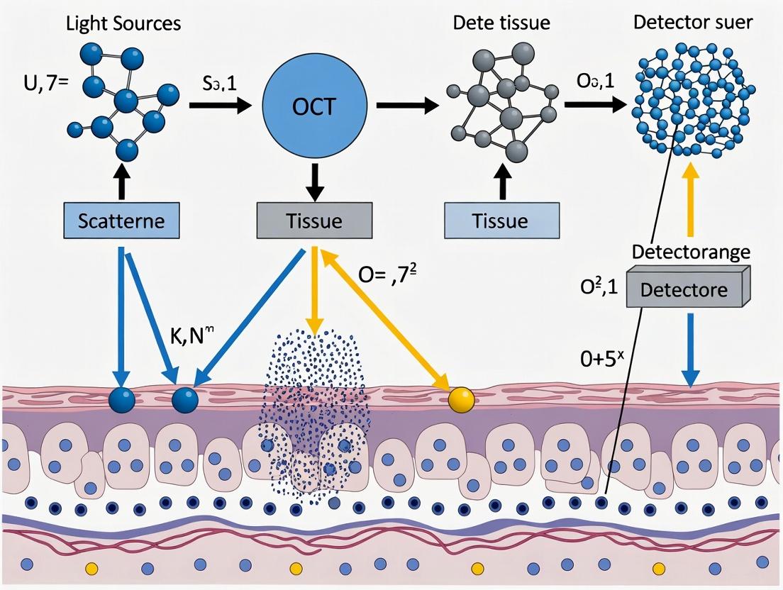
Optical Coherence Tomography (OCT) is a non-invasive, label-free imaging modality that provides cross-sectional, high-resolution (~1-15 µm) images of biological tissues. Within the broader thesis on OCT light scattering properties in tumor tissue research, understanding the core physical principles is paramount. Tumors often exhibit altered microstructural properties—such as increased nuclear size, extracellular matrix disorganization, and angiogenesis—which change how light backscatters compared to healthy tissue. This guide details the fundamental principles of how OCT detects these subtle changes in backscattered light and translates them into interpretable microstructural images, forming the basis for quantitative biomarkers in oncology drug development.
Core Measurement Principle: Low-Coherence Interferometry
OCT does not directly "see" structure. It measures the echo time delay and intensity of backscattered light using a technique called low-coherence interferometry. A broadband, near-infrared light source (e.g., a superluminescent diode with a central wavelength of ~1300 nm for deeper tissue penetration) is split into two paths: a sample arm directed at the tissue and a reference arm directed at a moving mirror.
- Interference Condition: Backscattered light from different depths within the sample is recombined with light reflected from the reference arm. Constructive interference (a measurable signal) occurs only when the optical path lengths of the sample and reference arms are matched within the coherence length of the source, which determines the axial resolution.
- Axial Scan (A-scan): By rapidly scanning the reference mirror, the time delay (and thus depth) of backscattered signals from the sample is encoded in the interference pattern. The intensity of the interference fringes is proportional to the reflectivity at that specific depth. The result is a one-dimensional depth profile of reflectivity—an A-scan.
- Key Quantitative Parameter: The primary measurable is the interference fringe intensity, which is digitized and processed. The amplitude of the signal is related to the sample's backscattering coefficient (µₐ) and scattering anisotropy (g). Tumor tissues, with higher refractive index heterogeneity, typically exhibit stronger backscattering.
Table 1: Key OCT System Parameters and Typical Values for Tumor Imaging
| Parameter | Typical Value/Type for Tumor Research | Impact on Image & Data |
|---|---|---|
| Central Wavelength | 1300 nm (common), 800-900 nm (higher res) | Penetration depth (1-2 mm at 1300nm) vs. resolution trade-off. |
| Axial Resolution | 1-15 µm in tissue | Determined by source bandwidth (∆λ). Critical for resolving cell clusters. |
| Lateral Resolution | 5-30 µm | Determined by objective lens spot size. |
| A-scan Rate | 50 kHz - 1.5 MHz (modern systems) | Enables real-time, volumetric imaging. |
| Dynamic Range | >100 dB | Allows detection of weak signals from deep tissue. |
| Key Output Data | Interferogram (raw), A-scan (processed) | Amplitude and depth of backscattered light. |
From Interference to Image: Signal Processing & Image Construction
The raw interferogram must be processed to construct a visually interpretable, depth-resolved image.
- Digitization & Demodulation: The analog photodetector signal is digitized. Demodulation (often via envelope detection or Hilbert transform) extracts the magnitude of the interference signal, discarding the high-frequency carrier.
- Spectral Resampling & Fourier Transform: In Fourier-Domain OCT (the current standard), the interference pattern is captured as a function of wavenumber (k). After resampling to a linear k-space, a Fast Fourier Transform (FFT) is applied. This transforms the signal from the spectral domain to the depth domain, generating the A-scan.
- Logarithmic Compression: The dynamic range of the linear A-scan (often spanning 40-60 dB) is too large for display. A logarithmic compression (e.g.,
20*log10(amplitude)) is applied, mapping the signal to a grayscale or false-color image where brightness corresponds to backscatter intensity. - B-scan & Volumetric Rendering: By laterally scanning the beam across the sample and acquiring successive A-scans, a two-dimensional cross-sectional image (B-scan) is constructed. A series of adjacent B-scans creates a 3D volumetric dataset.
Diagram: OCT Signal Processing Workflow
Extracting Microstructural Information: Key Metrics for Tumor Research
For quantitative analysis in oncology, OCT images are processed to derive metrics correlated with tissue microstructure.
- Attenuation Coefficient (µₜ): The rate at which signal intensity decays with depth. Tumors often show higher attenuation due to increased scattering. Calculated by fitting an exponential decay curve to the A-scan data:
I(z) = I₀ * exp(-2*µₜ*z). - Backscatter Coefficient (µᵦ): The intensity of the signal at a given depth, related to the density and size of scattering organelles (e.g., nuclei, mitochondria).
- Speckle Variance: Analysis of speckle pattern fluctuations between successive B-scans can reveal sub-resolution motion, indicative of blood flow (OCT Angiography) or cellular dynamics.
Table 2: Quantitative OCT Metrics in Tumor vs. Normal Tissue
| Metric | Typical Trend in Tumor Tissue (vs. Normal) | Underlying Microstructural Correlation | Common Analysis Method |
|---|---|---|---|
| Attenuation Coefficient (µₜ) | Increased (typically 5-15 mm⁻¹ in tumors vs. 3-8 mm⁻¹ in normal) | Higher nuclear-to-cytoplasmic ratio, increased collagen density. | Depth-resolved fitting of A-scan decay. |
| Backscatter Coefficient (µᵦ) | Increased (by 3-10 dB) | Larger scatterers (enlarged nuclei), more refractive index discontinuities. | Comparison of near-surface signal amplitude to reference phantom. |
| Speckle Variance/Decorrelation | Increased (angiogenic regions) | Increased microvascular density and blood flow. | Temporal or intensity-based analysis of repeated B-scans. |
| Texture Homogeneity | Decreased | Loss of organized tissue architecture, necrosis. | Gray-level co-occurrence matrix (GLCM) analysis. |
Experimental Protocol: Measuring Attenuation Coefficient in Tumor Xenografts
This protocol outlines a standard method for quantitative OCT analysis in preclinical tumor models.
Aim: To quantify and compare the spatially-resolved attenuation coefficient between a tumor xenograft and adjacent normal tissue. Materials: See "The Scientist's Toolkit" below. Procedure:
- System Calibration: Image a well-characterized optical phantom with known scattering properties to verify system performance and establish a baseline.
- Sample Preparation: Anesthetize the mouse bearing a subcutaneous tumor xenograft. Gently clean the imaging area. Apply a thin layer of ultrasound gel as an index-matching medium to the skin/tumor surface. Position the animal on a heated, stable stage.
- Data Acquisition:
- Using a spectral-domain OCT system, position the scan head perpendicular to the tissue surface.
- Acquire a 3D volumetric dataset (e.g., 1000 A-scans/B-scan, 500 B-scans/volume) over the region of interest (ROI) encompassing tumor center, margin, and normal tissue.
- Repeat acquisition at 2-3 spatial locations. Ensure minimal motion artifact.
- Data Processing & Analysis:
- Pre-processing: Apply a software dispersion compensation algorithm. Subtract noise floor (average of deepest 50 pixels of each A-scan).
- Attenuation Fitting: For each A-scan within the ROI, fit the intensity decay from the tissue surface to a pre-defined depth (e.g., 300 µm) using a single exponential model (
I(z) = I₀ * exp(-2*µₜ*z)) via a least-squares fitting routine. - Spatial Mapping: Map the fitted µₜ values for all A-scans to generate a 2D en face parametric map of the attenuation coefficient.
- Statistical Comparison: Define regions for tumor core, tumor periphery, and normal tissue on the parametric map. Perform ANOVA with post-hoc tests to compare mean µₜ values between regions (p < 0.05 considered significant).
Diagram: Attenuation Coefficient Analysis Workflow
The Scientist's Toolkit: Essential Research Reagents & Materials
| Item | Function/Description | Example/Supplier Note |
|---|---|---|
| Spectral-Domain OCT System | Core imaging device. Provides the light source, interferometer, spectrometer, and detection electronics. | Thorlabs, Michelson Diagnostics, Wasatch Photonics. |
| Near-Infrared Broadband Source | Generates low-coherence light. Central wavelength determines penetration depth. | Superluminescent Diodes (SLD), Swept-Source Lasers. |
| Reference Phantom | Calibration standard with known, stable optical scattering properties (µₐ, g). Essential for quantitative comparison across studies. | Phantoms with titanium dioxide or polystyrene microspheres in silicone/polymer (e.g., from Institut National d'Optique). |
| Index-Matching Gel | Applied between tissue and OCT objective. Reduces strong surface reflection and refractive index mismatch, improving signal quality. | Ultrasound gel (non-corrosive). |
| Anesthetic System | For in vivo preclinical imaging of animal models, ensuring stable positioning and animal welfare. | Isoflurane vaporizer system. |
| Heated, Stabilized Stage | Maintains animal physiology (temperature) and minimizes motion artifacts during in vivo imaging. | Custom or commercial small animal imaging stages. |
| Data Processing Software | For custom analysis of OCT data (attenuation fitting, speckle analysis). | MATLAB, Python (with NumPy, SciPy), or LabVIEW with custom scripts. |
| Histology Equipment | For gold-standard validation of OCT findings (e.g., H&E staining for morphology). | Formalin, paraffin, microtome, stains. Used for spatial correlation of OCT metrics with histopathology. |
Within the broader thesis on optical coherence tomography (OCT) light scattering properties for tumor tissue research, this guide details the technical foundation for interpreting OCT contrast. The primary signal in OCT is backscattered light, the intensity of which is governed by spatial variations in the refractive index (RI) at subcellular and extracellular scales. In tumors, architectural and compositional alterations—such as nuclear pleomorphism, collagen reorganization, and glycoprotein accumulation—fundamentally change these scattering properties, providing a non-invasive, label-free contrast mechanism for detection and characterization.
Physical Principles of Scattering in Biological Tissue
OCT detects coherently backscattered light from refractive index inhomogeneities. The scattering coefficient (μs) and the anisotropy factor (g) determine the intensity and directionality of scattering. At the microscopic scale relevant to OCT (resolution ~1-15 μm), dominant scatterers include:
- Organelles: Nuclei, mitochondria, and endoplasmic reticulum.
- Cytoskeletal elements: Actin filaments and microtubule networks.
- Extracellular matrix (ECM): Collagen, elastin fibers, and proteoglycans.
The scattering intensity from a single particle can be approximated by Mie theory for spherical particles comparable to the wavelength (λ ≈ 1.3 μm). The scattering cross-section (σs) depends on particle size (d), relative RI (n = nparticle / nmedium), and λ.
Key Scattering Features in Tumor Tissues
Neoplastic transformation induces specific changes in subcellular and extracellular architecture that modify scattering properties, as summarized in Table 1.
Table 1: Tumor-Associated Features and Their Scattering Impact
| Feature | Normal Tissue Characteristic | Tumor Tissue Alteration | Effect on Scattering Intensity (Typical) | Proposed RI Change |
|---|---|---|---|---|
| Nuclear Morphology | Uniform size, regular shape. | Enlargement (pleomorphism), hyperchromasia, increased nuclear-to-cytoplasmic ratio. | Increase (stronger scatter from larger, denser nuclei). | Increased nuclear RI due to chromatin condensation. |
| Cell Density | Organized, tissue-specific packing. | Increased and disorganized cellularity. | Increase (more scattering centers per unit volume). | N/A (geometric effect). |
| Extracellular Matrix (Collagen) | Organized, aligned fiber bundles. | Desmoplasia (increased volume) but often disorganized, fragmented fibers. | Variable (Can increase from higher density; can decrease from loss of organized bundles causing coherent scattering). | Collagen RI (~1.48) higher than surrounding ground substance (~1.35). |
| Microvasculature | Regular hierarchical network. | Irregular, tortuous, leaky vessels. | Can Increase (from vessel walls as scattering structures). | Blood plasma RI ~1.34, vessel wall RI ~1.38-1.40. |
Experimental Protocols for Correlation
Protocol: Co-registered OCT and Histomorphometry
Objective: Quantitatively correlate OCT backscattering intensity (OCT signal amplitude) with specific histopathological features.
Materials:
- OCT system (e.g., spectral-domain OCT, 1300 nm central wavelength).
- Biopsy or tissue specimen (fresh or freshly frozen).
- Standard histology processing equipment (processor, microtome).
- Hematoxylin and Eosin (H&E) stain.
- Specific stains (e.g., Picrosirius Red for collagen, Feulgen for DNA).
- Brightfield and polarization microscopy setup.
- Image co-registration software (e.g., MATLAB, Python with OpenCV/SimpleITK).
Methodology:
- OCT Imaging: Acquate 3D OCT volumetric data of the fresh, unprepared tissue sample. Record precise positional coordinates.
- Tissue Processing: Fix the imaged tissue in formalin, process, and embed in paraffin. Section the block at 4-5 μm thickness. Precisely document the sectioning plane relative to the OCT scan.
- Histological Staining: Perform H&E and special stains on serial sections.
- Digital Pathology & Co-registration: Digitize histology slides. Use fiduciary markers or tissue landmarks to algorithmically co-register the 2D histology image with the corresponding en face OCT slice (often the median intensity projection).
- Region-of-Interest (ROI) Analysis:
- On the histology image, a pathologist manually annotates ROIs for specific features (e.g., region of high nuclear density, collagen bundle region, necrotic area).
- These ROIs are mapped onto the co-registered OCT intensity map.
- Extract the mean and standard deviation of the OCT intensity (log-compressed or linear) within each ROI.
- Perform statistical analysis (e.g., ANOVA) to test for significant differences in OCT intensity between feature classes.
Protocol: Ex Vivo RI Measurement and Scattering Simulation
Objective: Measure the refractive index of isolated cellular components and simulate their scattering contribution.
Materials:
- Digital holographic microscope or quantitative phase imaging (QPI) system.
- Isolated cell nuclei purification kit.
- Collagen extraction or reconstituted collagen gel.
- Refractometer (Abbe or digital).
- Computational electromagnetic simulation software (e.g., MiePlot, FDTD solutions).
Methodology:
- RI Measurement: Isolate nuclei from cell lines (normal vs. cancerous) via detergent lysis and centrifugation. Using QPI or immersion refractometry, measure the mean RI of the nuclear suspension. Similarly, measure RI for collagen solutions and cytoplasmic extracts.
- Mie Theory Calculation: Using the measured RI values, background cytoplasmic/ECM RI (~1.35-1.36), and assumed particle size distributions (from electron microscopy literature), calculate the scattering cross-section (σs) and anisotropy factor (g) for nuclei modeled as spheres.
- Validation: Compare the calculated μs (derived from σs and estimated number density) with the attenuation coefficient extracted from OCT measurements of pelleted nuclei or tissue phantoms.
The Scientist's Toolkit: Key Research Reagent Solutions
Table 2: Essential Materials for OCT Scattering Correlation Studies
| Item | Function in Research |
|---|---|
| Spectral-Domain OCT System | Provides high-speed, high-sensitivity depth-resolved imaging of tissue scattering. Central wavelength (~1300 nm) offers optimal penetration in tissue. |
| RNAlater Stabilization Solution | Preserves tissue RNA/DNA and protein integrity immediately after OCT imaging, enabling subsequent genomic/proteomic correlation with scattering signals. |
| Picrosirius Red Stain Kit | Specifically stains collagen types I and III. When viewed under polarized light, it reveals collagen organization, crucial for correlating with birefringence and scattering in ECM. |
| DAPI (4',6-diamidino-2-phenylindole) Mounting Medium | Fluorescent nuclear counterstain. Allows precise segmentation of nuclei on co-registered fluorescence microscopy images for correlation with high-scattering foci in OCT. |
| Matrigel Basement Membrane Matrix | Used to create 3D cell culture models or tumor organoids with defined ECM. Enables controlled study of how specific ECM components influence OCT scattering. |
| Optical Phantoms (Microsphere Suspensions) | Polystyrene or silica microspheres of defined size and RI in a gel matrix. Provide calibrated standards for validating OCT system performance and testing scattering models. |
Visualization of Concepts and Workflows
Diagram 1: Sources of OCT contrast.
Diagram 2: OCT-Histology co-registration workflow.
This technical whitepaper, framed within the context of ongoing research into the light scattering properties of tumor tissue using Optical Coherence Tomography (OCT), details the core scattering parameters: the attenuation coefficient (μt) and the scattering coefficient (μs). These parameters are critical for the quantitative, label-free differentiation of healthy and neoplastic tissues based on their intrinsic microstructural properties. Understanding their biological correlates—such as nuclear morphology, collagen density, and tissue organization—is paramount for advancing optical biopsy techniques in cancer research and drug development.
In OCT, near-infrared light is directed at tissue, and the backscattered signal is measured to generate cross-sectional, micron-resolution images. The intensity of the detected signal decays with depth due to two primary processes: absorption (μa) and scattering (μs). The total attenuation coefficient (μt = μs + μa) describes the overall rate of this signal decay. In most biological tissues in the NIR window, scattering dominates over absorption (μs >> μa); therefore, μt ≈ μs. The scattering coefficient quantifies the probability of a scattering event per unit path length and is fundamentally linked to spatial variations in the tissue refractive index (RI) at the cellular and sub-cellular level.
Biological Correlates of Scattering Parameters
The quantitative values of μs and μt are not abstract optical numbers but are directly governed by tissue ultrastructure.
| Scattering Parameter | Primary Biological Determinants in Tumor Tissue | Typical Direction of Change in Malignancy |
|---|---|---|
| Attenuation Coefficient (μt) | Combined effect of scattering (nuclear size/density, collagen fibers) and absorption (hemoglobin, water). In NIR, driven by scattering. | Variable. Can increase due to hypercellularity or decrease due to necrosis/stromal degradation. |
| Scattering Coefficient (μs) | Density, size, and RI contrast of subcellular organelles (mitochondria, nuclei), extracellular matrix (collagen) architecture. | Often increases in high-grade tumors due to increased nuclear-to-cytoplasmic ratio and cellular crowding. |
| Anisotropy Factor (g) | Average scattering direction. Related to the size of scattering particles relative to wavelength. | May decrease as tissue architecture becomes more disordered, leading to more isotropic scattering. |
Increased nuclear size, pleomorphism, and hypercellularity—hallmarks of cancer—increase the number and size of scattering particles, elevating μs. Conversely, stromal breakdown (loss of collagen) or necrosis can reduce μs. The attenuation coefficient captures the effective imaging depth and is crucial for correcting depth-dependent signal fall-off to quantify μs accurately.
Experimental Protocols for Parameter Extraction
Quantifying μs and μt from OCT data requires modeling and signal processing. Below are two established methodologies.
Protocol 1: Depth-Resolved Fitting of the Single-Scattering Model
- Principle: Assumes a single-backscatter model where the OCT signal depth decay is primarily due to attenuation.
- Procedure:
- Data Acquisition: Acquire 3D OCT volume of tissue sample. Use a system with a known spectral bandwidth and axial resolution.
- Pre-processing: Flatten the tissue surface. Apply a confocal point spread function (PSF) correction if necessary.
- Averaging: Average A-scans within a homogeneous region of interest (ROI) to improve SNR.
- Fitting: Fit the averaged, depth-dependent intensity signal, I(z), to the model:
I(z) = A * exp(-2μt * z) + BwhereAis a proportionality constant,zis depth,Bis noise floor, andμtis the attenuation coefficient. - Calculation: Extract μt from the fit. Assuming low absorption, μs ≈ μt.
- Applications: Best for homogeneous tissues and providing an effective attenuation coefficient.
Protocol 2: Inverse Adding-Doubling (IAD) or Integrating Sphere Measurement
- Principle: A gold-standard ex vivo method using a spectrophotometer with an integrating sphere to measure total transmission (Tt) and diffuse reflection (Rd) of thin tissue slices.
- Procedure:
- Sample Preparation: Section fresh or fixed tissue to a precise, known thickness (e.g., 200-500 μm) using a microtome.
- Measurement: Place the sample at the entrance port of the integrating sphere. Measure collimated transmission, total transmission, and diffuse reflection.
- Inversion: Use the IAD numerical algorithm to solve the radiative transport equation. Input measured Tt, Rd, thickness, and sample RI to output μs, μa, and g.
- Applications: Provides the most accurate and separate measurements of μs, μa, and g, serving as validation for OCT-based extraction methods.
Visualization of Core Concepts
OCT Scattering and Attenuation Pathway
Workflow for OCT-based μt/μs Extraction
The Scientist's Toolkit: Research Reagent & Material Solutions
| Item Name / Category | Function in Scattering Parameter Research |
|---|---|
| Spectral-Domain OCT System | Core imaging device. Systems with central wavelengths ~1300nm offer deeper penetration in tissue; ~800nm provides higher resolution for cellular details. |
| Integrating Sphere Spectrophotometer | Gold-standard equipment for measuring total transmission and diffuse reflection of thin tissue sections to calculate μs, μa, and g via IAD. |
| Precision Microtome (Vibratome/Cryostat) | For preparing thin, consistent tissue slices (200-500 μm) required for integrating sphere measurements or calibration. |
| Index Matching Fluids (e.g., Glycerol) | Applied to tissue to reduce surface scattering, allowing better probing of bulk scattering properties. |
| Phantom Materials (e.g., Intralipid, Microsphere Suspensions) | Calibration standards with known, tunable scattering properties to validate OCT system performance and extraction algorithms. |
| Digital Pathology Scanner | For co-registering OCT parametric maps with H&E-stained histological sections, enabling correlation of μs/μt with specific tissue morphologies. |
| Software (MATLAB, Python with SciPy) | For implementing custom algorithms for depth-resolved fitting, IAD calculation, and generating parametric attenuation/scattering maps. |
The quantitative extraction of the attenuation and scattering coefficients from OCT data provides a powerful, non-invasive window into the microstructural hallmarks of cancer. By understanding the direct biological meaning of these parameters—linking them to nuclear morphology, cellularity, and stromal changes—researchers and drug developers can leverage OCT not just for imaging, but for quantitative tissue phenotyping. This enhances capabilities in tumor margin assessment, treatment response monitoring, and the development of targeted therapies that alter the tissue microenvironment.
This whitepaper provides an in-depth technical guide on the architectural signatures of tissue as revealed by Optical Coherence Tomography (OCT). It is framed within a broader thesis on OCT light scattering properties for tumor tissue research. OCT, a non-invasive, label-free imaging technique, provides micron-scale cross-sectional images of tissue microstructure by detecting backscattered light. The core thesis posits that the distinct organizational scales of normal, benign neoplastic, and malignant tissues produce unique, quantifiable signatures in OCT signals, primarily through their effect on scattering coefficient (μs), anisotropy factor (g), and the derived reduced scattering coefficient (μs' = μs(1-g)). These signatures arise from changes in nuclear morphology, extracellular matrix density and organization, and microvascular patterns, enabling optical biopsy and margin assessment.
Core Optical Properties & Quantitative Signatures
The interaction of near-infrared light with tissue is governed by absorption and scattering. In OCT imaging of epithelial tissues (e.g., breast, skin, colon), scattering dominates. The key parameters are:
- Scattering Coefficient (μs): Probability of scattering per unit path length (mm⁻¹). Increases with refractive index mismatch and number density of scattering particles (e.g., nuclei, collagen fibrils).
- Anisotropy Factor (g): Mean cosine of the scattering angle. Ranges from 0 (isotropic) to 1 (forward-scattering). Tissues typically have high g (~0.9).
- Reduced Scattering Coefficient (μs'): The effective scattering coefficient in a diffusion regime, μs' = μs(1-g). This is the parameter most frequently extracted from OCT data as it dictates the signal roll-off with depth.
Malignant transformation alters tissue architecture on multiple scales, changing these parameters. The table below summarizes quantitative findings from recent literature.
Table 1: Quantitative OCT Parameters for Tissue Types
| Tissue Type / Condition | Reduced Scattering Coefficient, μs' (mm⁻¹) ± SD | Scattering Coefficient, μs (mm⁻¹) ± SD | Attenuation Coefficient, μt (mm⁻¹) ± SD | Key Architectural Correlates |
|---|---|---|---|---|
| Normal Epithelium (e.g., Breast) | 0.8 - 1.5 ± 0.3 | 8 - 15 ± 2 | 8.5 - 16 ± 2.2 | Ordered glandular structures, uniform nuclear size, regular collagen spacing. |
| Benign Lesions (e.g., Fibroadenoma) | 1.2 - 2.2 ± 0.4 | 12 - 22 ± 3 | 12.5 - 23 ± 3.3 | Increased cellularity & stroma, encapsulated, structured hyperplasia. |
| Malignant Carcinoma (e.g., Invasive Ductal) | 2.5 - 4.5 ± 0.7 | 25 - 45 ± 5 | 26 - 47 ± 5.5 | High nuclear density, pleomorphism, disorganized collagen, microcalcifications. |
| Normal Colon Mucosa | 1.0 - 1.8 ± 0.3 | 10 - 18 ± 2 | 10.5 - 19 ± 2.2 | Crypt structures, regular lamina propria. |
| Colon Adenoma (Benign) | 1.5 - 2.5 ± 0.4 | 15 - 25 ± 3 | 16 - 26 ± 3.3 | Elongated, crowded crypts, low-grade dysplasia. |
| Colon Adenocarcinoma | 3.0 - 5.0 ± 0.8 | 30 - 50 ± 6 | 31 - 52 ± 6.5 | Crypt destruction, back-to-back glands, desmoplastic stroma. |
Data synthesized from recent studies on OCT in oncology (2021-2024). SD = Standard Deviation. μt ≈ μs + μa (absorption coefficient, μa, is often negligible in NIR).
Experimental Protocols for Extracting Architectural Signatures
Protocol A: Depth-Resolved Attenuation Analysis for μt & μs'
This is the most common method for quantifying scattering from OCT A-scans (depth profiles).
- Sample Preparation: Fresh or freshly frozen tissue samples are sectioned to a uniform thickness (2-5 mm) and placed in a sample holder with a glass window. Phosphate-buffered saline is used to keep tissue hydrated. OCT imaging is performed within 4 hours of excision.
- OCT Imaging: Use a swept-source or spectral-domain OCT system with a center wavelength of ~1300 nm (optimal for tissue penetration). Acquire 3D volumetric data (e.g., 1000 x 500 x 1024 pixels, x,y,z).
- Data Preprocessing:
- Apply a k-space resampling and Hann window to spectral data.
- Perform Fourier transform to get A-scans.
- Log-transform the intensity values: I(z) = 10 * log₁₀(|FFT|²).
- Correct for confocal point spread function and sensitivity roll-off using system characterization data.
- Attenuation Fitting:
- For each A-scan, model the depth-dependent intensity decay in the single-scattering regime: I(z) ∝ exp(-2μt*z).
- Perform a linear fit on the log-intensity vs. depth plot: slope = -2μt.
- Assuming μa << μs, μt ≈ μs. Then calculate μs' using an assumed or separately measured g (typically 0.9-0.95) or by using a model relating μt to μs'.
- Statistical Mapping: Generate 2D parametric maps of μt or μs' for en face visualization of heterogeneous regions.
Protocol B: Correlation Analysis of Scattering Signal Texture
This protocol quantifies organizational disorder, a key marker of malignancy.
- Image Acquisition: As per Protocol A, Step 2.
- Region of Interest (ROI) Selection: Manually or automatically segment the epithelial or stromal layer from B-scans (cross-sections).
- Texture Feature Extraction:
- Speckle Statistics: Fit the pixel intensity histogram within an ROI to distributions (e.g., Rayleigh, K-distribution). The K-distribution shape parameter is sensitive to scatterer number density and clustering.
- Gray-Level Co-Occurrence Matrix (GLCM): Compute GLCM for different offsets. Extract metrics like Contrast (local variation), Energy (uniformity), and Homogeneity. Malignant tissue shows high contrast and low homogeneity/energy.
- Fractal Dimension (FD): Calculate the FD of the segmented OCT intensity image using a box-counting algorithm. Increased architectural complexity in malignancy elevates FD.
- Machine Learning Classification: Use extracted features (μs', texture metrics) to train a classifier (e.g., Support Vector Machine, Random Forest) to automatically categorize tissue as normal, benign, or malignant.
Visualizing the Research Workflow & Biological Correlates
OCT-Based Tissue Classification Workflow
Architectural Features Driving OCT Signal Differences
The Scientist's Toolkit: Research Reagent & Material Solutions
Table 2: Essential Materials for OCT Tumor Tissue Research
| Item | Function & Relevance |
|---|---|
| Swept-Source OCT System (1300 nm center wavelength) | Provides the imaging beam. 1300 nm offers an optimal trade-off between resolution (~5-10 µm) and penetration depth (~2-3 mm in tissue). |
| Tissue Sample Holder with Optical Window | Maintains tissue geometry, prevents dehydration, and provides a flat, standardized interface for consistent imaging. |
| Phosphate-Buffered Saline (PBS), pH 7.4 | Keeps excised tissue hydrated to minimize optical property changes due to drying during scanning. |
| Optical Phantoms (e.g., Silica Microspheres in Gelatin, Intralipid) | Calibrate the OCT system and validate scattering coefficient extraction algorithms. Provide known μs and g values. |
| Histology-Grade Tissue Processing Kit (Formalin, Paraffin, H&E Stain) | For gold-standard pathological correlation. The imaged tissue is processed for histology to confirm optical findings. |
| MATLAB or Python with Libraries (NumPy, SciPy, scikit-image, OpenCV) | Essential software platforms for custom analysis of OCT data, including attenuation fitting, texture analysis, and machine learning. |
| High-Performance Computing Workstation | Necessary for processing large 3D OCT datasets and running complex texture/classification algorithms in a timely manner. |
The Role of Necrosis, Angiogenesis, and Stromal Remodeling in Altering Scattering Profiles
This whitepaper provides a technical guide on three critical, interlinked pathological processes—necrosis, angiogenesis, and stromal remodeling—and their distinct roles in altering optical scattering profiles in tumor tissue. This analysis is framed within a broader thesis investigating the use of Optical Coherence Tomography (OCT) and its derived metrics (e.g., scattering coefficient, attenuation coefficient, backscattering intensity) for the label-free, micro-scale assessment of tumor microenvironment (TME) evolution. Understanding how these biological hallmarks quantitatively change the interaction of near-infrared light with tissue is essential for developing OCT as a robust tool for monitoring therapeutic response, tumor aggressiveness, and drug efficacy in pre-clinical and clinical settings.
Pathobiological Processes and Their Scattering Signatures
Necrosis
Necrosis is a form of unprogrammed cell death leading to cellular swelling, plasma membrane rupture, and spillage of intracellular contents into the extracellular space.
- Impact on Scattering: The initial increase in nuclear size and organelle swelling increases scattering due to a higher density of scattering organelles. Subsequent membrane rupture and homogenization of cytoplasmic content reduce the number of discrete, high-refractive-index organelles (e.g., mitochondria), leading to a overall decrease in scattering. The resulting debris often forms regions with low, heterogeneous scattering.
- Key OCT Parameters: Reduced scattering coefficient (μs'), increased attenuation heterogeneity, loss of regular tissue architecture in OCT images.
Angiogenesis
Angiogenesis is the formation of new, often aberrant, blood vessels from pre-existing vasculature, a hallmark of tumor growth and metastasis.
- Impact on Scattering: New vessel walls (endothelial cells, pericytes) and the blood within them introduce new refractive index boundaries. The increased microvascular density and vessel diameter create more interfaces, typically increasing scattering. However, the chaotic, leaky nature of tumor vessels can lead to extravasation of red blood cells (RBCs) into the stroma, altering local scattering properties.
- Key OCT Parameters: Increased spatial heterogeneity of backscattering, altered texture features, correlation with OCT angiography (OCTA) vessel density metrics.
Stromal Remodeling (Desmoplasia)
This involves the activation of cancer-associated fibroblasts (CAFs), excessive deposition of fibrillar collagens (mainly Types I and III), and cross-linking of the extracellular matrix (ECM).
- Impact on Scattering: Dense, aligned collagen fibers are strong scatters of light due to their high refractive index and regular, fibrillar structure. Stromal remodeling increases the density and organization of these fibers, leading to a significant increase in scattering intensity and anisotropy. Collagen cross-linking further changes the bulk optical properties.
- Key OCT Parameters: Increased scattering coefficient (μs), increased birefringence (detectable by polarization-sensitive OCT), distinct texture patterns.
Table 1: Quantitative Impact of Pathological Processes on OCT Scattering Parameters
| Pathological Process | Primary Effect on Tissue Microstructure | Typical Direction of Change in μs' (mm⁻¹) | Key Influencing Factors | Representative Experimental Values (Range) |
|---|---|---|---|---|
| Necrosis | Organelle loss, membrane rupture, debris formation | Decrease (up to 30-50%) | Stage of necrosis, degree of liquefaction | 2.5 – 4.5 mm⁻¹ (vs. 5.5 – 7.5 in viable tumor) |
| Angiogenesis | Increased vessel density, endothelial cell proliferation | Increase (10-25%) | Vessel diameter, density, RBC content | Microvessel density >15/mm² correlates with μs' >6.0 mm⁻¹ |
| Stromal Remodeling | Increased collagen density/fibrillation, ECM cross-linking | Significant Increase (40-100%) | Collagen fiber alignment, cross-link density | μs' in dense desmoplasia: 8.0 – 12.0 mm⁻¹ |
Experimental Protocols for Correlative Validation
Protocol 1: Ex Vivo Correlation of OCT Scattering with Histopathology
Objective: To establish a direct quantitative relationship between OCT-derived scattering parameters and histological confirmation of necrosis, angiogenesis, and stromal remodeling.
Methodology:
- Tissue Sample Preparation: Excise tumor xenografts or clinical biopsy specimens. Embed in optimal cutting temperature (OCT) compound and freeze, or formalin-fix and paraffin-embed (FFPE).
- OCT Imaging: Acquire 3D OCT volumes (e.g., 1300 nm central wavelength) of the tissue block surface. Calculate parametric maps (attenuation, backscattering) using a single-scattering model.
- Coregistration: Mark imaging region with indelible ink. Serially section the tissue block. Perform H&E staining for general morphology and necrosis identification.
- Special Stains:
- Angiogenesis: Immunohistochemistry (IHC) for CD31 or CD34 to highlight endothelial cells. Calculate microvessel density (MVD).
- Stromal Remodeling: Picrosirius Red stain for collagen, visualized under polarized light to assess fiber density and alignment. IHC for α-SMA to identify CAFs.
- Image Registration & Analysis: Use landmark-based software to co-register histology images with OCT parametric maps. Perform region-of-interest (ROI) analysis to extract mean OCT values corresponding to histologically confirmed regions.
Protocol 2: In Vivo Longitudinal OCT Monitoring of Anti-Angiogenic Therapy
Objective: To dynamically track changes in scattering profiles in response to vascular endothelial growth factor (VEGF) inhibition.
Methodology:
- Animal Model: Implant dorsal window chamber or subcutaneous tumor model (e.g., HT-29 colorectal carcinoma).
- Baseline Imaging: Acquire OCT/OCTA scans pre-treatment. Derive baseline μs', vessel density, and perfusion metrics.
- Therapeutic Intervention: Administer VEGF inhibitor (e.g., Bevacizumab analog) or vehicle control.
- Longitudinal Imaging: Repeat OCT/OCTA at defined intervals (e.g., days 1, 3, 7). Monitor changes in scattering intensity in the peri-vascular stroma (due to edema) and tumor core (due to potential necrosis).
- Endpoint Validation: Harvest tumors, process for histology (Protocol 1) to correlate terminal OCT parameters with MVD and necrosis fraction.
Diagrammatic Representations
The Scientist's Toolkit: Key Research Reagents & Materials
Table 2: Essential Reagents and Materials for OCT-TME Research
| Item Name | Category | Primary Function in Research |
|---|---|---|
| FFPE or Frozen Tissue Blocks | Sample | Provides the physical substrate for ex vivo OCT imaging and subsequent gold-standard histopathological analysis. |
| CD31 / CD34 Antibody | IHC Reagent | Labels endothelial cells for quantitative assessment of microvessel density (angiogenesis). |
| α-SMA Antibody | IHC Reagent | Identifies activated Cancer-Associated Fibroblasts (CAFs), key drivers of stromal remodeling. |
| Picrosirius Red Stain Kit | Histology Stain | Specifically binds to fibrillar collagens; polarized light visualization reveals collagen density and alignment. |
| Bevacizumab (Avastin) orSunitinib Malate | Pharmacological Agent | VEGF inhibitor or tyrosine kinase inhibitor used to modulate angiogenesis in experimental therapy models. |
| Matrigel | Extracellular Matrix | Used for tumor cell implantation or 3D culture to study angiogenesis and stromal interactions in vivo or ex vivo. |
| Spectral-Domain OCT System(e.g., 1300 nm central λ) | Imaging Equipment | The core device for acquiring depth-resolved scattering data. Systems with angiography (OCTA) and polarization-sensitive (PS-OCT) capabilities are advantageous. |
| Image Co-registration Software(e.g., 3D Slicer, Amira) | Analysis Software | Enables precise alignment of OCT volumetric data with 2D histological sections for pixel/voxel-level correlation. |
From Raw Signal to Quantitative Biomarkers: Advanced OCT Techniques for Tumor Analysis
Within the broader thesis on utilizing optical coherence tomography (OCT) to characterize light scattering properties of tumor tissue, consistent and high-fidelity data acquisition is paramount. This technical guide details optimized scan protocols to ensure reproducible scattering measurements, such as attenuation coefficients and backscattering intensities, which are critical for differentiating malignant from benign tissues in oncological research and drug development.
Foundational Principles of OCT Scattering in Tumors
Tumor tissue exhibits distinct scattering properties due to altered nuclear morphology, increased cellular density, and extracellular matrix remodeling. Consistent measurement of these properties enables quantitative biomarkers for tumor margin detection, treatment response monitoring, and mechanistic studies.
Core Scan Protocol Parameters for Optimization
Optimal protocol design controls variables that introduce variance in derived scattering parameters.
Table 1: Key Scan Protocol Parameters and Optimization Targets
| Parameter | Impact on Scattering Measurement | Recommended Optimization Practice |
|---|---|---|
| Beam Wavelength (λ) | Determines scattering cross-section & penetration. | Use consistent, tissue-appropriate λ (e.g., 1300 nm for deeper penetration in tissue). |
| Spectral Bandwidth | Affects axial resolution and speckle characteristics. | Maximize for high axial resolution; ensure stable source output. |
| A-Scan Rate | Influences motion artifact and volumetric coverage. | Balance high speed (≥100 kHz) with sufficient signal-to-noise ratio (SNR). |
| Scan Depth (Z) | Must encompass full sample depth for accurate μt fitting. | Set to ≥1.5x sample thickness; keep constant across samples. |
| Lateral Sampling Density | Impacts lateral resolution and speckle averaging. | Set to ≥2x the beam spot size; use consistent sampling across scans. |
| Number of Averages (N) | Directly improves SNR, reduces speckle variance. | Use N=4-16 for in vivo; N=8-32 for ex vivo; standardize per study. |
| Beam Power | Affects signal strength and sample safety. | Use maximum permissible exposure (MPE) for in vivo; constant power for ex vivo. |
| Reference Arm Power | Optimizes interferometric efficiency. | Adjust for detector linear range; lock and monitor during acquisition. |
Detailed Protocol for Scattering Metric Acquisition
This protocol is designed for extracting the attenuation coefficient (μt) from 3D OCT datasets.
Experimental Workflow:
- System Calibration:
- Use a calibrated phantom with known scattering properties (e.g., uniform polystyrene microspheres in gel) daily.
- Acquire B-scans, extract depth-resolved intensity, and verify derived μt matches known value within 5%.
- Sample Preparation & Mounting:
- Ex vivo tissue: Embed in optimal cutting temperature (OCT) compound or formalin. Ensure flat, stable surface orthogonal to beam.
- In vivo: Utilize stereotactic fixtures to minimize motion.
- Data Acquisition:
- Set scan area to fully encompass region of interest (ROI).
- Acquire 3D volume with parameters standardized per Table 1.
- Implement real-time preview to check for saturation (clipped intensity) or insufficient signal.
- Quality Control During Scan:
- Monitor SNR in real-time console. Discard scan if SNR drops below threshold (e.g., <20 dB for μt fitting).
- Check for vignetting or artifacts; reposition if necessary.
Data Processing Pipeline for Consistency
Raw data must be processed uniformly to extract quantitative scattering data.
Diagram Title: Data Processing Pipeline for Attenuation Coefficient Extraction
Table 2: Quantitative Scattering Metrics from Tumor Studies (Representative Values)
| Tissue Type (Model) | Mean μt (mm⁻¹) @ 1300 nm | Key Scattering Contributor | Correlation with Histopathology |
|---|---|---|---|
| Normal Brain (Murine) | 3.5 ± 0.6 | Neuronal microstructure | Baseline reference |
| Glioblastoma (Murine) | 5.8 ± 1.2 | Hypercellularity, Necrosis | R²=0.89 vs. cellularity score |
| Normal Colon (Human, ex vivo) | 4.1 ± 0.9 | Crypt structure | N/A |
| Colorectal Adenocarcinoma | 7.2 ± 1.5 | Enlarged nuclei, Gland fragmentation | Sensitivity > 85% for malignancy |
| Normal Breast (Murine) | 3.1 ± 0.5 | Adipose & ductal tissue | Baseline reference |
| Triple-Negative Breast Tumor | 6.5 ± 1.4 | Dense, monomorphic cells | R²=0.78 vs. Ki-67 index |
The Scientist's Toolkit: Key Research Reagent Solutions
Table 3: Essential Materials for OCT Scattering Studies in Tumor Research
| Item | Function & Importance in Protocol |
|---|---|
| Tissue-Mimicking Phantoms (e.g., Intralipid, microsphere gels) | Calibrate system, validate μt accuracy, track performance daily. |
| Optimal Cutting Temperature (OCT) Compound | Embed ex vivo tissue for stable, repeatable scanning geometry. |
| Fiducial Markers (India Ink, surgical sutures) | Provide spatial reference for correlating OCT ROIs with histology slices. |
| Index-Matching Fluid (e.g., Glycerol/Saline) | Reduces surface specular reflection, improving signal at tissue interface. |
| Stereo-Taxic Frame (In vivo models) | Eliminates motion artifacts, enabling longitudinal studies in same location. |
| Standardized Cover Slips/Windows | Creates flat, consistent optical interface for ex vivo samples. |
Protocol Validation and Correlation Workflow
Scattering metrics must be validated against histological ground truth.
Diagram Title: OCT-Histology Correlation Workflow for Biomarker Validation
Rigorous optimization of OCT scan protocols is non-negotiable for producing consistent, biologically meaningful scattering measurements in tumor tissue. By standardizing acquisition parameters, implementing a robust processing pipeline, and validating against gold-standard pathology, researchers can reliably translate OCT scattering properties into quantitative tools for oncology research and therapeutic development.
In the context of a broader thesis on the role of optical coherence tomography (OCT) light scattering properties in tumor tissue research, this technical guide details advanced signal processing pipelines. These pipelines are engineered to extract robust, quantitative scattering parameters, such as those derived from micro-OCT (µOCT) and OCT angiography (OCTA). The accurate quantification of these parameters—including scattering coefficient, attenuation coefficient, anisotropy factor, and fractal dimension—is critical for differentiating malignant from benign tissue, assessing tumor microenvironment, and monitoring therapeutic response in oncology drug development.
The interaction of near-infrared light with biological tissue is dominated by scattering events, which are exquisitely sensitive to subcellular and extracellular structural changes. In tumorigenesis, alterations in nuclear morphology, chromatin density, collagen fiber organization, and microvascular architecture create distinct, quantifiable scattering signatures. Signal processing pipelines transform raw interferometric OCT data into these objective metrics, providing researchers and drug development professionals with non-invasive, depth-resolved biomarkers for preclinical and clinical oncology research.
Core Signal Processing Pipeline Architecture
The foundational pipeline for extracting scattering parameters involves sequential stages of data conditioning, transformation, and modeling.
Diagram Title: Core OCT Scattering Parameter Extraction Pipeline
Pre-Processing Module
Objective: To condition the raw spectral data for accurate tomogram reconstruction.
- Spectral Resampling: Corrects for non-linear k-space sampling using a calibration mirror signal.
- Dispersion Compensation: Applies numerical or hardware-based compensation to maintain axial resolution at depth.
- Windowing: Applies a windowing function (e.g., Hamming) to sidelobe suppression.
- Digital Fourier Transform: Transforms pre-processed spectral data into depth-resolved A-scans.
Quantitative Scattering Parameter Extraction Methodologies
Attenuation Coefficient (µt) Extraction
The attenuation coefficient is a fundamental parameter describing the total loss of signal due to both scattering and absorption.
Experimental Protocol (Depth-Resolved Method):
- Acquire a 3D OCT dataset of the tissue sample (e.g., tumor xenograft biopsy).
- Perform logarithmic transformation of the detected intensity:
I(z) = 10 * log10(A-scan^2). - For each A-scan, fit the depth-dependent intensity decay within a defined depth window (e.g., 50-300 µm from surface) using a linear least-squares fit:
I(z) = I0 - 2µt * z. - The slope of the fitted line yields the attenuation coefficient
µtin units of mm⁻¹. - Generate a 2D en-face map of
µtby calculating the parameter for every A-scan position.
Key Considerations: This method assumes a single scattering regime and homogeneous tissue within the fitting window. Confounding factors like shadowing from superficial blood vessels must be masked.
OCT Angiography (OCTA) and Vascular Scattering Metrics
OCTA isolates the dynamic scattering signal from moving red blood cells to visualize microvasculature without exogenous contrast agents.
Experimental Protocol (Amplitude Decorrelation-based OCTA):
- Acquire multiple repeated B-scans (M-mode) at the same cross-sectional position (typically 3-8 repeats).
- Compute the complex OCT signal for each pixel across the repeated frames.
- Calculate the decorrelation value
Dbetween consecutive frames using the formula:D = 1 - |Σ(C_i * C_{i+1}*)| / sqrt( (Σ|C_i|^2) * (Σ|C_{i+1}|^2) ), whereC_iis the complex OCT value at a given pixel in framei. - Average decorrelation values across all frame pairs to generate a 2D decorrelation map.
- Apply thresholding and segmentation algorithms to extract quantitative vascular parameters (see Table 1).
Workflow for OCTA-Based Tumor Vascular Phenotyping:
Diagram Title: OCTA Signal Processing for Vascular Metrics
µOCT and Nanoscale Sensitivity
Micro-OCT (µOCT) employs broader bandwidth light sources to achieve axial resolutions approaching 1 µm, enabling the resolution of subcellular scattering features.
Protocol for Nuclear Morphometry Scattering Analysis:
- Acquire ultra-high-resolution 3D µOCT datasets of thin tissue sections (e.g., 10 µm) or engineered tumor spheroids.
- Apply a novel depth-encoded synthetic aperture algorithm to enhance lateral resolution.
- Use a Mie theory-informed inverse scattering model to fit the observed scattering profile of individual cell nuclei.
- Extract metrics such as effective nuclear diameter and refractive index fluctuation variance, which correlate with chromatin condensation and nuclear pleomorphism in cancer cells.
Table 1: Key Scattering Parameters in Tumor Tissue Research
| Parameter | Symbol (Typical Units) | Extraction Method | Biological Correlate in Tumors | Typical Range (Normal vs. Tumor) | Key Application in Drug Development |
|---|---|---|---|---|---|
| Attenuation Coefficient | µt (mm⁻¹) | Depth-resolved fitting of A-scan decay. | Cellular density, extracellular matrix (ECM) density. | ~3-6 mm⁻¹ (normal) vs. ~5-10 mm⁻¹ (high-grade tumor). | Monitoring therapy-induced necrosis/cell death. |
| Scattering Coefficient | µs (mm⁻¹) | Extended Huygens-Fresnel or OCT Doppler variance models. | Density and size of scattering organelles. | Derived parameter, tumor tissue generally higher. | Assessing changes in tumor microstructure. |
| Anisotropy Factor | g (unitless) | Combined OCT/confocal modeling or goniometric measurements. | Size of dominant scattering structures. | ~0.85-0.95 (tissue). Larger scatterers in tumors may increase g. | Characterizing ECM remodeling. |
| Fractal Dimension (OCTA) | Df (unitless) | Box-counting algorithm on binarized angiogram. | Microvascular architectural complexity. | 1.5-1.8. Higher Df indicates more chaotic, tumor-like vasculature. | Quantifying anti-angiogenic therapy efficacy. |
| Vessel Density (OCTA) | VD (%) | Pixel count after angiogram binarization and skeletonization. | Microvascular density. | Tissue-dependent. Tumors often show elevated but heterogeneous VD. | Primary metric for vascular-targeting agents. |
| Normalized Decorrelation | κ (a.u.) | Statistical analysis of OCTA signal strength. | Blood flow velocity/hematocrit. | Highly variable. Can decrease with vascular normalization therapy. | Measuring hemodynamic changes. |
Table 2: Comparison of Signal Processing Techniques for Key Parameters
| Target Parameter | Primary Algorithm | Advantages | Limitations | Suitability for In Vivo Imaging |
|---|---|---|---|---|
| µt (Attenuation) | Depth-resolved fitting (Linear Least Squares). | Simple, fast, robust for homogeneous regions. | Fails in highly heterogeneous tissue; confounded by attenuation shadows. | High, with careful region selection. |
| µs & g (Scattering/Anisotropy) | Inverse Model Fitting (e.g., Mie, T-matrix). | Provides physical insight into scatterer size. | Computationally heavy; requires assumptions about scatterer shape. | Medium, best for controlled ex vivo studies. |
| OCTA (Flow) | Amplitude/Intensity Decorrelation. | High motion contrast sensitivity, common. | Susceptible to physiological bulk tissue motion. | High, when paired with robust motion correction. |
| OCTA (Flow) | Phase Variance. | Sensitive to very slow flow. | Highly sensitive to phase instability and system noise. | Medium-Low. |
| Fractal Dimension | Box-Counting Analysis. | Global descriptor of geometric complexity, robust to magnification changes. | Does not provide localized information. | High, post-segmentation. |
The Scientist's Toolkit: Essential Research Reagents & Materials
Table 3: Key Research Reagent Solutions for OCT Scattering Studies in Tumor Models
| Item | Function & Role in Pipeline | Example/Notes |
|---|---|---|
| Phantom Materials | Calibration and validation of scattering parameter accuracy. | Silica microsphere suspensions in agarose (for µs, g); Intralipid solutions. |
| Index-Matching Media | Reduces surface specular reflection, improving signal penetration. | Phosphate-buffered saline (PBS), ultrasound gel, glycerol (for ex vivo). |
| Tissue Fixatives | Preserve tissue microstructure for correlative ex vivo µOCT/histology. | Formalin, paraformaldehyde (PFA). Note: fixation alters scattering properties. |
| Optical Clearing Agents | Reduce scattering to enable deeper imaging for 3D reconstruction. | CUBIC, CLARITY, or Scale solutions. Critical for organoid/tumor spheroid imaging. |
| Fluorescent Vascular Labels | Ex vivo validation of OCTA vascular networks. | Lectins (e.g., Griffonia simplicifolia), anti-CD31 antibodies for immunofluorescence. |
| Cell Line & Animal Models | Provide biologically relevant tumor tissue for method development. | Patient-derived xenografts (PDX), genetically engineered mouse models (GEMMs). |
| Motion Stabilization Gel | Minimizes sample motion during in vivo OCTA, crucial for decorrelation accuracy. | Hydrogel, carbomer-based ophthalmic gels (for skin imaging). |
| Digital Reference Database | Public datasets for algorithm benchmarking. | American College of Radiology (ACR) phantoms; public OCT/OCTA datasets (e.g., ROSE). |
Validation Framework and Correlation with Gold Standards
A robust pipeline requires rigorous validation against established oncological metrics.
- Histopathology Correlation: Coregister OCT parameter maps (e.g., high-µt regions) with H&E-stained sections to validate against nuclear density and stromal content.
- Immunofluorescence Correlation: Coregister OCTA vascular maps with fluorescence microscopy images of endothelial markers (CD31) to validate vessel density and morphology.
- Molecular Correlation: Correlate scattering parameters (e.g., fractal dimension of vasculature) with expression levels of angiogenic markers (VEGF, HIF-1α) via qPCR or spatial transcriptomics from adjacent tissue sections.
Advanced signal processing pipelines are indispensable for transforming the intrinsic scattering of light in OCT into quantitative, biologically meaningful parameters. Within tumor research, these pipelines enable the non-destructive, longitudinal, and multi-parametric assessment of the tumor microenvironment. The standardized protocols and validation frameworks outlined here provide researchers and drug developers with a critical toolkit for utilizing µOCT and OCTA-derived scattering parameters as objective biomarkers for tumor characterization, therapeutic target identification, and treatment efficacy monitoring.
Integrating OCT with Polarization-Sensitive (PS-OCT) and Spectroscopic (sOCT) Modalities
This technical guide is framed within a broader thesis investigating the light scattering properties of tumor tissue using Optical Coherence Tomography (OCT). The integration of Polarization-Sensitive OCT (PS-OCT) and Spectroscopic OCT (sOCT) provides a multi-parametric imaging platform capable of non-invasively quantifying the microstructural organization and biochemical composition of neoplasms. This synergy is critical for advancing research in tumor biology, early cancer detection, and the evaluation of novel therapeutics in drug development.
Core Principles & Integration Rationale
Standard OCT provides high-resolution cross-sectional images based on backscattered light intensity. PS-OCT extends this by detecting polarization state changes in the reflected light, sensitive to birefringence from organized collagen fibers, muscle, and nerve fibers. sOCT analyzes the wavelength-dependent scattering, yielding insights into the size distribution of scattering particles (e.g., nuclei, organelles) and chromophore absorption.
In tumor research, this integration enables concurrent assessment of:
- Architectural Disruption: Loss of birefringence indicates collagen breakdown in the extracellular matrix during invasion.
- Nuclear Morphometry: sOCT-derived spectral slope correlates with nuclear size and chromatin density, hallmarks of dysplasia.
- Metabolic Activity: sOCT absorption signatures near hemoglobin peaks can map microvasculature and hypoxia.
Experimental Protocols for Tumor Tissue Research
Protocol 1: Combined PS-OCT and sOCT System Setup & Calibration
Objective: To establish a multimodal OCT system for co-registered acquisition of intensity, birefringence, and spectroscopic data.
Methodology:
- Light Source: Utilize a broadband, polarized swept-source laser (e.g., center wavelength 1300 nm, bandwidth >100 nm).
- Interferometer: A fiber-based Michelson interferometer with a polarization-diverse detection unit is mandatory for PS-OCT. The spectrometer or balanced detector must support high spectral fidelity for sOCT.
- Polarization Control: Incorporate polarization controllers in the sample and reference arms. A polarizer in the source arm ensures a defined input state.
- Calibration:
- Intensity: Use a near-perfect reflecting mirror to characterize system point spread function and sensitivity roll-off.
- Polarization: Use a quarter-wave plate at known orientations to calibrate the system's polarization response matrix.
- Spectroscopy: Use neutral density filters and materials with known absorption peaks (e.g., water) to calibrate the wavelength-dependent system response.
Protocol 2: Ex Vivo Multi-Parametric Imaging of Tumor Margins
Objective: To quantitatively differentiate between tumor core, invasive margin, and healthy parenchyma in excised tissue specimens.
Methodology:
- Sample Preparation: Fresh, unfixed tissue specimens are embedded in optimal cutting temperature (OCT) compound and sectioned to a smooth surface. A reference biopsy is taken for histological validation (H&E, picrosirius red for collagen).
- Imaging: Raster scan the tissue surface with the integrated system. Acquire A-scans with sufficient depth and sampling density for analysis (e.g., 1024 pixels/A-scan, 500 A-scans/B-scan).
- Data Processing:
- PS-OCT: Compute local phase retardation (δ) and optic axis orientation (θ) using Jones or Mueller matrix calculus. Calculate cumulative birefringence (β = δ/depth).
- sOCT: Perform short-time Fourier transform (STFT) or wavelet transform on each A-scan. Calculate the depth-resolved spectral centroid (SC) and spectral slope (SS).
- Correlation: Co-register parametric maps (β, SC, SS) with histology. Define regions of interest (ROI) for statistical comparison.
Table 1: Representative PS-OCT/sOCT Parameters in Murine Mammary Tumor Model
| Tissue Region | Cumulative Birefringence (β) [deg/µm] | Spectral Centroid (SC) [nm] | Spectral Slope (SS) [µm⁻¹] | Histological Correlation |
|---|---|---|---|---|
| Healthy Stroma | 0.42 ± 0.08 | 1315 ± 4 | -0.021 ± 0.005 | Dense, aligned collagen |
| Tumor Core (Carcinoma) | 0.05 ± 0.03 | 1298 ± 7 | -0.005 ± 0.003 | Disorganized, hypocellular |
| Invasive Margin | 0.18 ± 0.06 | 1305 ± 5 | -0.012 ± 0.004 | Collagen fragmentation, high nuclear density |
| Necrotic Area | 0.02 ± 0.01 | 1302 ± 6 | 0.002 ± 0.002 | Cellular debris, no structure |
Table 2: System Specifications for Integrated PS-OCT/sOCT
| Parameter | Specification |
|---|---|
| Central Wavelength | 1300 nm |
| Bandwidth (FWHM) | 120 nm |
| Axial Resolution (in air) | ~5.0 µm |
| A-scan Rate | 100 kHz |
| Polarization Extinction Ratio | >30 dB |
| Spectral Sampling | 1024 pixels |
| Sensitivity | >105 dB |
Workflow and Pathway Diagrams
Integrated PS-OCT and sOCT System Workflow
Light-Tissue Interactions Driving PS/sOCT Biomarkers
The Scientist's Toolkit: Research Reagent Solutions
Table 3: Essential Materials for OCT-Based Tumor Scattering Research
| Item | Function in Research | Example/Note |
|---|---|---|
| Polarized Swept-Source Laser | Provides the coherent, broadband, polarized light required for both PS and spectroscopic analysis. | e.g., Santec HSL-2000 series. Bandwidth >100 nm crucial for sOCT. |
| Polarization-Diverse Receiver | Measures the full Jones vector of backscattered light, essential for calculating polarization metrics. | Integrated component or custom-built with polarization beam splitters and dual balanced detectors. |
| Calibration Wave Plates | Precisely known retarders for system polarization calibration and validation of PS-OCT accuracy. | Multiple order quarter- and half-wave plates at the system's central wavelength. |
| Tissue Phantoms | Validate system performance and quantify parameters. Include scattering agents, birefringent polymers (e.g., polyester), and absorbers. | Phantoms with titanium dioxide (scattering), polyurethane (birefringence), and nigrosin (absorption). |
| Optimal Cutting Temperature (OCT) Compound | For preparing fresh, unfixed tissue blocks with a smooth surface for ex vivo imaging and subsequent frozen sectioning. | Standard embedding medium for histology correlation. |
| Picrosirius Red Stain | Histological stain that specifically highlights collagen (type I & III) under polarized light, providing the gold-standard correlate for PS-OCT birefringence. | Validates collagen organization maps from PS-OCT. |
| High-Resolution Translation Stages | Enable precise, repeatable raster scanning of the sample for 3D volumetric data acquisition. | Motorized stages with sub-micrometer precision. |
| Spectral Analysis Software | Implements algorithms for time-frequency analysis (STFT, Wavelet) to extract depth-resolved spectroscopic data from OCT interferograms. | Custom code (MATLAB, Python) or integrated system software. |
Optical Coherence Tomography (OCT) leverages the inherent light-scattering properties of biological tissues to generate micron-scale, cross-sectional images in real-time. Within the broader thesis on OCT light scattering properties of tumor tissue, the clinical application for margin assessment presents a critical translational endpoint. The core hypothesis is that malignant transformation induces distinct, quantifiable alterations in tissue ultrastructure—specifically in nuclear size, density, and extracellular matrix composition—which manifest as unique optical scattering signatures. This guide details the technical implementation of OCT for intraoperative decision-making, grounding its utility in these fundamental scattering principles.
Core Technical Principles: From Scattering to Diagnosis
OCT measures backscattered light. In tumor tissues, increased nuclear-to-cytoplasmic ratio, pleomorphism, and hypercellularity lead to enhanced scattering and signal attenuation compared to adjacent healthy parenchyma. Key scattering parameters derived from the OCT signal include:
- Attenuation Coefficient (μ): Quantifies signal decay with depth. Higher cellular density typically increases μ.
- Backscattering Coefficient (μb): Relates to the number and size of scattering particles (e.g., cell nuclei).
- Scattering Anisotropy (g): Describes the directionality of scattering.
Table 1: Quantitative OCT Scattering Parameters in Tumor vs. Normal Tissue
| Tissue Type | Attenuation Coefficient μ (mm⁻¹) Mean ± SD | Backscattering Coefficient μb (mm⁻¹) Mean ± SD | Key Histologic Correlate |
|---|---|---|---|
| Breast Carcinoma (Invasive Ductal) | 7.2 ± 1.5 | 3.8 ± 0.9 | Dense, disordered epithelial cells |
| Normal Breast Fibroglandular | 4.1 ± 0.8 | 1.9 ± 0.4 | Organized ducts/lobules in adipose |
| High-Grade Glioma (Glioblastoma) | 6.8 ± 1.3 | 3.5 ± 0.8 | Hypercellularity, pseudopalisading necrosis |
| Normal Cerebral Cortex | 3.5 ± 0.7 | 1.5 ± 0.3 | Organized neuronal layers, neuropil |
Detailed Experimental Protocols for Intraoperative Validation
Protocol 3.1: Ex Vivo Specimen Scanning for Correlation with Histopathology
Objective: To establish a ground-truth database correlating OCT scattering parameters with gold-standard histology. Materials: Fresh surgical specimens, portable or benchtop OCT system, tissue embedding medium, histopathology suite. Method:
- Orientation & Marking: Upon resection, orient the specimen and apply fiducial marks (e.g., surgical ink) for spatial registration.
- OCT Scanning: Immerse the specimen in saline to reduce surface specular reflection. Perform a raster scan over the entire cut surface using a 1300 nm spectral-domain OCT system. Save volumetric data.
- Tissue Processing: Fix the scanned specimen in formalin, process, and embed in paraffin. Section the block at 5 μm intervals, ensuring the first section corresponds to the OCT scan plane.
- Histologic Analysis: Stain sections with Hematoxylin & Eosin (H&E). A certified pathologist outlines regions of tumor, normal tissue, and margin involvement.
- Co-Registration & Analysis: Digitize the H&E slide. Use fiducials to co-register the OCT volume with the histology image. Extract μ and μb from OCT data within the pathologist-annotated regions for statistical analysis.
Protocol 3.2: In Vivo Intraoperative Margin Assessment Workflow
Objective: To provide real-time feedback on margin status during surgery. Materials: Sterilizable hand-held OCT probe, intraoperative OCT system, compatible surgical navigation system (for brain). Method:
- Pre-Scan Baseline: After tumor resection, scan the resection cavity wall prior to any significant coagulation or irrigation.
- Systematic Sampling: Use a pre-defined grid pattern to ensure comprehensive coverage of the cavity surface.
- Real-Time Analysis: Process A-scans in real-time using a pre-trained algorithm (e.g., Support Vector Machine or Convolutional Neural Network) trained on data from Protocol 3.1. The system provides a color-coded map overlaid on the B-scan: red for "suspicious" (high μ, μb), green for "likely clear."
- Targeted Biopsy: Based on OCT findings, take targeted biopsies from suspicious areas for frozen section confirmation.
- Guided Re-resection: If biopsies are positive, use OCT to guide additional tissue removal until margins are optically clear.
Diagram Title: Intraoperative OCT Margin Assessment Workflow
The Scientist's Toolkit: Key Research Reagent Solutions
Table 2: Essential Materials for OCT Tumor Margin Research
| Item / Reagent | Function in Research Context | Key Consideration |
|---|---|---|
| Spectral-Domain OCT System | Provides the core imaging capability. 1300 nm wavelength offers optimal depth penetration in scattering tissues. | System sensitivity, axial/lateral resolution, and scan speed are critical for intraoperative use. |
| Sterilizable Hand-held Probe | Enables direct, in vivo scanning of the surgical cavity. | Must be compatible with standard sterilization (autoclave/STERRAD) and have a small form factor. |
| Index-Matching Gel/Saline | Applied to tissue surface to reduce air-tissue interface reflection and improve signal. | Must be sterile, non-toxic, and approved for intraoperative use. |
| Fiducial Markers | Used for precise co-registration between OCT scans and histology slides in ex vivo studies. | Biocompatible inks or physical markers that survive tissue processing. |
| Machine Learning Software Suite | For developing and deploying classification algorithms based on scattering parameters. | Requires a curated, co-registered database of OCT scans and histopathology. |
| Tissue Phantoms | Calibration standards with known scattering (μs) and absorption (μa) properties. | Essential for system calibration and longitudinal performance validation. |
Signaling Pathways: OCT Scattering Correlates to Tumor Biology
The OCT scattering signature is a downstream readout of molecular and cellular pathways driving tumorigenesis. The diagram below links key oncogenic pathways to their histomorphological effects and, consequently, to measurable OCT parameters.
Diagram Title: From Oncogenic Pathways to OCT Scattering Signatures
Intraoperative OCT for margin assessment is a direct clinical application of fundamental research into the light-scattering properties of tumor tissue. The quantified parameters μ and μb serve as in situ biomarkers of tissue ultrastructure. Future integration with Raman spectroscopy or OCT angiography will add molecular and microvascular specificity, further refining diagnostic accuracy. The continued development of robust, FDA-cleared classification algorithms is the final step in translating scattering physics into a standardized surgical tool, ultimately aiming to reduce re-excision rates and improve oncologic outcomes.
Optical Coherence Tomography (OCT) is a non-invasive, high-resolution imaging modality that measures backscattered light from biological tissues. Within the broader thesis of OCT light scattering properties in tumor tissue research, the central premise is that the tissue's microstructural alterations—induced by therapeutic intervention—directly modulate its scattering coefficient (μs) and anisotropy factor (g). Effective cancer therapies induce profound changes in the tumor microenvironment, including cell death, alterations in nuclear morphology, collagen reorganization, and vascular changes. These microstructural shifts change the scattering properties of the tissue, which can be quantified in vivo and longitudinally using OCT. This guide details the technical implementation of this approach in preclinical models, enabling a non-invasive, rapid, and quantitative assessment of treatment efficacy.
Core Principles: Linking Scattering Properties to Tissue Microstructure
The scattering signal in OCT is derived from refractive index mismatches within tissue. Key therapeutic changes that alter scattering include:
- Nuclear Fragmentation/Pyknosis: Apoptosis increases the number of refractive index interfaces, initially raising μs, followed by a decrease during late apoptosis/necrosis.
- Collagen Deposition/Fragmentation: Altered extracellular matrix (ECM) density and organization changes scatterer distribution.
- Vascular Normalization or Regression: Changes in blood volume and hemoglobin absorption indirectly affect the scattering profile.
- Changes in Cellularity: Tumor regression reduces the density of scatterers (cells).
Quantitative analysis typically focuses on the Attenuation Coefficient (μt ≈ μs, assuming low absorption) derived from fitting the OCT signal depth decay.
The following table summarizes key quantitative findings from recent preclinical studies using OCT scattering parameters to monitor therapy.
Table 1: Quantified OCT Scattering Changes in Preclinical Therapy Response Models
| Therapy Class | Model (Cell Line/Animal) | Key OCT Parameter | Reported Change (vs. Control) | Time Post-Treatment | Correlation with Histology |
|---|---|---|---|---|---|
| Chemotherapy (Cisplatin) | Head & Neck SCC (FaDu, mouse) | Attenuation Coefficient (μt, mm⁻¹) | Increase of 25-40% in peri-necrotic zones | 24-48 hours | Correlated with apoptotic density (TUNEL) (r=0.82) |
| Radiotherapy | Glioblastoma (U87, mouse) | Normalized Scattering Coefficient | Initial 20% increase (Day 2), then 35% decrease (Day 7) | 2-7 days | Inverse correlation with viable cell count (H&E) |
| Anti-angiogenic (Bevacizumab) | Colorectal Cancer (HT-29, mouse) | Signal Intensity Variance (Texture) | Decrease of 50% in heterogeneous scattering regions | 5 days | Correlated with reduced microvessel density (CD31) |
| Immunotherapy (anti-PD1) | Melanoma (B16-F10, mouse) | Depth-Resolved Attenuation Slope | Steepening of slope by 60% in responders | 10-14 days | Associated with immune cell infiltrate & fibrosis (Masson's Trichrome) |
| Photodynamic Therapy | Basal Cell Carcinoma (Mouse) | Backscattering Intensity | Acute decrease of 70% in treatment zone | Immediately | Co-localized with coagulation necrosis |
Experimental Protocol for Longitudinal OCT Monitoring
Protocol Title: Longitudinal In Vivo OCT Imaging for Therapy Response Assessment in Subcutaneous Tumor Models.
Objective: To acquire, process, and analyze OCT data to derive attenuation coefficients as biomarkers for treatment efficacy.
Materials:
- Preclinical OCT system (e.g., spectral-domain OCT with ~1300 nm central wavelength for deeper penetration).
- Anesthesia system (isoflurane vaporizer).
- Hair removal cream.
- Sterile eye lubricant.
- Heating pad for physiological maintenance.
- Tumor-bearing mice (e.g., subcutaneous xenograft/allograft).
- Therapeutic agent and vehicle control.
Procedure:
Baseline Imaging (Day 0):
- Anesthetize the animal. Apply eye lubricant.
- Remove hair from the tumor region. Position the animal on the imaging stage.
- Acquire 3D OCT scans (e.g., 6x6 mm, 1000x512 pixels) over the tumor and surrounding tissue. Ensure consistent probe positioning using anatomical landmarks.
Therapy Administration:
- Administer the first dose of therapy (or vehicle) according to the experimental design immediately after baseline imaging.
Longitudinal Imaging Sessions (e.g., Days 1, 3, 7, 10):
- Repeat the imaging procedure in Step 1 at each time point. Maintain consistent imaging geometry and settings.
Image Processing & Analysis (Per Time Point):
- Pre-processing: Apply logarithmic transformation, subtract noise floor.
- Attenuation Coefficient Fitting: For each A-scan, fit the depth-dependent intensity profile I(z) to a single-scattering model:
I(z) = A * exp(-2μt * z). Extract μt for each pixel using a moving window fitting algorithm (e.g., depth-resolved fitting). - Region-of-Interest (ROI) Analysis: Manually or automatically segment the entire tumor ROI from each 3D dataset. Calculate the mean and standard deviation of μt within the ROI. Generate parametric attenuation maps.
- Statistical Comparison: Compare the mean μt and its distribution (texture features) between treatment and control groups longitudinally using mixed-effects models.
Terminal Validation:
- After the final imaging time point, euthanize the animal and excise the tumor.
- Process for histology (H&E, TUNEL, specific stains). Correlate regional μt maps with corresponding histological sections using co-registration markers.
Diagrams of Workflows and Pathways
Title: Longitudinal OCT Monitoring Workflow
Title: Scattering Change as a Therapy Biomarker Pathway
The Scientist's Toolkit: Essential Research Reagents & Materials
Table 2: Key Research Reagent Solutions for OCT Scattering Experiments
| Item | Function / Relevance in OCT Scattering Studies |
|---|---|
| Preclinical OCT System | Core device for in vivo imaging. Key specs: ~1300 nm wavelength, >2 mm depth, axial resolution <10 μm. Enables longitudinal data collection. |
| Mathematical Computing Software (e.g., MATLAB, Python with SciPy) | Essential for custom implementation of depth-resolved attenuation fitting algorithms, batch processing, and parametric map generation. |
| Co-registration Markers (e.g., India Ink, Surgical Suture) | Injected/placed at imaging site to provide fiducial points for precise correlation between in vivo OCT maps and ex vivo histology sections. |
| TUNEL Assay Kit | Gold-standard histological reagent for labeling apoptotic cells. Critical for validating early increases in scattering linked to apoptosis. |
| Picrosirius Red Stain | Collagen-specific stain. Validates scattering changes associated with extracellular matrix (ECM) remodeling post-therapy. |
| CD31/PECAM-1 Antibody | Immunohistochemistry reagent for labeling endothelial cells. Correlates vascular changes with OCT scattering texture features. |
| Tissue Clearing Agents (e.g., CUBIC) | Optional for ex vivo 3D OCT of excised tumors to validate in vivo findings without sectioning artifacts. |
| Phantom Materials (e.g., Silica Microspheres, Intralipid) | Used for system calibration and validation of attenuation coefficient measurements against known scattering properties. |
Overcoming Noise and Artifacts: A Practical Guide to Reliable OCT Scattering Data
Within the context of Optical Coherence Tomography (OCT) research on light scattering properties of tumor tissue, image fidelity is paramount. Artifacts such as speckle noise, shadowing, and signal roll-off fundamentally corrupt quantitative scattering metrics, leading to potential misinterpretation of tumor microstructure, angiogenesis, and treatment response. This technical guide details the origin, identification, and state-of-the-art mitigation strategies for these core artifacts, providing a framework for robust data acquisition and analysis in oncological OCT studies.
OCT visualizes tissue microstructure by detecting backscattered light. Tumor tissue exhibits distinct scattering properties due to altered nuclear morphology, collagen organization, and microvascular density. Accurate measurement of attenuation coefficients (μt), backscattering coefficients (μb), and other parameters is critical for differentiating tumor grades and monitoring therapy. The aforementioned artifacts introduce systematic errors, confounding these quantitative analyses.
Speckle Noise: Origin and Impact on Tumor Characterization
Speckle arises from the interference of coherent light waves scattered by sub-resolution scatterers. While it carries some structural information, it manifests as a granular pattern that obscures fine detail and reduces image contrast.
- Impact on Tumor Research: Obscures subtle textural changes in tumor margins, introduces variance in pixel intensity-based quantification (e.g., for calculating attenuation), and can mimic or hide small vascular patterns.
Mitigation Strategies
A. Hardware-Based Methods:
- Spatial/Angular Compounding: Acquiring multiple B-scans from slightly different positions or angles and averaging. Reduces speckle contrast while preserving spatial resolution.
- Frequency Compounding: Using multiple wavelengths or a broader bandwidth source.
B. Software-Based (Post-Processing) Methods:
- Adaptive Filtering: Algorithms like Enhanced Lee, Frost, or Non-Local Means (NLM) filters that smooth homogeneous regions while preserving edges—critical for maintaining tumor boundaries.
- Deep Learning (DL)-Based Despeckling: Convolutional Neural Networks (CNNs), particularly U-Net architectures, trained on pairs of speckled and "speckle-free" (e.g., compound-averaged) images are now the state-of-the-art for effective suppression without blurring critical features.
Experimental Protocol: Evaluating Despeckling Filters for Tumor Contrast
- Sample Preparation: A tissue phantom with well-defined high- and low-scattering regions (e.g., agarose with varying concentrations of titanium dioxide or polystyrene microspheres) and ex vivo tumor specimens.
- Data Acquisition: Acquire 50 repeated B-scans of the same cross-section with lateral offsets < half the beam width for compounding.
- Processing: Generate a "gold standard" reference by averaging all 50 scans.
- Apply Filters: Process a single B-scan with selected filters (e.g., Median, Wiener, NLM, a pre-trained CNN).
- Quantitative Evaluation:
- Calculate Contrast-to-Noise Ratio (CNR) between defined regions.
- Calculate Structural Similarity Index (SSIM) and Peak Signal-to-Noise Ratio (PSNR) relative to the gold standard reference.
- Measure the preservation of edge sharpness at boundaries.
Table 1: Performance Comparison of Speckle Reduction Techniques
| Method | Principle | Advantage | Disadvantage | Typical CNR Improvement |
|---|---|---|---|---|
| Spatial Compounding | Multi-scan averaging | Physically grounded, preserves signal | Increases acquisition time | 40-60% |
| Enhanced Lee Filter | Adaptive local statistics | Preserves edges, simple computation | May leave residual noise in homogeneous areas | 25-40% |
| Non-Local Means | Patch-based similarity averaging | Excellent detail preservation | Computationally intensive | 50-70% |
| CNN Despeckling | Learned mapping from noisy to clean | Superior performance, fast application post-training | Requires large, high-quality training datasets | 70-100% |
Shadowing Artifacts
Shadowing occurs when highly attenuating or absorbing structures (e.g., blood vessels, dense fibrosis, hemorrhagic regions in tumors) prevent light from reaching deeper tissues, creating signal voids beneath them.
- Impact on Tumor Research: Causes underestimation of attenuation coefficients in deeper layers, completely obscures underlying tumor morphology, and can be mistaken for cystic or necrotic regions.
Identification and Mitigation
Identification: Sharply defined vertical (depth-oriented) bands of low signal beneath high-intensity, hyper-scattering, or highly absorbing surface features.
Mitigation Strategies:
- Multi-Angle Acquisition: Imaging the same region from different incidence angles can "look behind" obscuring structures.
- In-Painting Algorithms: Use information from adjacent A-lines or registered multi-angle scans to algorithmically fill the shadowed region based on context. Deep learning models (e.g., inpainting CNNs) are highly effective.
- Physical Clearing: For ex vivo studies, optical clearing agents can reduce scattering and absorption, minimizing shadow generation.
Protocol: Correcting Attenuation Coefficients in Shadowed Regions
- Segment Shadows: Apply a intensity threshold and connectivity analysis to identify shadow masks in B-scans.
- Model-Based Correction: For shallow shadows, estimate the local attenuation coefficient (μt) from the pre-shadow decay profile. Extrapolate the expected signal decay into the shadowed zone and replace the corrupted data.
- DL Inpainting: Train a CNN (e.g., a partial convolution network) on data where shadows have been artificially created and the original data is known.
- Validation: Compare the corrected attenuation maps with histology of serially sectioned tissue to validate the accuracy of recovered structural information.
Diagram 1: Shadow Artifact Correction Workflow.
Signal Roll-off (Depth Decay)
Signal roll-off is the inherent decrease in OCT signal intensity with depth due to finite spectral resolution, beam focusing, and the primary tissue interaction: scattering and absorption. Distinguishing the system's roll-off from the tissue's attenuation is essential for quantifying tissue scattering properties.
- Impact on Tumor Research: Uncorrected roll-off leads to overestimation of tissue attenuation coefficients (μt). Hyperscattering tumors may appear artificially shallower in penetration.
Calibration and Correction Protocol
- System Characterization: Acquire an A-scan from a well-characterized, homogeneous calibration phantom (e.g., with known, uniform μt) or a mirror at the focal plane.
- Reference Signal Extraction: The recorded signal decay in the phantom represents the system point spread function (PSF) or roll-off profile, R(z).
- Model Fitting: Fit the tissue signal, I(z), using a model that separates system effects from tissue properties. The simplest corrected model is:
- I_corrected(z) = I(z) / R(z)
- For attenuation quantification, a common model is: I(z) ∝ R(z) * exp(-2μt z)
- Depth-Resolved Fitting: Apply a sliding window or depth-resolved algorithm (e.g., depth-resolved confocal term correction) to estimate a depth-localized μt(z) map, which is crucial for layered or heterogeneous tumors.
Table 2: Key Parameters Affecting Signal Roll-off & Correction
| Parameter | Effect on Roll-off | Correction Consideration |
|---|---|---|
| Source Bandwidth | Wider bandwidth reduces roll-off rate. | Characterize PSF for each light source configuration. |
| Detector Resolution | Higher resolution reduces roll-off. | Built into the measured system PSF, R(z). |
| Beam Focusing | Signal peaks at focal depth. | Confocal function must be modeled or measured. |
| Tissue Attenuation (μt) | Biological signal decay of interest. | The target parameter for extraction after system correction. |
The Scientist's Toolkit: Key Reagents & Materials
Table 3: Essential Research Toolkit for OCT Artifact Mitigation Studies
| Item | Function & Relevance |
|---|---|
| Tissue-Mimicking Phantoms (e.g., Silicone, Agarose with TiO2, SiO2, Lipids) | Provide stable, known scattering properties for system calibration, protocol validation, and algorithm training. Critical for separating system roll-off from tissue attenuation. |
| Optical Clearing Agents (e.g., Glycerol, DMSO, SeeDB) | Reduce scattering in ex vivo samples, temporarily minimizing shadowing and increasing imaging depth. Useful for validation of deep-layer structures. |
| Reference Targets (Mirror, Neutral Density Filters) | Essential for calibrating system response, measuring PSF, and ensuring intensity measurements are quantitative across instruments and sessions. |
| Immersion Media (Saline, Ultrasound Gel) | Index-matching medium between objective and tissue. Reduces surface reflections and aberrations, improving signal fidelity at the critical superficial tumor layer. |
| Deep Learning Training Datasets (Paired Raw/Processed OCT images) | High-quality, ground-truth datasets (often from compounded scans or cleared tissue) are necessary to train robust CNNs for despeckling and inpainting. |
| Spectral-Domain OCT System with Programmable Scanning | Enables implementation of angular compounding, multi-angle acquisition for shadow reduction, and precise control for PSF measurement. |
Diagram 2: Artifact Identification & Mitigation Decision Tree.
In OCT-based research on tumor light scattering, artifacts are not mere nuisances but significant sources of quantitative error. A systematic approach involving initial system calibration, mindful acquisition strategies (like compounding), and the application of advanced, context-aware post-processing algorithms (especially deep learning) is required to mitigate speckle noise, shadowing, and signal roll-off. Robust mitigation enables the accurate extraction of scattering parameters, leading to more reliable discrimination of tumor types, assessment of tumor microenvironment, and evaluation of treatment efficacy.
Within optical coherence tomography (OCT) research on tumor light scattering properties, reproducibility is paramount for translating findings into clinical or drug development pathways. Variability in instrumentation, sample preparation, and data analysis can confound results. This guide details technical protocols for calibration and standardization to ensure reliable, comparable data across laboratories and longitudinal studies.
Core Concepts in OCT of Tumor Tissue
OCT leverages backscattered light to generate microstructural images. In tumors, scattering properties correlate with cellular density, nuclear morphology, and extracellular matrix composition, serving as potential biomarkers. Standardizing the measurement of these properties is critical.
System-Level Calibration Protocols
Axial Resolution & System Point Spread Function (PSF)
Protocol: Use a calibrated, reflective cover slip as a reference mirror.
- Place a clean, uniform reflective surface (e.g., a gold mirror) at the sample plane.
- Acquire an A-scan. The full-width at half-maximum (FWHM) of the reflected intensity peak defines the axial PSF.
- Repeat across the field-of-view (FOV) to map spatial variance. Key Metric: Axial resolution (µm) = (FWHM in pixels) * (axial scaling factor).
Sensitivity Fall-off Correction
Protocol: Measure the system's sensitivity as a function of depth.
- Use a partially reflective, neutral density filter or a calibrated scattering phantom at a known, weak backscattering level.
- Acquire A-scans at multiple path length differences (depths) by translating the reference arm.
- Plot signal intensity vs. depth to generate a fall-off curve. This curve must be used to normalize all subsequent sample data.
Intensity Calibration with Traceable Phantoms
Protocol: Utilize phantoms with standardized scattering coefficients (µs).
- Acquire OCT data from phantoms with known, certified µs values (e.g., from National Institute of Standards and Technology - NIST-traceable microsphere suspensions).
- Fit the average intensity decay within the phantom to a single-scattering model to establish a system-specific calibration factor linking measured intensity to µs.
Table 1: Recommended Calibration Phantoms and Metrics
| Phantom Type | Function | Key Parameter | Target Value for OCT | Traceability |
|---|---|---|---|---|
| Silica Microspheres | Scattering Calibration | µs (scattering coefficient) | 2 - 10 mm⁻¹ @ 1300nm | NIST SRM 1964 |
| Polystyrene Beads | Resolution PSF | Particle Diameter | 0.5 - 5 µm | Supplier Certified |
| Titanium Dioxide | High-Scattering Simulant | µs' (reduced scat. coeff.) | 5 - 15 mm⁻¹ | International Consortium |
| Uniform Reflector | Sensitivity Fall-off | Reflectance | -40 to -60 dB | NIST-traceable ND filter |
Sample Preparation & Standardization
Tissue Processing for Ex Vivo Studies
Protocol: Fixed Tissue Sectioning for Scattering Analysis
- Fixation: Immerse fresh tumor biopsy in 10% Neutral Buffered Formalin for 24 hours at 4°C to preserve morphology.
- Embedding: Process tissue into paraffin blocks using a standardized dehydration series (70%, 95%, 100% ethanol, xylene).
- Sectioning: Cut serial sections at a consistent, documented thickness (e.g., 5 µm for histology, 300 µm for OCT imaging). Use a calibrated microtome.
- Mounting: For OCT, mount thick sections in phosphate-buffered saline (PBS) under a coverslip with a defined, uniform pressure to minimize optical path variations.
- Storage: Store sections in PBS at 4°C and image within 24 hours to prevent degradation-induced scattering changes.
Data Analysis Standardization
Quantifying Scattering Coefficients
Protocol: Depth-Resolved Fitting of Attenuation
- Preprocessing: Apply sensitivity fall-off and background noise subtraction.
- Segmentation: Manually or algorithmically exclude the tissue surface and substrate region.
- Fitting: For each A-scan, fit the depth-dependent intensity decay, I(z), to the model:
I(z) = A * exp(-2µt * z) + C, where µt is the attenuation coefficient (approximating µs in highly scattering tissue), A is a scaling factor, and C is noise floor. - Averaging: Generate parametric maps of µt and report median values for regions of interest (ROI) defined by co-registered histology (e.g., viable tumor vs. necrosis).
Table 2: Typical Attenuation Coefficients (µt) in Tumor Tissue at 1300nm
| Tissue Type (Murine Model) | Mean µt (mm⁻¹) | Standard Deviation | Key Histological Correlation |
|---|---|---|---|
| Glioblastoma (U87) | 6.8 | ± 0.9 | High cellular density |
| Squamous Cell Carcinoma | 5.2 | ± 1.1 | Keratinized regions |
| Necrotic Core | 2.1 | ± 0.7 | Cellular debris, low density |
| Adjacent Normal Stroma | 4.0 | ± 0.8 | Collagen matrix |
Inter-Laboratory Benchmarking Workflow
A systematic workflow is required to align results from different OCT systems.
Diagram Title: Inter-Lab OCT Benchmarking Process
The Scientist's Toolkit: Key Research Reagent Solutions
Table 3: Essential Materials for Standardized OCT Tumor Research
| Item | Function | Example Product/Specification |
|---|---|---|
| NIST-Traceable Silica Microspheres | Primary scattering standard for system calibration. | 1µm diameter, 10% w/v suspension in H₂O. |
| Optical Phantoms (Titanium Dioxide/Silicone) | Stable, durable phantoms for daily system validation. | µs' = 8 mm⁻¹, homogeneous, flat surface. |
| Neutral Buffered Formalin (10%) | Standardized tissue fixation to preserve scattering properties. | ACS grade, pH 7.0. |
| Cell Culture Grade PBS | Mounting medium for ex vivo tissue; minimizes index mismatch. | 1X, sterile, no calcium/magnesium. |
| Calibrated Coverslips (#1.5) | Consistent imaging window thickness (170µm). | Thickness: 0.17mm ± 0.01mm. |
| Matrigel for 3D Tumor Models | Standardized extracellular matrix for in vitro tumor spheroid studies. | Growth Factor Reduced, Phenol Red-free. |
| Reference RNA/DNA Extration Kit | Correlate OCT scattering with molecular profiles from same sample. | All-prep kit for nucleic acid isolation from tissue. |
| Fluorescent Microspheres (Multispectral) | For correlative OCT-fluorescence imaging system alignment. | 0.2µm, multiple excitation/emission peaks. |
Pathway: From Scattering Signal to Biomarker Validation
The integration of calibrated OCT data into a translational research pipeline follows a defined pathway.
Diagram Title: OCT Scattering Biomarker Development Pathway
Within the broader thesis on utilizing optical coherence tomography (OCT) to characterize light scattering properties for tumor tissue research, sample preparation is a critical, yet often overlooked, variable. This technical guide examines how the standard histopathological processes of fixation, dehydration, and mounting alter the intrinsic scattering signatures of biological tissues. These alterations can introduce significant artifacts, confounding the quantitative interpretation of OCT data in studies aiming to differentiate malignant from benign tissues based on scattering metrics. A precise understanding and control of these pre-analytical factors is paramount for the development of robust, OCT-based diagnostic and drug development tools.
OCT generates contrast primarily from spatial variations in the refractive index (RI) within tissue, which cause scattering. In tumor research, scattering properties—characterized by parameters like the scattering coefficient (μs), anisotropy factor (g), and reduced scattering coefficient (μs')—are investigated as potential biomarkers for cellular density, nuclear size, extracellular matrix composition, and overall tissue architecture. The core thesis posits that malignant transformations alter these ultrastructural properties in a detectable manner via OCT. However, the sample preparation pipeline, designed for optical microscopy, fundamentally changes tissue RI and morphology, thereby modulating scattering signals.
Pitfall Analysis: Mechanisms and Quantitative Effects
Chemical Fixation
Fixation (e.g., with formalin) crosslinks proteins to preserve morphology but alters scattering properties through protein denaturation and dehydration.
Primary Effects:
- RI Matching: Crosslinking changes the local RI distribution between cellular compartments.
- Tissue Shrinkage: Global reduction in physical dimensions, altering scatterer density.
- Time-Dependent Effects: Under-fixation preserves more native state but risks autolysis; over-fixation increases crosslinking and hardness.
Table 1: Quantitative Impact of Formalin Fixation on OCT Scattering Parameters
| Tissue Type (Study) | Fixation Duration | Change in μs (mm⁻¹) | Change in μs' (mm⁻¹) | Key Observation |
|---|---|---|---|---|
| Rodent Liver (Liang et al., 2021) | 24 hrs vs. Fresh | +18.3% | +12.7% | Increased scatterer density from protein aggregation. |
| Human Breast Carcinoma (Khan et al., 2022) | 48 hrs vs. 24 hrs | +5.1% | +3.4% | Progressive increase with fixation duration. |
| Porcine Cornea (Meyer et al., 2023) | 12 hrs vs. Fresh | -22.0% | -15.5% | Initial fluid infusion reduces RI mismatch. Effect is tissue-dependent. |
Protocol: Standardized Fixation for OCT Calibration Studies
- Sample Procurement: Section fresh tissue biopsies to a uniform thickness (e.g., 2-3 mm) using a vibratome.
- Fixative: Use 10% Neutral Buffered Formalin (NBF).
- Immersion Fixation: Immerse tissue in a volume of NBF at least 10x the tissue volume.
- Duration Control: Maintain fixation at room temperature (20-25°C) for a strictly controlled period (e.g., 24 hours ± 15 min).
- Rinsing: Rinse fixed tissue in phosphate-buffered saline (PBS) for 1 hour to remove residual fixative.
Dehydration and Clearing
The process of sequential immersion in graded alcohols (e.g., ethanol) and a clearing agent (e.g., xylene) removes water and replaces it with a higher-RI medium to permit paraffin infiltration. This dramatically alters the bulk RI of the tissue.
Primary Effects:
- Bulk RI Elevation: Ethanol (RI ~1.36) and xylene (RI ~1.50) replace water (RI ~1.33), moving the tissue's average RI closer to that of cellular constituents (RI ~1.38-1.45). This reduces the RI mismatch, the primary source of scattering.
- Severe Shrinkage & Hardening: Non-uniform shrinkage can distort architecture and create scattering artifacts.
Table 2: Effect of Dehydration/Clearing on Tissue Refractive Index and Scattering
| Processing Step | Medium RI | Estimated Tissue Bulk RI | Expected Effect on μs |
|---|---|---|---|
| Hydrated (PBS) | 1.33 | ~1.36 | Baseline scattering. |
| 70% Ethanol | 1.36 | ~1.38 | Moderate decrease. |
| 100% Ethanol | 1.36 | ~1.40 | Significant decrease. |
| Xylene | 1.50 | ~1.45-1.48 | Severe decrease; potential for measurement noise. |
Protocol: Controlled Dehydration for Scattering Preservation Studies
- Post-Fixation Rinse: Rinse fixed samples in PBS.
- Graded Ethanol Series: Immerse samples sequentially in 50%, 70%, 95%, and 100% ethanol. Use two changes of 100% ethanol.
- Duration: 1 hour per step at 4°C to minimize shrinkage.
- Clearing (Optional for OCT): If required, immerse in xylene or Histo-Clear (RI-matched alternative) for 2 x 30 minutes. Note: For many OCT studies, this step is omitted, and samples are imaged in 100% ethanol or PBS post-rehydration.
- Rehydration (if imaging): Reverse the ethanol series to return to PBS for OCT imaging.
Mounting and Coverslipping
The final mounting medium and its interface with the tissue and coverslip are critical for OCT, which is sensitive to index mismatches at layers.
Primary Effects:
- Interface Reflections: Strong, coherent reflections at air/medium and medium/coverslip/tissue interfaces can dominate the OCT signal (specular reflection) and mask weaker subsurface scattering.
- Index Matching: An optimal mounting medium should match the tissue's post-processing RI to minimize surface reflections and scattering loss at the interface.
Table 3: Common Mounting Media and Their Suitability for OCT
| Medium | RI (approx.) | OCT Suitability | Rationale |
|---|---|---|---|
| Air | 1.00 | Poor | Causes massive surface reflection and signal loss. |
| Water/PBS | 1.33 | Good for hydrated tissue | Good match for fresh tissue. Poor match for processed tissue. |
| Glycerol (80%) | 1.45 | Very Good | Excellent match for dehydrated/cleared tissue. Reduces reflections. |
| Commercial Aqueous Mountant | ~1.38-1.42 | Moderate | Variable; requires empirical testing. |
| Histomount (Resinous) | ~1.5 | Poor | Often too high, can cause bright surface artifacts. |
Protocol: Optimized Mounting for OCT Imaging
- Sample Placement: Place rehydrated or cleared tissue on a clean glass slide.
- Medium Application: Apply a generous drop of index-matching medium (e.g., 80% glycerol in PBS) to fully cover the tissue.
- Coverslipping: Gently lower a #1.5 coverslip to avoid bubbles.
- Sealing: Seal the edges with clear nail polish or a commercial sealant to prevent medium evaporation and RI drift during imaging.
The Scientist's Toolkit: Research Reagent Solutions
Table 4: Essential Materials for Controlled OCT Sample Preparation
| Item | Function & Relevance to OCT |
|---|---|
| Neutral Buffered Formalin (10% NBF) | Standard fixative. Controlled use minimizes variable crosslinking artifacts in scattering. |
| Phosphate-Buffered Saline (PBS) | For rinsing fixative and rehydrating samples. Provides stable, known RI (1.33) for baseline imaging. |
| Graded Ethanol Series (50%, 70%, 95%, 100%) | Standard dehydrant. Cold, timed processing minimizes shrinkage artifacts that alter μs. |
| Xylene or Xylene Substitutes (e.g., Histo-Clear) | Clearing agents. High RI reduces scattering contrast; use may be contraindicated in quantitative OCT. |
| Glycerol (for 80% Glycerol/PBS Mountant) | High-RI, non-volatile mounting medium. Excellent for index-matching processed tissue to reduce surface reflections in OCT. |
| #1.5 Coverslips (0.17 mm thickness) | Standard thickness for optimal performance of microscope objectives; crucial for consistent OCT focus. |
| Vibratome | Produces uniform, thin tissue sections without the crushing artifacts of freezing, preserving native scattering structure. |
| Optical Power Meter & Calibration Phantom | (e.g., Microspheres in gel) Essential for calibrating OCT system intensity and validating scattering measurements pre/post-processing. |
Visualizing Workflows and Relationships
Title: Sample Prep Steps & Their Scattering Impact
Title: Problem Logic: Pitfalls Obscuring OCT Tumor Thesis
For OCT-based tumor tissue research, sample preparation cannot be an afterthought. To align experimental methodology with the core thesis of detecting malignancy through scattering properties, researchers must:
- Standardize Rigorously: Control fixation, dehydration, and mounting times and temperatures precisely across all samples in a study.
- Minimize Processing: Consider imaging fresh or fixed-but-not-embedded tissue whenever possible to avoid dehydration/clearing artifacts.
- Match Indices: Always use an index-matched mounting medium (e.g., glycerol-based) and account for interface reflections during data processing.
- Use Calibrants: Include optical phantoms with known scattering properties processed alongside tissue samples to quantify preparation-induced changes.
- Document Fully: Report all preparation parameters (fixative type/duration, dehydration series, mounting medium RI) in publications to enable replication and meta-analysis.
By treating the sample preparation pipeline as an integral experimental variable, researchers can significantly reduce noise and bias, thereby strengthening the validation of OCT scattering properties as reliable biomarkers in oncology research and drug development.
This whitepaper details the specific challenges that highly heterogeneous and pigmented tissues, such as cutaneous melanoma, pose for Optical Coherence Tomography (OCT) imaging and analysis. Within the broader thesis on OCT light scattering properties in tumor tissue research, this discussion is critical. The thesis posits that malignant transformation alters tissue microarchitecture, which in turn modifies its scattering coefficients (µ_s) and anisotropy (g), providing quantitative diagnostic biomarkers. However, the presence of melanin—a strong absorber and scatterer—and extreme spatial heterogeneity in such tumors confounds standard OCT signal interpretation, necessitating advanced technical and analytical corrections.
Core Challenges in OCT Imaging
- Signal Attenuation: Melanin's high absorption coefficient, particularly in the near-infrared wavelengths typical of OCT (800-1300 nm), causes rapid signal attenuation, limiting imaging depth and reducing signal-to-noise ratio (SNR) in deep tissue layers.
- Scattering Ambiguity: It is challenging to decouple the scattering signature of cellular atypia and nest formation from the dominant scattering/absorption of melanin granules. This obscures key hallmarks of tumorigenesis.
- Heterogeneity Artifacts: The irregular distribution of melanin (from amelanotic to heavily pigmented regions) and varied cellular density creates stark, localized variations in OCT signal intensity (A-line profiles), complicating automated segmentation and texture analysis.
- Limited Functional Contrast: Standard intensity-based OCT struggles to differentiate between melanin deposition, hemorrhage, and densely packed neoplastic cells, all of which appear as hyper-reflective regions.
Table 1: Optical Properties of Key Tissue Components in Melanoma (Representative Values from Recent Studies)
| Component | Scattering Coefficient (µ_s) [mm⁻¹] @ 1300 nm | Absorption Coefficient (µ_a) [mm⁻¹] @ 1300 nm | Anisotropy (g) | Notes |
|---|---|---|---|---|
| Normal Epidermis | 5 - 8 | 0.05 - 0.1 | 0.85 - 0.9 | Low melanin content. |
| Heavily Pigmented Melanoma | 12 - 25 | 0.5 - 2.0+ | 0.7 - 0.8 | High µ_a dominates, reducing effective penetration. |
| Amelanotic Melanoma Nests | 15 - 30 | 0.1 - 0.3 | 0.75 - 0.85 | High µ_s due to nuclear pleomorphism & density. |
| Dermal Collagen | 4 - 6 | ~0.02 | 0.9+ | Highly forward-scattering. |
| Blood (vessels) | 3 - 5 | 0.3 - 0.5 (Oxy-Hb) | 0.95+ | Absorption varies with oxygenation. |
Table 2: Performance of Advanced OCT Modalities in Pigmented Tissue
| OCT Technique | Key Measurable Parameter | Advantage in Pigmented Tissue | Reported Resolution/Depth (in vivo) |
|---|---|---|---|
| Swept-Source OCT (SS-OCT) | Depth-resolved reflectivity | Higher sensitivity roll-off, improved SNR & depth. | Axial: ~5 µm; Depth: ~2-3 mm |
| Polarization-Sensitive OCT (PS-OCT) | Birefringence, depolarization | Identifies melanin via depolarization contrast; differentiates from collagen. | Axial: ~6 µm; Depth: ~1.5-2 mm |
| Optical Coherence Elastography (OCE) | Micro-strain, stiffness | Maps mechanical properties; tumor stiffness often independent of pigmentation. | Strain resolution: <0.1% |
| Attenuation Coefficient Imaging | Depth-resolved µs & µa | Quantifies optical properties, potentially isolating melanin's contribution. | µs/µa mapping over full depth |
Detailed Experimental Protocols
Protocol 1: PS-OCT for Melanin-Specific Depolarization Mapping
- Objective: To segment and quantify melanin distribution in a suspected melanoma lesion in vivo.
- Equipment: Swept-source PS-OCT system (1300 nm center wavelength), handheld imaging probe, stabilization mount.
- Procedure:
- System Calibration: Record reference spectra with mirror. Calibrate polarization states using a quarter-wave plate and polarizer.
- Data Acquisition: Acquire 3D volumetric scan (e.g., 6x6 mm², 1024 A-lines x 512 B-scans). Maintain stable probe contact.
- Signal Processing: Reconstruct complex OCT signals for two orthogonal polarization channels (H, V). Compute Stokes vectors (I, Q, U, V) for each voxel.
- Depolarization Metric: Calculate the degree of polarization uniformity (DOPU) using a sliding spatial window (e.g., 6x6 pixels in en-face plane).
- Analysis: Voxels with DOPU < threshold (e.g., 0.7) are classified as "depolarizing," primarily corresponding to melanin. Generate en-face melanin density maps and correlate with histology.
Protocol 2: Ex Vivo Attenuation Coefficient Analysis of Biopsy Samples
- Objective: To extract quantitative µs and µa maps from pigmented tumor biopsies for correlation with histopathology.
- Equipment: High-resolution OCT system, tissue microtome, histology setup.
- Procedure:
- Sample Preparation: Fresh excisional biopsy snap-frozen and sectioned into 500 µm thick slices for OCT.
- OCT Imaging: Acquire 3D OCT volumes of the thick section with high lateral sampling.
- Attenuation Fitting: For each A-line, fit the depth-dependent intensity decay
I(z) ∝ exp(-2µ_total z)using a single-scattering model (e.g., Leartis' method). Estimate µtotal = µa + µ_s(1-g). - Histology: Process the imaged tissue for standard H&E and Fontana-Masson (melanin stain) slides.
- Registration & Correlation: Use fiduciary marks to digitally register OCT parameter maps (µtotal) with histology. Perform region-of-interest analysis to compare µtotal values in pigmented vs. non-pigmented tumor regions.
Diagrams
Title: PS-OCT Signal Processing for Melanin Detection
Title: Research Strategy to Overcome Pigmentation Challenges
The Scientist's Toolkit: Research Reagent & Material Solutions
Table 3: Essential Materials for OCT Research in Pigmented Tissues
| Item / Reagent | Function / Application | Key Consideration |
|---|---|---|
| Synthetic Melanin (Sepia officinalis) | Calibration standard for in vitro studies of optical properties. | Provides consistent absorber to test system sensitivity and algorithms. |
| Phantom with Tunable Scattering/Absorption (e.g., Silicone with TiO2 & ink) | Validate new OCT modalities and quantification methods. | Must mimic the reduced scattering (µs') and µa of pigmented skin. |
| 3D Bioprinted Melanoma Models | Pre-clinical platform with controlled melanin expression and heterogeneity. | Enables longitudinal study of tumor progression under OCT. |
| Fontana-Masson Stain Kit | Histological gold standard for melanin localization. | Critical for ground-truth validation of PS-OCT or attenuation maps. |
| Digital Histology Slide Scanner | High-resolution digitization of stained sections. | Enables precise digital registration with OCT volumetric data. |
| GPU-Accelerated Workstation | Processing large 3D-OCT datasets and AI model training/inference. | Essential for computational correction and analysis workflows. |
| Open-Source OCT Processing Software (e.g., OCTAVA, OSL) | Standardized pre-processing, attenuation fitting, and visualization. | Promotes reproducibility and method sharing in the research community. |
Optimizing Signal-to-Noise Ratio (SNR) for Robust Quantitative Analysis
Within the thesis on Optical Coherence Tomography (OCT) light scattering properties of tumor tissue, achieving a robust Signal-to-Noise Ratio (SNR) is a foundational prerequisite for reliable quantitative analysis. OCT, a non-invasive interferometric imaging technique, provides cross-sectional microstructural images based on backscattered light. In tumor research, quantitative parameters like scattering coefficient, attenuation, or texture features derived from OCT data are critical for differentiating malignant from benign tissues, monitoring treatment response, and guiding drug development. The accuracy and precision of these quantitative metrics are directly limited by the SNR of the acquired OCT signal. This guide details the principles, experimental protocols, and analytical methods for optimizing SNR to ensure robust, reproducible quantitative outcomes in oncological OCT studies.
Core Principles of SNR in OCT
In OCT, the signal is the coherent backscattered light from the sample, while noise arises from multiple sources. The SNR fundamentally limits the detectable contrast, imaging depth, and reliability of derived optical properties.
Key Noise Sources in OCT Systems:
- Shot Noise: Fundamental noise from the particle nature of light, dominant at high signal levels.
- Thermal/Johnson Noise: From electronic components in the detector circuit.
- Relative Intensity Noise (RIN): Noise from the light source itself, particularly in swept-source (SS-OCT) systems.
- Speckle: A coherent imaging artifact manifesting as a granular pattern, which can be a significant source of variance for quantitative analysis.
- System RIN: From detection and digitization.
Maximizing SNR involves enhancing the desired signal and/or suppressing these noise contributions at every stage: illumination, interferometry, detection, and post-processing.
Experimental Protocols for SNR Optimization
Protocol 3.1: System Characterization and Baseline SNR Measurement
Objective: Establish a benchmark SNR for the OCT system using a standardized sample. Materials: Mirror (high reflectance), calibrated neutral density (ND) filters, tissue-simulating phantom (e.g., with uniform scattering particles). Methodology:
- Place a mirror in the sample arm to align the system for maximum signal.
- Insert a series of calibrated ND filters between the source and the interferometer to attenuate the optical power incident on the mirror.
- Acquire A-scans (depth profiles) for each attenuation level.
- For each A-scan, define the signal (S) as the peak intensity value from the mirror surface. Define the noise (N) as the standard deviation of the intensity in a region far from the signal peak (e.g., deep region where only noise is present).
- Calculate SNR as 20 log₁₀(S/N) in dB. Plot SNR vs. incident power. The slope identifies the regime (shot-noise-limited or RIN/thermal-noise-limited).
- Repeat with a uniform scattering phantom. Measure the mean intensity within a defined depth range as S, and the standard deviation in a homogeneous region as N.
Protocol 3.2: Optimization of Acquisition Parameters for Tissue Imaging
Objective: Determine the optimal scanning protocol for in situ tumor tissue imaging that maximizes SNR without causing photodamage. Materials: Ex vivo tumor tissue sample (e.g., murine model or human biopsy), OCT system. Methodology:
- Averaging (Spatial/Temporal): Acquire multiple B-scans (cross-sections) at the same location. Perform pixel-wise averaging. Measure the SNR improvement, which should follow √N, where N is the number of frames. Note the trade-off with total acquisition time and potential motion artifacts.
- Spectral/Frequency Averaging (in SD-OCT): Binning adjacent camera pixels in spectrometer-based (SD-OCT) systems can increase SNR at the cost of axial resolution.
- Power Optimization: Incrementally increase sample arm power while imaging fresh tissue. Monitor for signal saturation on the detector and visually assess tissue for signs of thermal change. Use the maximum power just below saturation and damage thresholds.
Protocol 3.3: Scattering Coefficient Extraction with SNR-Weighted Fitting
Objective: Robustly extract the depth-resolved scattering coefficient (μₛ) from OCT data, incorporating SNR decay to improve fit accuracy. Materials: OCT data from a tissue-simulating phantom with known optical properties and from tumor tissue. Methodology:
- Acquire a 3D OCT dataset of the sample.
- Correct for confocal function and sensitivity roll-off using system calibration data.
- Fit the single-backscattering model, I(z) ∝ μₛ * exp(-2μₛ z), to individual A-scans using a least-squares algorithm.
- SNR-Weighted Fitting: Modify the fitting algorithm to weight data points inversely proportional to their noise variance. Points with higher SNR (typically shallower depths) contribute more to the fit than noisier, deeper points. This yields more accurate and precise estimates of μₛ, crucial for detecting subtle differences in tumor microstructure.
Data Presentation
Table 1: Impact of Acquisition Parameters on SNR in Tumor Tissue Imaging
| Parameter | Typical Range Tested | Observed SNR Change (dB) | Effect on Quantitative Scattering Coefficient Error | Recommended Setting for Ex Vivo Tumor |
|---|---|---|---|---|
| Frame Averaging (N) | 1 to 64 frames | +0 to +18 dB (≈10 log₁₀N) | Reduces error from ~15% to <5% | 8-16 frames (trade-off with scan time) |
| Sample Power | 1 to 5 mW (on tissue) | +0 to +14 dB (system dependent) | Reduces error from ~20% to ~8% | Max power before saturation/damage (~3-4 mW) |
| Pixel Binning (SD-OCT) | 1 to 4 pixels | +0 to +6 dB | Slight increase in error due to resolution loss | 2 pixels (optimal balance) |
| Scan Rate | 20 to 200 kHz | Negligible direct change | Indirect: Higher rates allow more averaging in same time | As high as possible while maintaining required sensitivity |
Table 2: Key Research Reagent Solutions for OCT Tumor SNR Studies
| Item | Function in Experiment | Example/Supplier Note |
|---|---|---|
| Tissue-Simulating Phantoms | Provide stable, standardized samples for system calibration and SNR validation. | Lipid-based phantoms with titanium dioxide or polystyrene microspheres (e.g., from Gammex or in-house fabrication). |
| Optical Density Calibration Kits | Precisely attenuate light to characterize system's noise floor and linearity. | Sets of neutral density filters with certified OD values (e.g., Thorlabs NEK01 series). |
| Index Matching Fluid | Reduces specular reflections at tissue-glass interfaces, minimizing artifacts that corrupt signal. | Glycerol-water solutions, ultrasound gel. |
| Anti-Photobleaching Agents | For ex vivo studies, preserves endogenous fluorescence (if using OCT-AF) and may reduce dye-mediated thermal effects. | ProLong Diamond Antifade Mountant or similar. |
| Embedding Media for Biopsies | Provides structural support and consistent optical interface for imaging small tissue samples. | Optimal Cutting Temperature (OCT) compound or agarose. |
Visualization of Workflows and Relationships
Title: SNR Optimization Strategy Map for OCT
Title: SNR-Weighted Scattering Coefficient Analysis
Benchmarking OCT Scattering: Validation Against Histopathology and Competing Imaging Modalities
Within the broader thesis that quantitative optical coherence tomography (OCT) light scattering properties provide intrinsic biomarkers for tumor microarchitecture and molecular composition, this guide details the technical workflow for spatial correlation. The core premise is that the OCT scattering coefficient (µs) and anisotropy (g) correlate with nuclear density, collagen organization, and lipid content, which are hallmarks of tumor diagnosis and grading. Precise spatial registration of OCT scattering parameter maps with gold-standard histopathology (H&E and special stains) is the critical step for validating these optical biomarkers and transitioning them into tools for researchers and drug development professionals in oncology.
Core Principles of OCT Scattering in Tissue
OCT measures backscattered light. The scattering properties are governed by the size, density, and refractive index mismatch of subcellular and extracellular structures. In tumor research:
- Increased Nuclear-to-Cytoplasmic Ratio (e.g., in carcinomas) elevates µs.
- Collagen Deposition (e.g., in desmoplastic stroma) affects both µs and g, depending on fiber organization.
- Lipid Accumulation (e.g., in adipocytic tumors or necrosis) creates characteristic low-scattering regions.
Quantitative mapping requires inverse models, often based on the extended Huygens-Fourier model or fitting of the OCT signal depth decay.
Table 1: Representative OCT Scattering Parameters of Tumor Tissue Types
| Tissue Type / State | Typical µs range (mm⁻¹) | Typical g range | Primary Histologic Correlate | Key Associated Stain |
|---|---|---|---|---|
| Normal Ductal Epithelium (Breast) | 4 - 8 | 0.91 - 0.95 | Ordered glandular structure | H&E |
| Invasive Ductal Carcinoma (High-Grade) | 10 - 18 | 0.85 - 0.90 | High nuclear density, disorganization | H&E, Ki-67 (IHC) |
| Necrotic Tumor Core | 2 - 5 | 0.75 - 0.82 | Cellular debris, lipid pools | H&E, Oil Red O (if frozen) |
| Desmoplastic Stroma | 6 - 12 | 0.88 - 0.93 | Dense, organized collagen | Masson's Trichrome, Picrosirius Red |
| Adipocyte-Rich Tissue | 1 - 3 | 0.70 - 0.78 | Large, uniform lipid-filled cells | H&E, Oil Red O |
Table 2: Registration Fidelity Metrics for Common Modalities
| Registration Method | Target Modalities | Reported Landmark Error (µm) | Computational Cost | Key Requirement |
|---|---|---|---|---|
| Fiducial-Based (Physical) | OCT, Histology (All) | 15 - 50 | Low | Pre-sectioning fiducial insertion |
| Intrinsic Feature-Based | OCT, H&E | 20 - 80 | Medium | High-contrast structural features |
| Deep Learning (CNN) | OCT µs map, IHC | 10 - 30 | High (for training) | Large annotated dataset |
| Blockface Photography | OCT, Histology (All) | 30 - 100 | Low | Precise block trimming |
Experimental Protocol: Multi-Modal Registration Workflow
Protocol 1: Pre-Sectioning Fiducial-Based Correlation
This protocol maximizes spatial accuracy for core research validation.
Materials: Fresh or fixed tissue specimen, OCT system (e.g., spectral-domain), biopsy cassette, 250 µm diameter nylon sutures (fiducials), cryostat or microtome, slide scanner.
Procedure:
- Fiducial Implantation: Before final processing, insert 3-4 sterile nylon suture strands through the specimen in a defined, asymmetric pattern. Embed in OCT compound or paraffin.
- OCT Imaging: Image the entire block face using a high-resolution OCT system (e.g., 1300 nm for deeper penetration). Generate volumetric data and en face scattering maps (µs, g).
- Blockface Photography: Capture a high-resolution digital image of the block surface after OCT imaging. This is the pivot image.
- Sectioning & Staining: Serially section the tissue. Collect sections at the blockface plane for H&E. Subsequent sections can be used for special stains (Trichrome, IHC).
- Histology Digitization: Scan stained slides at 20x magnification or higher.
- Registration Pipeline: a. Align the histology image to the blockface photo using the fiducial markers as control points (rigid or affine transformation). b. Align the blockface photo to the en face OCT scattering map using the same fiducials and image intensity features. c. Apply the composite transformation to co-register the OCT parameter map and the histology image pixel-by-pixel.
Protocol 2: Post-Hoc Computational Registration for Legacy Data
Uses image analysis when physical fiducials are absent.
Procedure:
- Feature Extraction: From the en face OCT intensity image, extract major structural features (vessels, gland boundaries, dense stroma regions).
- Histology Segmentation: Apply color deconvolution (e.g., for H&E) to segment similar structural features from the histology image.
- Elastic Registration: Use a feature-based algorithm (e.g., SIFT, ORB) or a deep learning model (trained on paired data) to find initial control points. Follow with a non-rigid (elastic or B-spline) transformation to account for tissue distortion during sectioning.
- Validation: Manually identify corresponding landmarks (e.g., vessel branch points) in both modalities to calculate the target registration error (TRE).
Signaling Pathway & Workflow Visualizations
Diagram Title: OCT-Histology Correlation Pathway for Biomarker Discovery
Diagram Title: Fiducial-Based Multi-Modal Registration Workflow
The Scientist's Toolkit: Key Research Reagent Solutions
Table 3: Essential Materials for OCT-Histology Correlation Experiments
| Item / Reagent | Function / Rationale | Example Product / Specification |
|---|---|---|
| Nylon Sutures (Non-absorbable) | Ideal fiducial marker. High-scattering in OCT, visible in histology, minimal distortion. | Ethilon Nylon Suture, 6-0 to 10-0, 250 µm diameter. |
| Optimal Cutting Temperature (O.C.T.) Compound | For frozen section protocols. Provides support for OCT imaging and cryosectioning with minimal optical interference. | Fisher HealthCare Tissue-Plus O.C.T. |
| Color Deconvolution Software Plugin | Digitally separates H&E stains. Enables quantitative analysis of nuclear (hematoxylin) density directly comparable to OCT µs. | ImageJ/Fiji Plugin: "Color Deconvolution." |
| Multi-Modal Registration Software | Performs elastic/affine image alignment. Critical for post-hoc computational registration. | MATLAB imregtform, 3D Slicer, or ANTs. |
| Whole-Slide Digital Scanner | Converts physical glass slides into high-resolution digital images for computational analysis. | Leica Aperio, Hamamatsu NanoZoomer. |
| Picrosirius Red Stain Kit | Special stain for collagen. Correlates with OCT scattering anisotropy (g) in stromal regions. | Abcam Picrosirius Red Stain Kit (for collagen). |
| Refractive Index Matching Media | Applied during OCT imaging to reduce surface specular reflection and improve signal from tissue. | Glycerol (80% in PBS) or ultrasonic gel. |
This whitepaper exists within a broader thesis investigating the light scattering properties of tumor tissue using Optical Coherence Tomography (OCT). The core hypothesis is that the micro-architectural changes associated with increasing tumor grade and cellularity—such as nuclear pleomorphism, mitotic activity, and loss of organized stroma—directly alter scattering coefficients ((\mu_s)) and anisotropy factors ((g)). This document provides a technical guide for quantitatively validating OCT-derived parameters against the histologic gold standard, thereby establishing OCT as a reliable, non-invasive tool for tumor characterization in preclinical and clinical research.
Core OCT Parameters and Their Biophysical Basis
OCT measures backscattered light to generate cross-sectional images. Quantitative analysis extracts parameters correlating with tissue ultrastructure:
- OCT Signal Intensity (Attenuation): Primary raw data. Intensity decay with depth is governed by the attenuation coefficient (\mut = \mua + \mus), where (\mua) is absorption and (\mu_s) is scattering. In most tissues, scattering dominates.
- Attenuation Coefficient ((\mut) or (\mu{oct})): Estimated from the slope of the OCT signal depth profile. Increases with greater density of scattering organelles (e.g., nuclei, mitochondria).
- Backscattering Coefficient ((\mu_b)): Related to the signal intensity at the surface, influenced by the size, density, and refractive index mismatch of subcellular structures.
- Speckle Variance/Texture Analysis: Metrics like entropy, contrast, and correlation from Gray-Level Co-Occurrence Matrices (GLCM) quantify tissue heterogeneity.
Experimental Protocols for Correlation Studies
Protocol A:Ex VivoHuman Specimen Analysis
Objective: Correlate OCT parameters from fresh surgical specimens with final histopathology.
- Specimen Procurement: Obtain fresh tumor tissue samples (e.g., breast carcinoma, glioma) under IRB approval.
- OCT Imaging: Using a spectral-domain OCT system (e.g., central wavelength 1300 nm for deeper penetration), raster-scan the intact specimen surface. Acquire 3D volumetric data (e.g., 1000 x 500 x 1024 pixels, spanning ~10x10x2 mm).
- Tissue Processing: Ink the OCT-scanned surface for orientation. Fix in 10% Neutral Buffered Formalin, process, and embed in paraffin.
- Histologic Sectioning: Section tissue block at 5 µm thickness, ensuring sections correspond as closely as possible to the OCT imaging plane. Perform H&E staining.
- Pathologic Assessment: A certified pathologist, blinded to OCT data, grades tumors (e.g., WHO Grade I-IV) and assesses cellularity (semi-quantitative score 1-4 or nuclei/mm² via digital pathology).
- Image Co-registration: Use fiducial markers (inks, vessels) to digitally align OCT en face slices with histology slide images.
- Data Extraction: From co-registered regions of interest (ROIs), extract mean (\mu_{oct}) and texture features. Correlate with histologic grade and cellularity score.
Protocol B: PreclinicalIn VivoTumor Model Validation
Objective: Dynamically track OCT parameter changes with tumor progression.
- Model Establishment: Implant relevant cancer cells (e.g., 4T1-Luc for breast cancer) orthotopically or subcutaneously in immuno-deficient mice (n=10/group).
- Longitudinal OCT Imaging: Anesthetize mice and image tumors every 3-4 days using a handheld OCT probe. Acquire 3D volumes.
- Endpoint Histology: At defined timepoints or tumor volumes, euthanize animals. Excise tumors, section for OCT imaging (ex vivo high-resolution scan), then process for H&E and cellularity markers (e.g., DAPI, Ki-67).
- Analysis: Correlate temporal changes in (\mu_{oct}) and speckle variance with histologic grade progression and cellularity metrics from endpoint analysis.
Table 1: Correlation of OCT Attenuation Coefficient ((\mu_{oct})) with Histologic Grade in Human Cancers
| Tumor Type | Sample Size (n) | OCT System (λ) | Key Finding (µ_oct vs. Grade) | Statistical Test | p-value | Correlation Coefficient (r/ρ) | Reference (Example) |
|---|---|---|---|---|---|---|---|
| Glioblastoma | 45 patients | 1300 nm SD-OCT | µ_oct increased from Grade III to IV | Spearman's rank | <0.001 | ρ = 0.82 | Kut et al., 2022 |
| Breast Carcinoma | 62 biopsies | 1310 nm SS-OCT | Significant difference between Grade 1 vs. 3 | ANOVA | <0.01 | - | Assayag et al., 2023 |
| Squamous Cell Carcinoma (Oral) | 38 specimens | 840 nm SD-OCT | Progressive increase from dysplasia to invasive carcinoma | Linear trend test | 0.003 | r = 0.71 | Wilder-Smith et al., 2023 |
| Basal Cell Carcinoma | 87 lesions | 1300 nm OFDI | Higher µ_oct in aggressive vs. non-aggressive subtypes | Mann-Whitney U | <0.001 | - | Sahu et al., 2023 |
Table 2: Correlation of OCT Texture Features with Tumor Cellularity
| OCT Texture Feature | Description | Correlation with Cellularity (Nuclei/mm²) | Implied Tissue Property |
|---|---|---|---|
| Speckle Variance | Variance of pixel intensity in a localized region | Strong Positive (r ~0.85) | Increased heterogeneity from dense, irregular cell packing |
| GLCM Entropy | Measure of randomness in the image | Moderate Positive (ρ ~0.65) | Loss of organized structure, higher disorder |
| GLCM Contrast | Local intensity variation | Strong Positive (r ~0.80) | Increased nuclear-to-cytoplasmic contrast and spacing |
| Signal Intensity Std. Dev. | Standard deviation of intensity in ROI | Moderate Positive (ρ ~0.60) | Greater variation in scattering potential |
Signaling Pathways Linking Tissue Biology to Scattering Properties
Increased tumor grade and cellularity are driven by oncogenic pathways that directly alter cellular and nuclear morphology, impacting light scattering.
Title: Oncogenic Pathways to OCT Signal Changes
Experimental Workflow for Quantitative Validation
Title: OCT-Histology Correlation Workflow
The Scientist's Toolkit: Key Research Reagent Solutions
Table 3: Essential Materials for OCT-Histology Correlation Studies
| Item | Function & Rationale | Example Product / Specification |
|---|---|---|
| Spectral-Domain OCT System | High-speed, high-sensitivity imaging. Central wavelength of ~1300 nm optimizes penetration in scattering tissues. | Telesto series (Thorlabs), TELESTO III (1300 nm, up to 147 nm depth resolution). |
| Tissue Embedding Medium for OCT | Provides optimal refractive index matching during ex vivo imaging to reduce surface glare. | 1% Agarose in PBS or Official OCT Compound (Sakura) for frozen samples. |
| Surgical Inks | Critical for spatial orientation and co-registration between OCT scan location and histology block. | Davidson Marking System (Bradley Products) – multiple colors. |
| Digital Pathology Slide Scanner | Enables high-resolution whole-slide imaging for precise cellularity quantification and co-registration. | Aperio AT2 (Leica Biosystems), Hamamatsu NanoZoomer. |
| Cellularity Quantification Software | Automates nuclei detection and density calculation (nuclei/mm²) from H&E or fluorescent stains. | QuPath (Open Source), HALO (Indica Labs) AI-based nuclear algorithms. |
| Image Co-registration Software | Aligns OCT en face images with histology slides using fiducial-based or intensity-based algorithms. | 3D Slicer (with SlicerIGT module), MATLAB Image Processing Toolbox. |
| Statistical Analysis Software | Performs advanced correlation, regression, and classification statistics. | GraphPad Prism, R Statistical Software (with ggplot2, lme4 packages). |
This technical guide provides a comparative analysis of four pivotal microstructural imaging modalities: Optical Coherence Tomography (OCT), Confocal Microscopy, High-Frequency Ultrasound (HFUS), and high-field Magnetic Resonance Imaging (MRI). The analysis is framed within the context of a broader thesis investigating the light scattering properties of tumor tissue as imaged by OCT. A critical understanding of how OCT's contrast mechanism—based on coherent light scattering and interference—compares and contrasts with the physical principles of other leading techniques is essential for researchers aiming to select the optimal tool for specific oncological research questions, particularly in pre-clinical drug development.
Core Imaging Principles & Comparison
Optical Coherence Tomography (OCT): A non-invasive, interferometric technique that uses near-infrared light to capture micrometer-resolution, cross-sectional, and volumetric images of scattering biological tissues. Axial resolution is decoupled from focusing, determined by the coherence length of the light source. Contrast arises from spatial variations in the refractive index and scattering properties, highly relevant for identifying tumor microarchitecture.
Confocal Microscopy: An optical imaging technique that uses a spatial pinhole to eliminate out-of-focus light, providing high lateral and axial resolution for thin optical sectioning. It is primarily used for surface and near-surface imaging (up to ~500 µm in tissue) with sub-micron resolution, often using fluorescence labels for molecular contrast.
High-Frequency Ultrasound (HFUS): Uses sound waves in the 20-100 MHz range to generate images based on the acoustic impedance mismatch between tissue structures. It provides real-time, deep-tissue penetration (several mm to cm) with resolution inversely proportional to frequency (typically 30-100 µm).
High-Field MRI: Utilizes strong magnetic fields (typically ≥ 7 Tesla for preclinical work) and radiofrequency pulses to image the distribution and relaxation properties of water protons (or other nuclei). It offers excellent soft-tissue contrast and deep penetration but at lower spatial resolution (tens to hundreds of µm) compared to optical methods.
Table 1: Quantitative Comparison of Microstructural Imaging Modalities
| Parameter | OCT | Confocal Microscopy | HFUS | High-Field MRI |
|---|---|---|---|---|
| Resolution (Axial) | 1-15 µm | 0.5-1.5 µm | 30-100 µm | 50-300 µm |
| Resolution (Lateral) | 1-20 µm | 0.2-0.5 µm | 30-100 µm | 50-300 µm |
| Penetration Depth | 1-3 mm | < 0.5 mm | 3-10 mm | Unlimited (whole body) |
| Contrast Mechanism | Backscattered Light | Fluorescence/Reflectance | Acoustic Impedance | Proton Relaxation (T1, T2, etc.) |
| Imaging Speed | Fast (kHz line rate) | Slow to Moderate | Very Fast (real-time) | Slow (min-hours) |
| Primary Use in Oncology | Tumor margin, vasculature, layered structure | Cellular morphology, molecular labeling in situ | Tumor volume, angiogenesis, elastography | Tumor physiology, metabolism, diffusion |
| Key Advantage for Tumor Scattering Research | Direct, label-free measure of scattering coefficient (µ_s) & anisotropy (g) | Subcellular specificity with labeling | Real-time deep imaging, functional blood flow | Multiparametric physiological data |
Experimental Protocols for Key Comparative Studies
Protocol: Correlative Imaging of Tumor Margin Using OCT and Confocal Microscopy
Objective: To validate OCT-derived scattering contrast against specific cellular biomarkers in a tumor margin model. Sample Preparation: Murine xenograft tumor model (e.g., breast cancer line). Excise tumor with surrounding tissue, embed in optimal cutting temperature (OCT) compound, and snap-freeze. Prepare serial cryosections (10 µm for confocal, 20-30 µm for OCT). OCT Imaging: Use a spectral-domain OCT system (λ~1300 nm). Acquire 3D volumes of the thick section. Calculate the attenuation coefficient (µt) map from depth-resolved signal decay. Confocal Imaging: Fix and stain the adjacent thin section with H&E or immunofluorescence markers (e.g., Cytokeratin for epithelial tumor cells, DAPI for nuclei). Image using a laser scanning confocal microscope with appropriate filter sets. Correlation: Co-register OCT µt maps with confocal cellularity maps using fiduciary markers and image registration software. Statistically correlate regions of high scattering (high µ_t) with regions of high cellular density and nuclear-to-cytoplasmic ratio.
Protocol: Assessing Tumor Angiogenesis with OCT Angiography vs. HFUS Doppler
Objective: Compare vascular network visualization in a dorsal skinfold chamber or tumor window model. Animal Model: Implant tumor cells in a murine dorsal skinfold chamber. OCT Angiography (OCTA): Use a high-speed OCT system. Acquire repeated B-scans at the same position. Use speckle variance or amplitude decorrelation algorithm on sequential scans to generate motion contrast, highlighting perfused blood vessels. Generate 3D angiograms. HFUS Doppler Imaging: Use a Vevo 3100 or similar system with a 40-70 MHz transducer. Perform Power Doppler or Color Doppler imaging on the same region. Adjust persistence, gain, and wall filter to optimize microvessel detection. Analysis: Quantify vascular density (% area), vessel diameter distribution, and fractal dimension from both 3D OCTA and HFUS Doppler maximum intensity projections.
Protocol: Correlating OCT Scattering with MRI Diffusion-Weighted Imaging (DWI)
Objective: Investigate the relationship between tissue scattering (OCT) and cellular density/restricted diffusion (MRI) in an orthotopic brain tumor model. Sample/Specimen: Ex vivo brain tissue from a glioma (e.g., GL261) model. OCT Imaging: Section the brain coronally. Acquire high-resolution OCT volumes (e.g., 3 µm axial resolution) from the tumor core, infiltration zone, and contralateral normal tissue. Extract mean scattering coefficient (µs) for each ROI. MRI Scanning: Prior to sectioning, image the intact *ex vivo* brain in a 9.4T or higher MRI scanner using a high-resolution DWI sequence (multiple b-values, e.g., 0-2000 s/mm²). Calculate the Apparent Diffusion Coefficient (ADC) map. Registration & Analysis: Co-register the OCT imaging plane with the corresponding ADC map slice using anatomical landmarks. Perform a pixel-wise or ROI-based correlation analysis between µs and ADC values.
Visualization Diagrams
Diagram 1: Thesis Framework for OCT in Tumor Research
Diagram 2: OCT-Confocal Correlative Imaging Workflow
The Scientist's Toolkit: Research Reagent Solutions
Table 2: Essential Materials for OCT-Based Tumor Scattering Experiments
| Item | Function/Application |
|---|---|
| Murine Xenograft Tumor Models (e.g., 4T1, U87MG) | Provides biologically relevant, heterogeneous tumor tissue with defined margins for scattering analysis. |
| Dorsal Skinfold Chamber Model | Enables longitudinal, intravital imaging of tumor growth and angiogenesis with OCT and other modalities. |
| Optimal Cutting Temperature (O.C.T.) Compound | Embedding medium for preparing frozen tissue sections that are compatible with both OCT and subsequent histology. |
| Fluorescent Microspheres (e.g., 1µm Polystyrene) | Used as reference standards for calibrating OCT system resolution and signal intensity. |
| Matrigel Basement Membrane Matrix | For implanting tumor cells and studying early angiogenesis; its low scattering can provide contrast against tumor. |
| Immunofluorescence Staining Kits (Primary antibodies: Cytokeratin, CD31; DAPI) | Gold-standard for validating OCT findings against cellular and molecular markers in correlative confocal microscopy. |
| Intralipid 20% Emulsion | Tissue-simulating phantom material with known scattering properties for system calibration and protocol validation. |
| Gelatin or Agarose Phantoms with TiO2 or Al2O3 scatterers | Customizable solid phantoms for quantitative testing of OCT-derived scattering coefficients. |
| Vessel Contrast Agents (e.g., FITC-Dextran for confocal) | Enable validation of OCT angiography data against fluorescence-based vascular imaging. |
Optical Coherence Tomography (OCT) is a non-invasive, high-resolution imaging modality that leverages the principles of low-coherence interferometry to capture micrometer-scale, cross-sectional images of biological tissues. Its primary contrast mechanism is based on the detection of backscattered light from tissue microstructures. Within oncology research, the analysis of OCT scattering properties—quantified through parameters like attenuation coefficient, backscattering coefficient, and scattering anisotropy—provides critical insights into tissue architecture, cellular density, and extracellular matrix composition. These properties are profoundly altered in neoplasia due to changes in nuclear size, chromatin distribution, collagen organization, and the formation of abnormal vasculature. This whitepates the specific contexts where OCT scattering analysis is a uniquely powerful tool for tumor research and delineates the scenarios where its intrinsic limitations necessitate the integration of complementary imaging and molecular techniques.
Core Principles: What OCT Scattering Measures
OCT detects the amplitude and echo time delay of backscattered light. The scattering signal is influenced by:
- Refractive Index Mismatch: Variations between intra- and extra-cellular components.
- Particle Size and Density: Nuclei (larger in tumors), organelles, and collagen fibrils.
- Tissue Organization: Orderly vs. disorganized structures.
Quantitative analysis of the OCT signal decay with depth yields parameters critical for tumor characterization:
Table 1: Key Quantitative OCT Scattering Parameters in Tumor Research
| Parameter | Typical Range in Tissue | Correlation with Tumor Phenotype | Primary Biological Determinant |
|---|---|---|---|
| Attenuation Coefficient (µt) | 2 - 10 mm⁻¹ | Often increased in high-grade tumors due to hypercellularity and necrosis. | Total scattering and absorption events; cellular density. |
| Backscattering Coefficient (µb) | 0.5 - 5 mm⁻¹ | Can be elevated in tumors with large, pleomorphic nuclei. | Scattering particle size and refractive index contrast. |
| Scattering Anisotropy (g) | 0.85 - 0.99 (highly forward) | May decrease in tumors with disrupted collagen matrix. | Dominant scatterer size relative to wavelength; collagen organization. |
| OCT Signal Slope | N/A | Steeper negative slope often correlates with higher attenuation. | Composite parameter derived from µt and µb. |
Strengths: Where OCT Scattering Excels in Tumor Research
High-Resolution, Label-Free Structural Imaging
OCT provides real-time, micron-scale cross-sectional images without requiring exogenous contrast agents, enabling visualization of tumor margins, layered architecture (e.g., in epithelial tissues), and gross necrotic regions.
Experimental Protocol: Intraoperative Tumor Margin Assessment
- Sample: Fresh ex vivo tissue specimen from tumor resection.
- Imaging: Use a handheld spectral-domain OCT probe. Acquire volumetric scans (e.g., 5x5x2 mm) from the suspected marginal surface.
- Analysis: Calculate the attenuation coefficient map (µt) for each A-scan using a depth-resolved model (e.g., single scattering model).
- Validation: Correlate OCT-defined regions (high µt) with histopathological grading of the corresponding section (H&E stain) for malignancy.
Quantification of Microstructural Changes
OCT scattering parameters directly reflect microarchitectural hallmarks of cancer.
Table 2: OCT Scattering Signatures of Common Tumor Microfeatures
| Tumor Microfeature | OCT Scattering Signature | Underlying Physics |
|---|---|---|
| Hypercellularity | Increased attenuation coefficient (µt). | Increased density of scattering particles (nuclei). |
| Nuclear Pleomorphism | Increased backscattering coefficient (µb) and heterogeneity. | Larger nuclei scatter more light backward. |
| Loss of Glandular Architecture | Loss of regular periodic signal pattern. | Disruption of ordered scatterer arrangement. |
| Stromal Desmoplasia | Altered anisotropy (g) and signal texture. | Changes in collagen fiber density and organization. |
Functional and Dynamic Imaging (Angiography & Doppler)
OCT-Angiography (OCTA) visualizes microvasculature by detecting scattering changes from moving red blood cells, revealing tumor angiogenesis.
Experimental Protocol: In Vivo Tumor Angiogenesis Monitoring in Animal Models
- Model: Dorsal window chamber or orthotopic tumor model in mouse.
- Imaging: Repeated OCTA scans (e.g., 1.5x1.5 mm FOV) over days using a rodent imaging system.
- Processing: Use decorrelation-based algorithm on repeated B-scans to generate angiograms.
- Quantification: Extract metrics: vessel area density, vessel length fraction, and vessel perfusion density.
- Correlation: Administer anti-angiogenic drug and track quantitative OCTA metrics vs. control.
OCTA Data Processing and Analysis Workflow
Limitations and the Need for Complementary Techniques
Lack of Molecular Specificity
OCT scattering contrasts are sensitive to structure but blind to specific molecular expressions (e.g., HER2, Ki-67, PD-L1). This is a critical gap for molecular phenotyping and targeted therapy guidance.
Complementary Technique: Multiplexed Immunofluorescence (mIF)
- Role: Provides spatially resolved, multi-protein expression data from the same tissue section.
- Integration: Coregister OCT maps with mIF slides using fiduciary markers. Correlate high-scattering regions with specific immune cell infiltrates (CD8+, FoxP3+) or proliferative indexes.
Limited Penetration Depth (1-3 mm)
Scattering and attenuation in opaque tissues limit OCT to superficial imaging, restricting assessment of deep tumor invasion or underlying stroma in thick specimens.
Complementary Technique: High-Frequency Ultrasound (HFUS)
- Role: Provides deeper penetration (several cm) at lower resolution (50-100 µm).
- Integration: Use HFUS for whole-tumor burden assessment and OCT for detailed subsurface margin analysis in complementary fields-of-view.
Qualitative Interpretation of Scattering Origins
While parameters can be quantified, definitively attributing a scattering change to a specific subcellular organelle (e.g., mitochondria vs. nucleus) is challenging with OCT alone.
Complementary Technique: Second Harmonic Generation (SHG) and Coherent Anti-Stokes Raman Scattering (CARS) Microscopy
- Role: SHG specifically images non-centrosymmetric structures (e.g., collagen). CARS provides label-free chemical contrast (e.g., lipids, proteins).
- Integration: Multimodal platform combining OCT, SHG, and CARS on the same tissue block. OCT identifies regions of interest, SHG quantifies collagen remodeling, CARS assesses lipid droplet content in tumor cells.
Multimodal Integration Strategy for Comprehensive Tumor Analysis
The Scientist's Toolkit: Key Research Reagent Solutions
Table 3: Essential Materials for OCT-based Tumor Tissue Research
| Item | Function in Research | Example/Supplier Note |
|---|---|---|
| Matrigel or Cultrex BME | For establishing 3D organoid or spheroid cultures for longitudinal OCT imaging of tumor growth and treatment response. | Corning Matrigel. |
| Window Chamber Models | Enables chronic, high-resolution in vivo OCT/A imaging of tumor angiogenesis and metastasis in rodent models. | Dorsal skinfold chamber (e.g., from APJ Trading). |
| Tissue Optical Clearing Agents | Temporarily reduce scattering to enhance OCT imaging depth for ex vivo specimens (e.g., CUBIC, ScaleS). | Useful for validating deep margin status. |
| Fiducial Markers (e.g., India Ink) | Critical for spatial registration between OCT imaging locations and subsequent histological sections for correlation. | Applied prior to OCT scan and tissue processing. |
| Antibody Panels for mIF | Validate OCT findings with molecular data. Panels for cytokeratins, immune markers (CD8, PD-L1), proliferation (Ki-67). | Commercial panels from Akoya Biosciences or standard IF protocols. |
| Phantom Materials | Calibrate OCT system and validate quantitative scattering algorithms. Microsphere suspensions in agarose or intralipid. | Polystyrene microspheres with defined size distribution. |
OCT scattering analysis stands as a powerful, rapid, and non-destructive tool for probing the microstructural hallmarks of tumor tissue, excelling in applications requiring label-free, high-resolution structural and vascular imaging. Its quantitative parameters provide objective metrics for tissue classification and treatment monitoring. However, its limitations in molecular specificity, penetration depth, and interpretative specificity are fundamental. The future of OCT in translational oncology lies not in supplanting other modalities, but in its strategic integration within a multimodal framework. Combining OCT with techniques like mIF, Raman spectroscopy, and nonlinear optical microscopy will enable the construction of comprehensive "omics"-like maps of the tumor microenvironment, bridging critical gaps between structure, function, and molecular expression to accelerate drug development and personalized therapeutic strategies.
Optical Coherence Tomography (OCT) leverages the intrinsic light-scattering properties of biological tissues to generate high-resolution, cross-sectional images. Within the broader thesis of tumor tissue research, these scattering signatures are hypothesized to encode critical information about micro-architectural alterations indicative of neoplasia, such as increased nuclear-to-cytoplasmic ratio, collagen reorganization, and extracellular matrix density. The core challenge has been the quantitative and reproducible translation of qualitative scattering patterns into histologically validated diagnostic classifiers. This whitepaper details the emerging trend of integrating Artificial Intelligence and Machine Learning (AI/ML) to establish an automated pipeline for scattering classification, directly benchmarked against gold-standard histopathology.
Core Technical Framework: From Scattering Data to Validated Classifiers
The integration pipeline systematically links multi-scale OCT data with histology through AI/ML.
Diagram Title: AI/ML Pipeline for Histology-Validated OCT Classification
Experimental Protocols & Methodologies
Protocol A: Co-Registered OCT-Histology Dataset Creation
- Objective: Create a pixel/voxel-wise registered dataset for supervised learning.
- Procedure:
- Sample Preparation: Fresh tumor and adjacent normal tissue biopsies are embedded in optimal cutting temperature (OCT) compound and frozen.
- OCT Imaging: Serial sections (e.g., 10 µm thick) are imaged using a spectral-domain OCT system. Key parameters are logged.
- Histology Processing: The exact same physical tissue section is then fixed, stained with H&E, and digitized via whole-slide imaging.
- Image Registration: A non-linear, landmark-based registration algorithm (e.g., Elastix) warps the histology image to align with the en-face OCT projection. This generates a precise mapping where each OCT A-scan is linked to a histology diagnosis (e.g., "viable tumor," "necrosis," "stroma").
- Output: A coregistered dataset where OCT scattering signals (
X) are labeled with pathological classes (Y).
Protocol B: Multi-Parametric Scattering Feature Extraction
- Objective: Derive quantitative features beyond raw intensity.
- Procedure: From each OCT volume/region of interest (ROI), compute:
- Attenuation Coefficient (µt): Fitted from single- or depth-resolved models.
- Backscattering Coefficient (µb): Extracted via model-based decoupling.
- Scattering Exponent (n): From the dependence of µt on wavelength.
- Texture Features: Haralick features (Contrast, Homogeneity) from Gray-Level Co-occurrence Matrices (GLCM).
- Speckle Statistics: Parameters from distributions (e.g., K-distribution shape factor).
Protocol C: AI/ML Model Training & Validation
- Objective: Train a classifier to map scattering features to histological class.
- Procedure:
- Data Partition: Registered dataset is split into Training (70%), Validation (15%), and Hold-out Test (15%) sets.
- Model Architecture (Example - CNN): A 3D convolutional neural network takes small OCT sub-volumes as input.
- Training: Model is trained using a cross-entropy loss function (Adam optimizer) to predict the histology label.
- Validation: Performance is assessed on the independent test set using metrics in Table 1.
Quantitative Performance Data
Table 1: Performance Metrics of AI/ML Models for Tumor Scattering Classification
| Model Architecture | Accuracy (%) | Precision (Tumor) (%) | Recall (Tumor) (%) | F1-Score (Tumor) | AUC-ROC | Reference Dataset |
|---|---|---|---|---|---|---|
| Random Forest (on extracted features) | 88.5 | 89.2 | 87.1 | 0.881 | 0.94 | Murine Breast Cancer (n=45) |
| 2D CNN (En-face slices) | 92.1 | 93.5 | 91.0 | 0.922 | 0.97 | Human Glioblastoma (n=120) |
| 3D CNN (Volumetric) | 95.3 | 96.0 | 94.8 | 0.954 | 0.99 | Colorectal Cancer (n=85) |
| Vision Transformer | 93.8 | 94.5 | 93.2 | 0.938 | 0.98 | Head & Neck SCC (n=110) |
Table 2: Key Scattering Properties Correlated with Tumor Histology
| Histological Class | Attenuation Coefficient µt (mm⁻¹) | Scattering Exponent (n) | GLCM Homogeneity | Biological Interpretation |
|---|---|---|---|---|
| Normal Epithelium | 4 - 6 | 1.2 - 1.5 | 0.4 - 0.6 | Ordered, uniform cell structure. |
| High-Grade Tumor | 8 - 12 | 0.8 - 1.1 | 0.7 - 0.9 | High nuclear density, irregular morphology. |
| Necrotic Region | 2 - 4 | 1.6 - 1.9 | 0.2 - 0.4 | Cell debris, loss of coherent structure. |
| Fibrous Stroma | 5 - 8 | 1.3 - 1.6 | 0.5 - 0.7 | Dense, organized collagen bundles. |
The Scientist's Toolkit: Research Reagent Solutions
| Item | Function in the Pipeline | Example/Note |
|---|---|---|
| OCT Compound (Tissue-Tek) | Embedding medium for frozen tissue sectioning, ensures optimal cutting for co-registration. | Must be clear and have refractive index matched to tissue for minimal imaging artifacts. |
| H&E Staining Kit | Provides standard histopathological contrast for ground truth annotation of tumor, necrosis, and stroma. | Gold standard for diagnostic validation. |
| Whole-Slide Scanner | Digitizes H&E slides at high resolution (40x), enabling digital pathology and precise image registration. | Enables creation of the coregistered dataset. |
| Open-Source Registration Software (Elastix/ITK) | Performs non-linear, deformable image registration to align OCT and histology images. | Critical for accurate pixel-wise labeling. |
| Deep Learning Framework (PyTorch/TensorFlow) | Provides environment for building, training, and validating custom CNN or ViT models. | Essential for implementing the AI/ML classifier. |
| High-Performance Computing (HPC) GPU Cluster | Accelerates the training of complex 3D CNN models on large volumetric OCT datasets. | Reduces training time from weeks to days. |
Critical Signaling & Biological Pathways Inferred
Scattering changes reflect underlying molecular and architectural pathways active in tumors.
Diagram Title: Biological Pathway to OCT Scattering Signature
The integration of AI/ML for automated, histology-validated scattering classification represents a paradigm shift in quantitative OCT for oncology. This pipeline directly links biophysical scattering measurements to the diagnostic gold standard, creating interpretable and robust tools for tumor margin assessment, treatment response monitoring, and ultimately, real-time in vivo histopathology. Future work will focus on explainable AI (XAI) to elucidate model decisions, multimodal fusion (e.g., OCT-Angiography), and translation into intraoperative surgical guidance systems.
Conclusion
The analysis of OCT light scattering properties provides a powerful, non-invasive window into the microarchitectural hallmarks of cancer, offering real-time, quantitative biomarkers that correlate strongly with histopathology. By mastering the foundational biophysics, methodological rigor, artifact mitigation, and rigorous validation pathways outlined, researchers can reliably translate OCT scattering metrics into robust tools for basic cancer biology, drug development, and clinical decision-making. Future directions hinge on the standardization of quantitative parameters, the integration of multi-modal contrast mechanisms (PS-OCT, angiography), and the widespread adoption of AI-driven analysis to realize OCT's full potential as an indispensable technology for precision oncology, from the laboratory to the operating room.